
Aorta

Aortic Aneurysm with Endoleak - Measurement
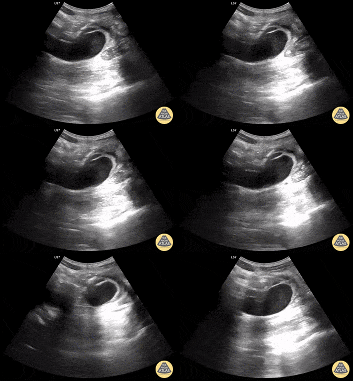
A patient in their 80s with PMH of abdominal aortic aneurysm s/p grafting with type 2 endoleak presented to the ED for dizziness. Bedside ultrasound demonstrated an 8.55 cm aortic aneurism with large thrombus. Endoleak is the hypoechoic area seen above the hyperechoic endograft.
Mehtab Galeh, MD; Bayley Espinoza, MD

Aortic Aneurysm with endoleak
A patient in their 80s with PMH of abdominal aortic aneurysm s/p grafting with type 2 endoleak presented to the ED for dizziness. Bedside ultrasound demonstrated an 8.55 cm aortic aneurism with large thrombus. Endoleak is the hypoechoic area seen above the hyperechoic endograft.
Mehtab Galeh, MD; Bayley Espinoza, MD

Suprasternal view of the aorta
Suprasternal notch view of the aortic arch with brachiocephalic artery (BCA), left common carotid artery (LCA), and left subclavian artery (LSA) branching off. Right pulmonary artery is also visible adjacent to the ascending aorta.
Charles Jang, EM PGY-3

Suprasternal view of the aorta with labels
Suprasternal notch view of the aortic arch with brachiocephalic artery (BCA), left common carotid artery (LCA), and left subclavian artery (LSA) branching off. Right pulmonary artery is also visible adjacent to the ascending aorta.
Charles Jang, EM PGY-3

Saccular AAA with Dissection Flap
Saccular AAA with Dissection Flap.
Contributor: Daniel Ostapowicz, MD
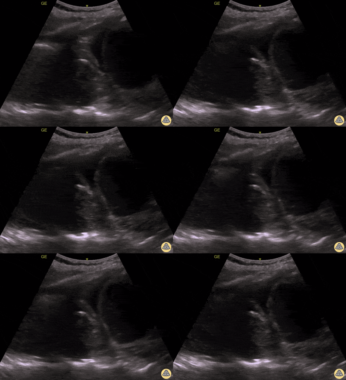
Thoracic Aortic Aneurysm Rupture Seen on PLAPS View
An elderly woman presented to the ED with dyspnea and left mid back pain for 1 week. Left PLAPS (Posterolateral Alveolar and/or Pleural Syndrome) view revealed a round-shaped pulsatile mass surrounded by pleural effusion & atelectatic lung. CTA later confirmed a partially ruptured, thrombosed thoracic aorta aneurysm. This case illustrates the possibility of quickly diagnosing life-threatening conditions with POCUS.
Contributed by: Caio Sangirardi, Emergency Medicine Resident Quinta D'Or Hospital, Rio de Janeiro - Brazil, @SangirardiMD

Aortic Dissection Flap in Desceding Aorta
Flap seen in the descending aorta consistent with aortic dissection
Contributed by: Dimitri Livshits DO, Ultrasound Fellow; Jane Belyavskaya MD, Ultrasound Fellow; Chris Hanuscin MD, Ultrasound Division Director (Kings County/SUNY Downstate)

AAA in Undifferentiated Shock
70s-year-old male brought in for altered mental status and hypotension.
As part of the initial exam, POCUS was performed. This clip, obtained using the curvilinear probe in transverse orientation over the mid-epigastric region, revealed a AAA measuring 8 cm in AP diameter at the largest point with heterogeneous echogenic material consistent with thrombus. CT abdomen/pelvis with IV contrast confirmed ruptured AAA with thrombosis and only minor extravasation.
In this case, POCUS played a critical role in the rapid diagnosis of a patient with undifferentiated shock, particularly in a patient who was unable to provide any history.
Katherine Spencer, MD
Guy Youngblood, MD, FACEP, FAAEM

Bilateral Iliac Artery Aneurysms
This patient originally complained of right back pain as well as numbness along the thigh and knee. Starting from the abdominal artery, gliding inferiorly will eventually reveal the bifurcation of the abdominal aorta into the left and right common iliac arteries. In this scan, you can see both iliac arteries have enlarged diameters, indicating aneurysms in both.
Image courtesy of Robert Jones DO, FACEP @RJonesSonoEM
Director, Emergency Ultrasound; MetroHealth Medical Center; Professor, Case Western Reserve Medical School, Cleveland, OH
View his original post here

Faint Dissection Flap in Descending Aorta
Patient presented with severe chest pain and concerning ECG without STEMI. A faint dissection flap can be seen in the aorta on this transverse view in the subcostal region.
Image courtesy of Robert Jones DO, FACEP @RJonesSonoEM
Director, Emergency Ultrasound; MetroHealth Medical Center; Professor, Case Western Reserve Medical School, Cleveland, OH
View his original post here

Sagittal View of Abdominal Aortic Aneurysm
Sagittal view of large abdominal aortic aneurysm incidentally found after accidental fall.
Image courtesy of Robert Jones DO, FACEP @RJonesSonoEM
Director, Emergency Ultrasound; MetroHealth Medical Center; Professor, Case Western Reserve Medical School, Cleveland, OH
View his original post here

Abdominal Aortic Aneurysm Presented As Cough
Here is another example of an abdominal aortic aneurysm that measured approximately 10.6 cm. The patient originally presented to the clinic complaining of a cough and was eventually admitted for surgical operation.
Image courtesy of Robert Jones DO, FACEP @RJonesSonoEM
Director, Emergency Ultrasound; MetroHealth Medical Center; Professor, Case Western Reserve Medical School, Cleveland, OH
View his original post here

AAA with Intramural Thrombus
The patient presented to the ED with a known abdominal aortic aneurysm (AAA). A point-of-care ultrasound (POCUS) examination of the abdominal aorta was performed using a curvilinear probe (longitudinal view shown). An infrarenal AAA was noted adjacent and superior to the bifurcation and measured a maximum diameter of approximately 5cm. Heterogeneous echogenic material was seen within the lumen of the aneurysm in keeping with intramural thrombosis, along with a hyperechoic line representing a chronic dissection.
This demonstrates the utility of POCUS in the rapid diagnosis of AAA and evaluation of size and intraluminal features such as intramural thrombus and dissection.
Andrew Namespetra, MB BCh BAO MSc
PGY-1 Emergency Medicine Resident, Central Michigan University (CMU)
Chad Bambrick, MD
PGY-2 Emergency Medicine Resident, CMU
Ryan Davis
MS4, CMU College of Medicine

Large AAA Rupture
This is a clip demonstrating a large abdominal aortic aneurysm with significant intramural thrombus. Signs of rupture can be appreciated near the right lower portion of the screen as there is disruption of the aorta wall. Heterogeneity within the intramural thrombus can also be an indicator of rupture.
Michael Macias, MD

Ruptured AAA
Sagittal view of an abdominal aortic aneurysm with a hemorrhage anterior to the aorta. Be sure to distinguish a AAA from a dissection by assessing the abnormal wall motion seen at the site of rupture.
Image courtesy of Robert Jones DO, FACEP @RJonesSonoEM
Director, Emergency Ultrasound; MetroHealth Medical Center; Professor, Case Western Reserve Medical School, Cleveland, OH
View his original post here

Aortic Dissection
A chest pain received an echo and showed evidence of a dissection in the PLAX, PSAX, and AP4C views within the descending aorta.
Image courtesy of Robert Jones DO, FACEP @RJonesSonoEM
Director, Emergency Ultrasound; MetroHealth Medical Center; Professor, Case Western Reserve Medical School, Cleveland, OH
View his original post here

Aortic Dissection
Patient presented with symptoms of stroke. Echo revealed pericardial effusion and aortic US showed a dissection flap emphasizing the importance of POCUS prior to TPA.
Image courtesy of Robert Jones DO, FACEP @RJonesSonoEM
Director, Emergency Ultrasound; MetroHealth Medical Center; Professor, Case Western Reserve Medical School, Cleveland, OH
View his original post here

AAA
Emergency response was called for an 88-year-old who experienced sudden onset thoracic and back pain. PMH significant for AAA (previously measuring 5.8 cm diameter on surveillance imaging 1 year ago). Mid-abdominal US view was obtained (shown here) and revealed AAA now measuring 7.2 cm diameter.
Wolfgang Geisser
@fentanyl05

Aortic Dissection Flap
61 year-old male presented with one day history of generalized abdominal pain, nausea, and vomiting. He was hemodynamically stable (BP 130/59); EKG notable for the presence of U waves and lateral ST depressions; labs revealed a negative troponin, potassium 3.1, lactic acid 5.4. Abdominal POCUS seen here revealed an abdominal aortic dissection flap as his unifying diagnosis.
Richard Cunningham, MD. Emergency Medicine

Normal Aortic Arch - Suprasternal View
This clip represents a normal suprasternal view. The brachiocephalic artery (BCA), left carotid artery (LCA) and left subclavian artery (LSA) emerge from the aortic arch (AA). The left subclavian artery (LSA) marks the division between the proximal thoracic aorta and the distal descending aorta (DA).
Dr. Felipe Urriola P., Emergency Unit, Puerto Aysen Hospital, Chilean Patagonia.

Normal Celiac Trunk & SMA - Longitudinal
At the center of the screen, the proximal aorta can be identified by the emergence of the coeliac trunk and the superior mesenteric artery. This clip is taken at the subxiphoid level, longitudinal to the body’s axis, and with the probe marker oriented towards the head.
Dr. Felipe Urriola P., Emergency Unit, Puerto Aysen Hospital, Chilean Patagonia.

Normal Aorta & Iliac Arteries - Transverse
At the center of the screen, the distal aorta lies anterior to a vertebral body. Sliding the probe caudally reveals the emergence of the iliac arteries. This clip is taken at the umbilicus level, transverse to the body’s axis, and with the probe marker oriented to the patient’s right.
Dr. Felipe Urriola P., Emergency Unit, Puerto Aysen Hospital, Chilean Patagonia.

Normal IVC and Aorta - Transverse view
The two round, anechoic structures laying on top of a hyperechoic vertebra are the IVC to the left of the screen (patient’s right), and the thicker, contractile aorta to the right (patient’s left). Deep inspiration or a sniff test demonstrates a collapsible IVC in normal subjects. Consider, however, that visualization of both these vessels may certainly be obscured by very obese patients or those with subcutaneous emphysema, significant bowel gas, ascites, or a large ventral hernia.
This clip is taken over the subxiphoid region, transverse to the body’s axis, and with the probe marker oriented to the patient’s right.
Dr. Felipe Urriola P., Emergency Unit, Puerto Aysen Hospital, Chilean Patagonia.

Normal IVC to AA lateral drag - Longitudinal View
While maintaining its orientation, the probe is dragged to the left of the patient, revealing the abdominal aorta (AA) and the emergence of the coeliac trunk. Notice the contractile, thicker aortic walls and how both the common hepatic artery and splenic artery form the “seagull sign”. Also notice that a normal IVC frequently demonstrates transmitted pulsations from the RA; hence, pulsation is not the proper method for differentiating it from the aorta.
This clip is taken over the subxiphoid region, longitudinally to the body’s axis, and with the probe marker oriented to the patient’s head.
Dr. Felipe Urriola P., Emergency Unit, Puerto Aysen Hospital, Chilean Patagonia.

Long Axis View of Large Abdominal Aortic Aneurysm
Long axis view of large abdominal aortic aneurysm containing intramural thrombus without evidence of the iliac arteries involvement.
Image courtesy of Giovanni Battista Fonsi
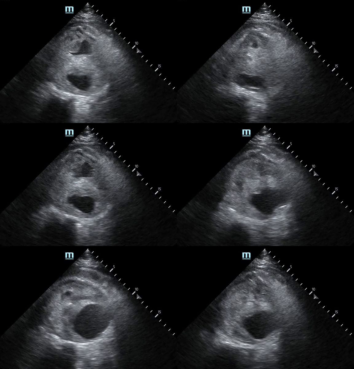
AAA Rupture: Not Your Typical Flank Pain
A 61-year-old male was brought in via EMS after a syncopal episode. He was diaphoretic and hypotensive, complaining of severe right flank pain.
While IV access was being obtained a bedside ultrasound was performed demonstrating a large abdominal aortic aneurysm with significant heterogeneous intraluminal clot. There is also appreciable focal hypoechoic disruption of the wall of the aneurysm consistent with rupture.
The patient was resuscitated in the ED and taken emergently to the OR to surgical repair.
Michael Macias, MD
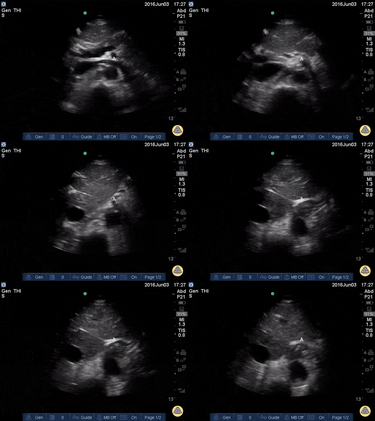
Proximal Aorta Anatomy
In the beginning of this clip we see several major structures in one view. From superficial to deep: liver, pancreas, splenic vein draining into the portal confluence, superior mesenteric artery in transverse (surrounded by a hyperechoic layer of fat creating the “mantle clock sign”), IVC with the left renal vein branching off, and the abdominal aorta. A vertebral body is visible at the bottom of the screen. As the probe is moved caudally, most of these vessels move out of plane and we are left following the course of the IVC and aorta inferiorly.
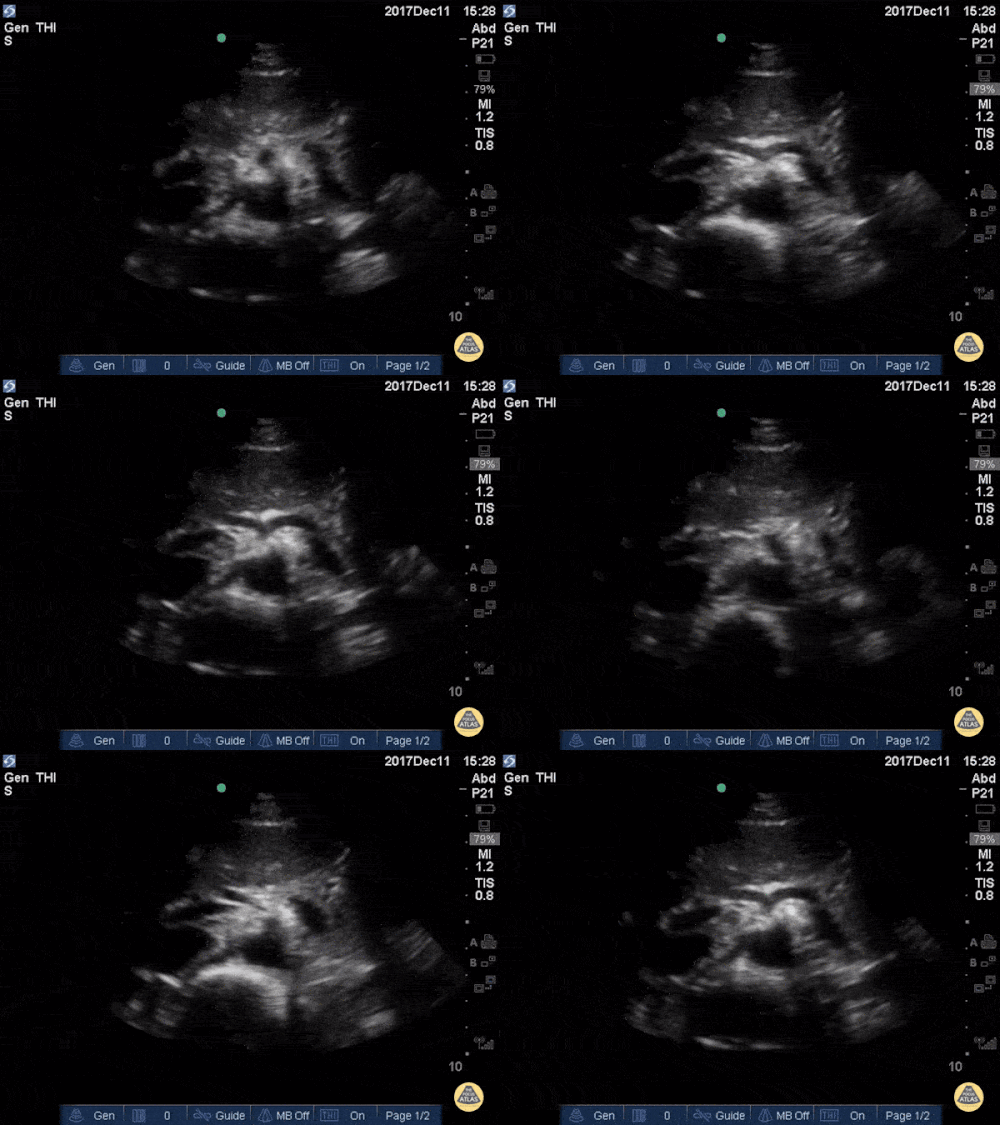
Seagull Sign
The vertebrae, seen here as the deepest structure as a hyperechoic arch with posterior shadowing (horseshoe sign), is a key anatomic reference when scanning the aorta. The aorta is located immediately anterior to the vertebrae and to the right side on the screen (patient’s left). Contrast this with the inferior vena cava, which is seen to the left of the aorta, often in an oval or teardrop shape. In the center of the screen we see the celiac trunk branching off the proximal abdominal aorta. The Y-shaped “seagull sign” is created by the celiac trunk as it branches into the hepatic artery (left) and splenic artery (right). The portal venous confluence is visible between the IVC and the hepatic artery.
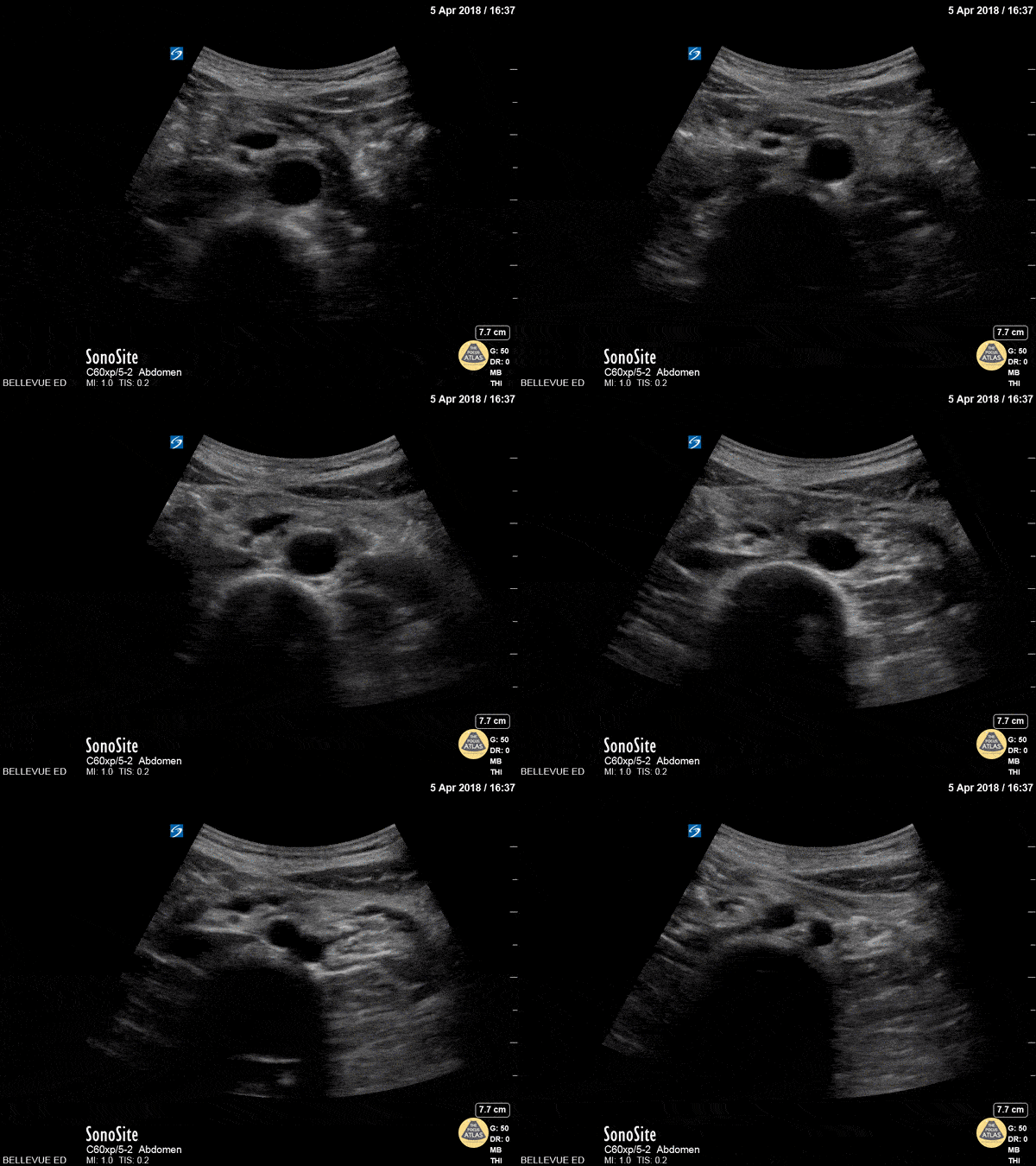
Distal Aorta
In the center of the screen we see the distal aorta in transverse view. As the probe is moved caudally, the aorta bifurcates into the two common iliac arteries. Deep to the aorta we see the round hyperechoic edge of the vertebral body and to the left of the aorta we see the more compressible inferior vena cava.

Smokin' AAA
A 70-year-old male presented as a trauma alert and was found to have this 10 cm abdominal aortic aneurysm (AAA) with associated echocardiographic smoke! Echogenic smoke is produced by the intersection of RBC’s and plasma proteins at low shear rate conditions.
Brian Toston, Internist. Adventura, FL
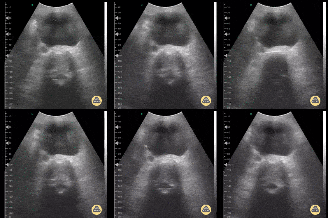
Abdominal Aortic Aneurysm
This image was taken from a pocket wireless device in the mesogastric region. We can see the short-axis aorta with its increased diameter and neighboring anatomical references such as the dorsal spine and inferior vena cava laterally.
Image courtesy of Dr. Renato Tambelli

Dissection Flap in Abdominal Aorta
An elderly male with hypertension and DM presents with C/O chest pain. Bedside ultrasound performed demonstrating a dissection flap in the lumen of the abdominal aorta. A subsequent parasternal long axis show extension into descending thoracic aorta as well.
Image courtesy of Robert Jones DO, FACEP @RJonesSonoEM
Director, Emergency Ultrasound; MetroHealth Medical Center; Professor, Case Western Reserve Medical School, Cleveland, OH
View his original post here.
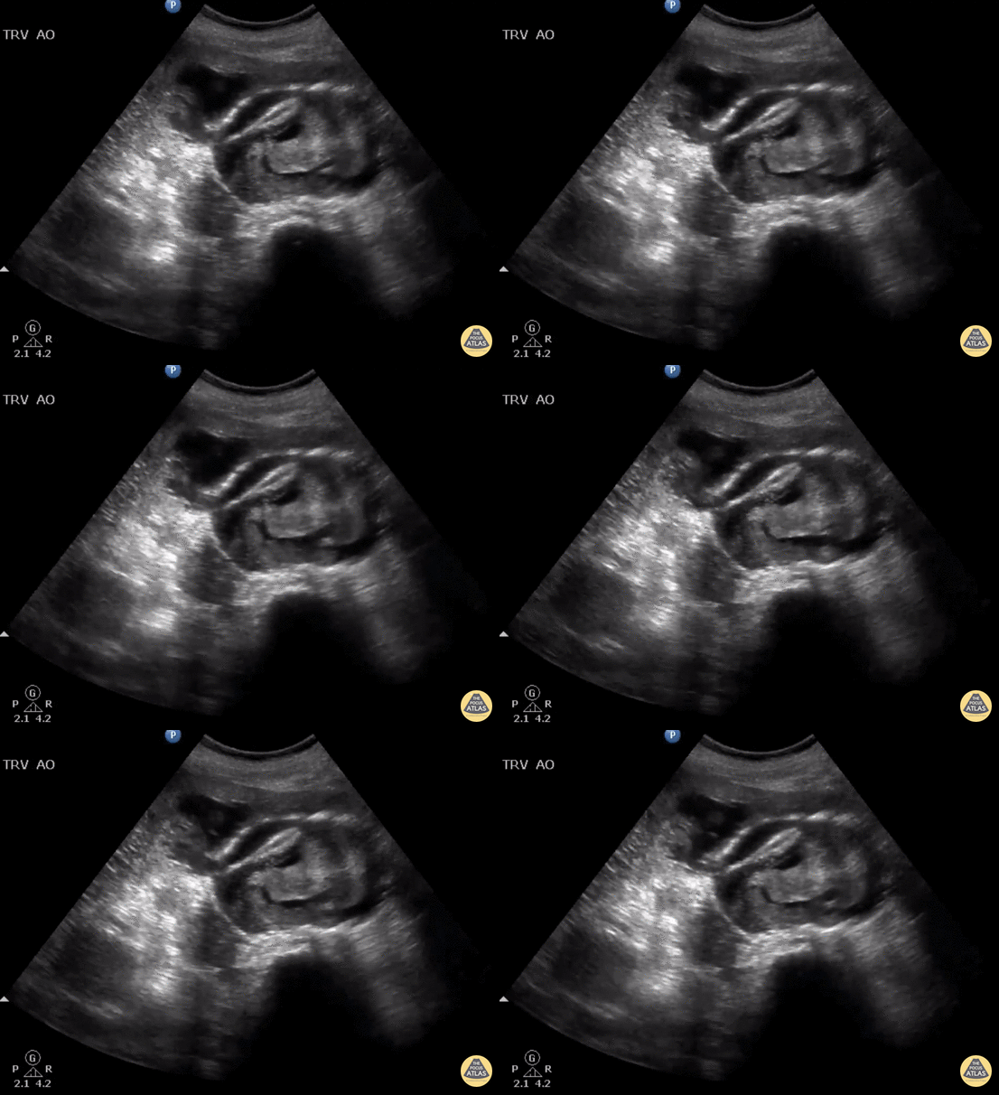
Ruptured AAA with Active Hemorrhage
Elderly female presents to ED with back pain and hypotension. Bedside US shows a ruptured AAA with active hemorrhage in the 9 o’clock position.
Image courtesy of Robert Jones DO, FACEP @RJonesSonoEM
Director, Emergency Ultrasound; MetroHealth Medical Center; Professor, Case Western Reserve Medical School, Cleveland, OH
View his original post here

Subacute Aortic Trauma
Patient with a subacute history of chest trauma (occurred approximately 30 days prior) presented reporting intermittent chest pain. He was noted in the ED to have persistent pain and to be sweating. Bedside evaluation included lung ultrasound that identified the cystic structure seen here with a hypoechoic interior with visible swirling. Chest CT shown confirmed this to be sequelae of aortic trauma.
Renato Melo, Emergency Physician Brazil. PocusJedi affiliated.
@Renato_Melo_

Incidental AAA
Routine abdominal evaluation using a Sonosite X-Porte curvilinear ultrasound probe revealed this 6.0cm abdominal aortic aneurysm (without rupture or dissection) in a patient with risk factors for the same. This enabled our middle-aged diabetic male with 80 pack/yr smoking history to receive timely evaluation by vascular surgery and subsequent appropriate serial monitoring and follow-up.
Siang-Chean Kua MS-4
Central Michigan University College of Medicine
Additional contributors: Ryan Shelby, MD, Thomas Ferreri, MD, Therese Mead, DO, RDMS, FACEP
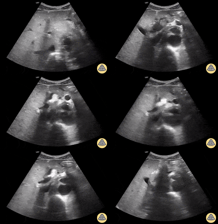
Aortic Endograft Leak - Transverse
Aortic Endograft Leak - Dr. Lindsay Howe Dr. Tim Scheel Dr. Paul Pelletier - Denver Health
Aortic Endograft Leak Long Axis
Aortic Endograft Leak - Dr. Lindsay Howe Dr. Tim Scheel Dr. Paul Pelletier
Extensive Type B Aortic Dissection
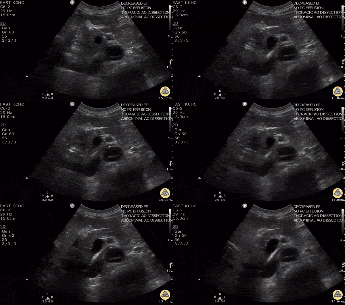
A 60 year old man was transferred to a trauma facility after presumed mechanical ground-level fall. He was only able to answer yes/no questions, vital signs were normal and stable upon arrival. He denied abdominal or back pain.
Upon arrival to receiving facility, POC ultrasound revealed intimal flap within the abdominal aorta extending from the subxiphoid region to the common iliac arteries. Bedside echo revealed no aortic root dilatation, pericardial effusion, or evidence of tamponade. CT scan confirmed thoracic and abdominal aortic dissection. Cardiothoracic surgery was notified immediately.
POCUS can play a critical part in allowing for rapid diagnosis and can expedite patient-care, particularly in patients with altered mental status who cannot provide a more robust history.
Quinn Fujii, DO
Desert Regional Medical Center, Emergency Medicine

Abdominal Aortic Dissection Flap
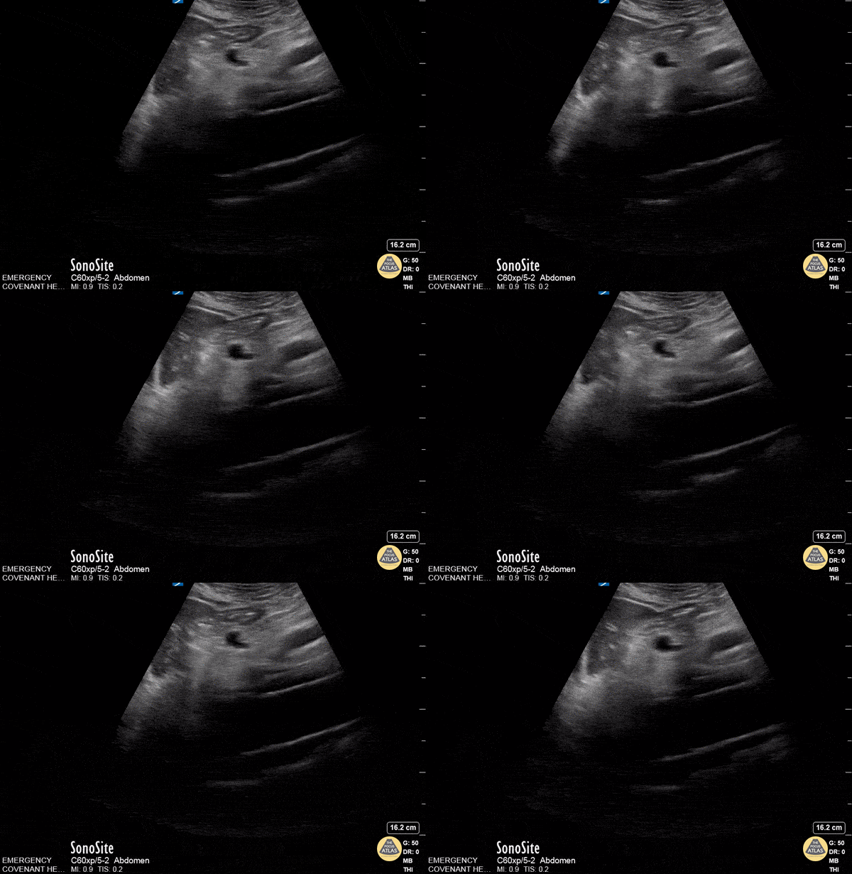
Patient with abrupt onset chest pain radiating to back. Normal ECG and Troponin. POCUS revealed dissection flap within abdominal aorta.
Nishant Cherian
Emergency Medicine Registrar
Aortic Dissection
50 y/o M w/ hx of HTN p/w sudden onset upper back pain. POCUS found dissection flap in the descending aorta in both parasternal long view and abdominal aorta. The diagnosis of aortic dissection was quickly confirmed by CT. Given the importance of timely diagnosis with aortic dissection, POCUS allowed rapid and non-invasive diagnosis of a potentially tricky diagnosis, and facilitated expedited treatment and transfer to a cardiothoracic surgery center.
Dr. Robert Allen - Kings County Emergency Medicine
Thoracic Aortic Dissection
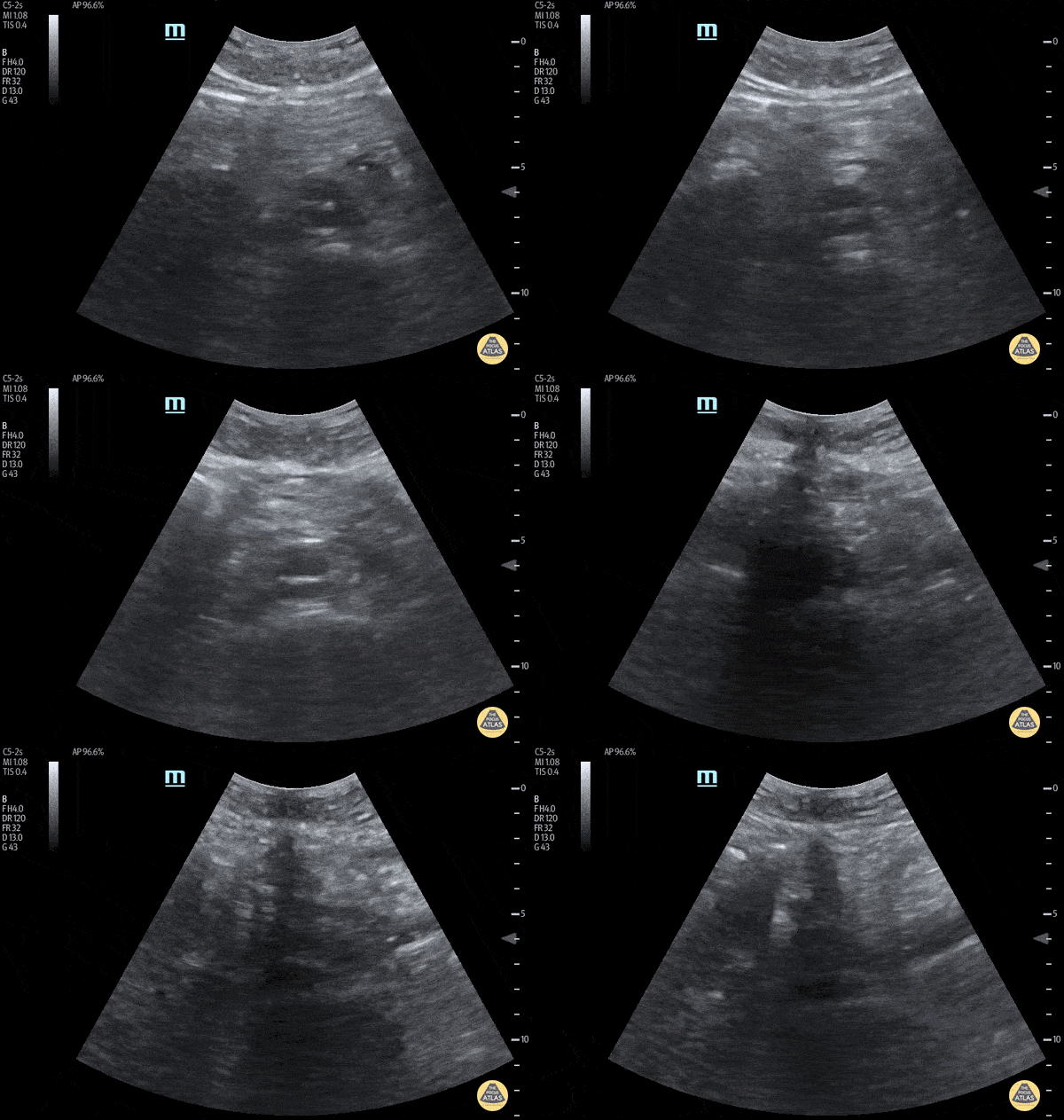
Approximately 70 year old male with history of AAA repair presented after an episode of syncope. POCUS performed in the ED revealed a dissected intimal flap and false lumen. CT confirmed the finding of thoracoabdominal aortic dissection.
David Hansen, DO
Matthew Wolf, MD
Therese Mead, DO, RDMS, FACEP
Central Michigan University
Aortic Graft Endoleak
Active extravasation is seen through endograft into false lumen. Known chronic endoleak, but CT 30 minutes prior to this study showed no active extravasation or impending rupture. Keep an open mind when reassessing your patients, and try not to anchor too much on prior results or others' opinions!
Dr. Jaffa
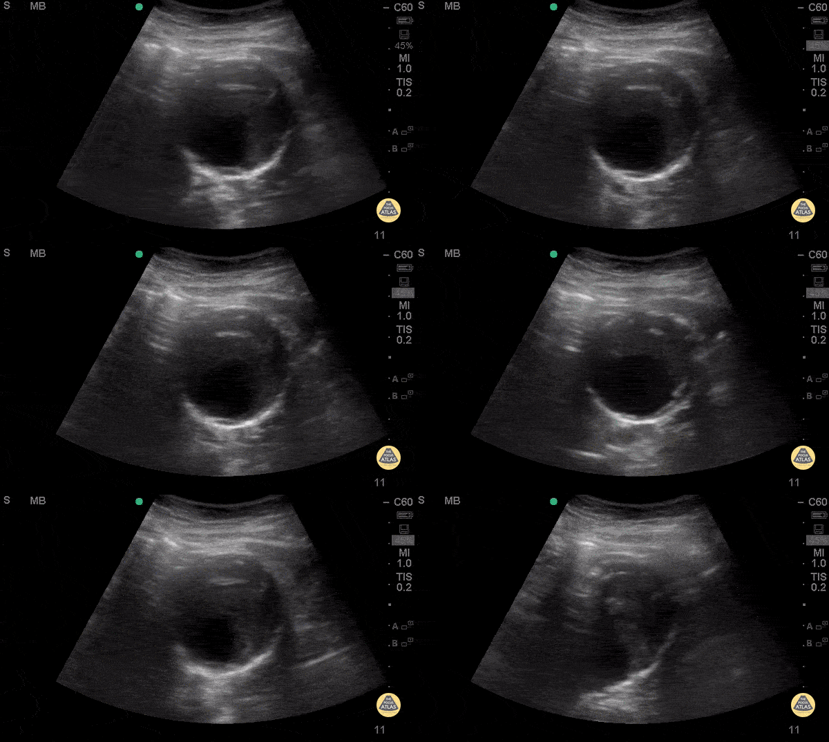
Mural Thrombus in AAA
74 y/o F hx stage 4 lung cancer, AAA, presented with chest pain and SOB x 1 day. POCUS shows the aorta is larger than the normal diameter of 3 cm, representing an abdominal aortic aneurysm, and measuring approximately 5.6 cm at its largest point. On cross section you are able to see a large mural thrombus with decreased diameter of blood flow through the center.
Juliana Jaramillo MD - Kings County Emergency Medicine
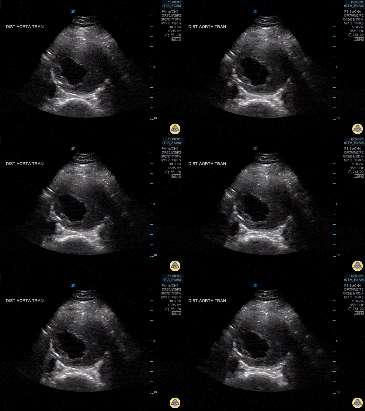
Abdominal Aortic Aneurysm with Thrombus
Approximately 6 cm abdominal aortic aneurysm with intramural thrombus.
Frances Russell, MD, RDMS
Assistant Professor of Emergency Medicine Division Chief, Ultrasound Fellowship Director, Ultrasound

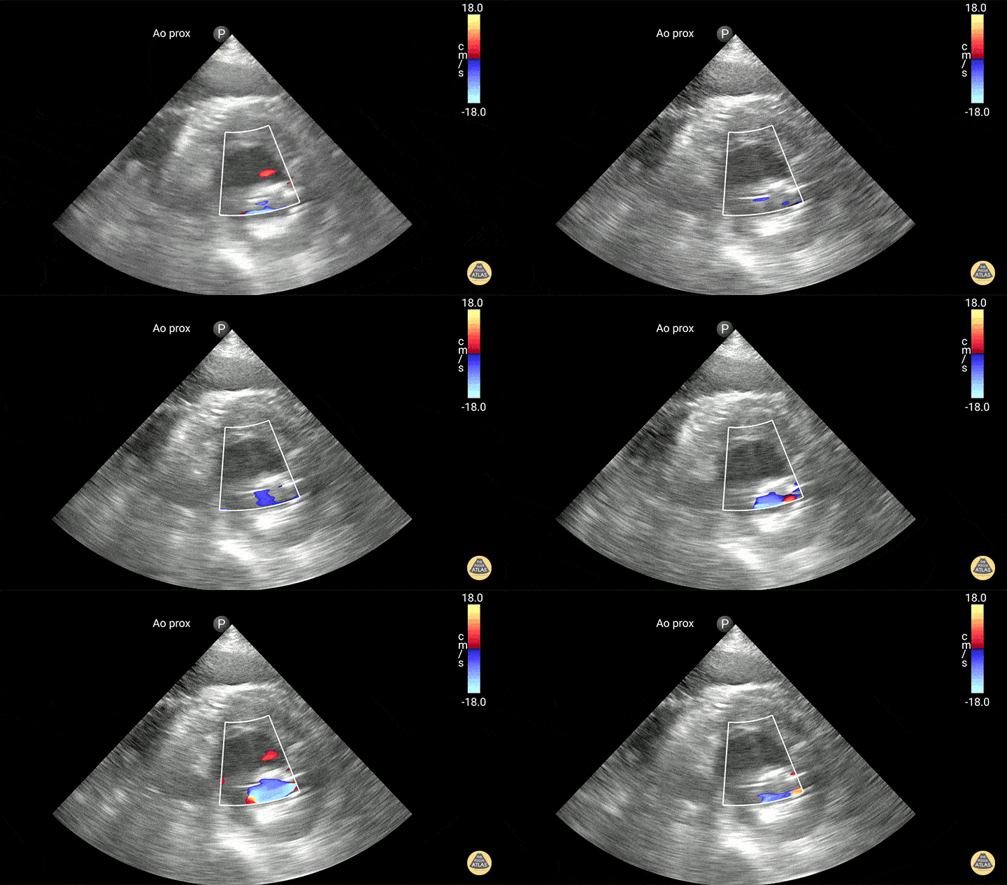
Mural Thrombus in AAA - Doppler
74 y/o F hx stage 4 lung cancer, AAA, presented with chest pain and SOB x 1 day. POCUS shows the aorta is larger than the normal diameter of 3 cm, representing an abdominal aortic aneurysm, and measuring approximately 5.6 cm at its largest point. On cross section you are able to see a large mural thrombus with decreased diameter of blood flow through the center.
Juliana Jaramillo MD - Kings County Emergency Medicine
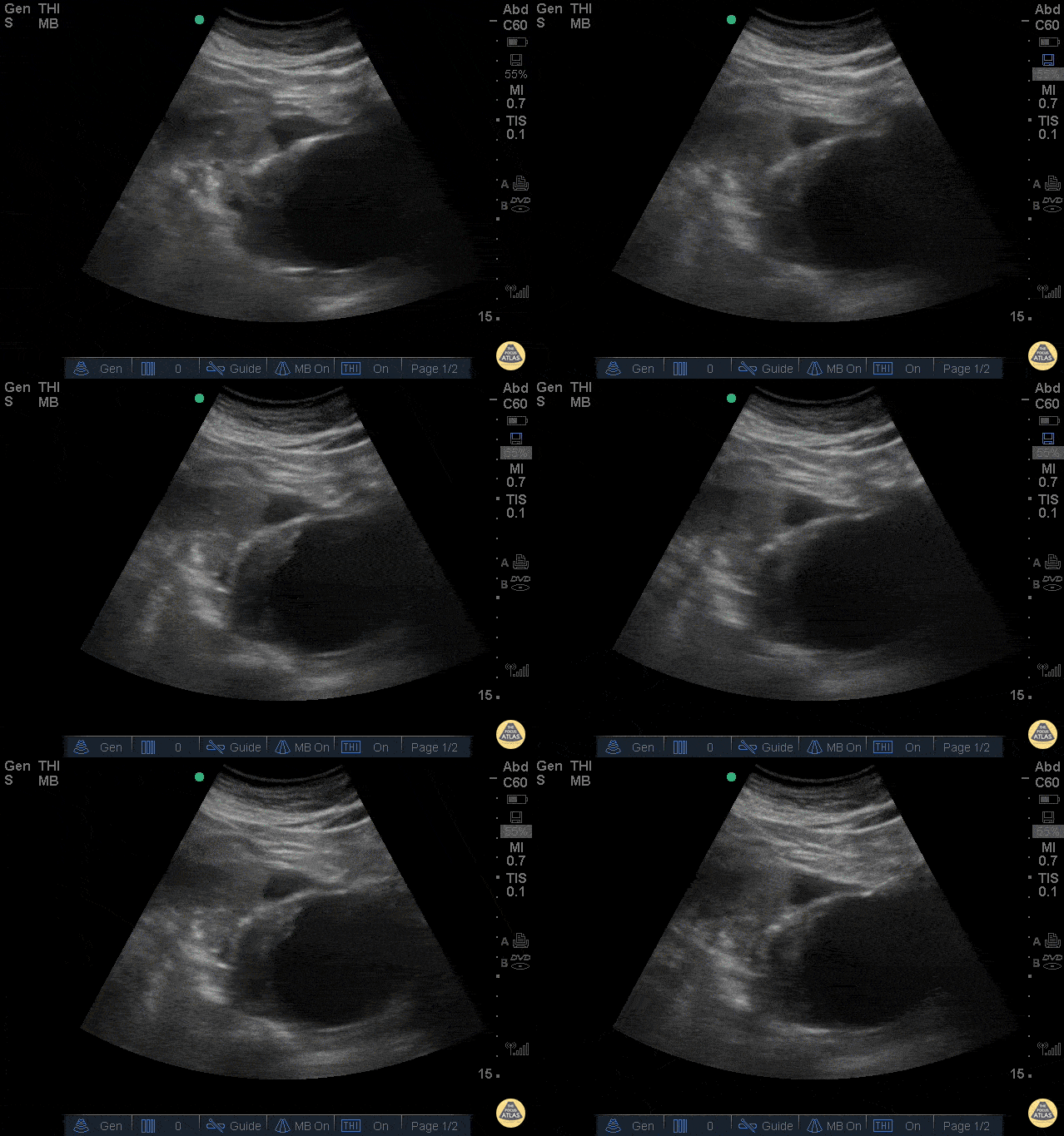
Ruptured AAA
The clip above is from a patient who presented with abdominal pain and syncope. His vitals were notable for tachycardia and hypotension. The image demonstrates a very large abdominal aortic aneurysm with anterior free fluid suggestive of rupture. The patient was taken emergently to the operating room for endograft repair and did well.
Jason Tanguay, DO
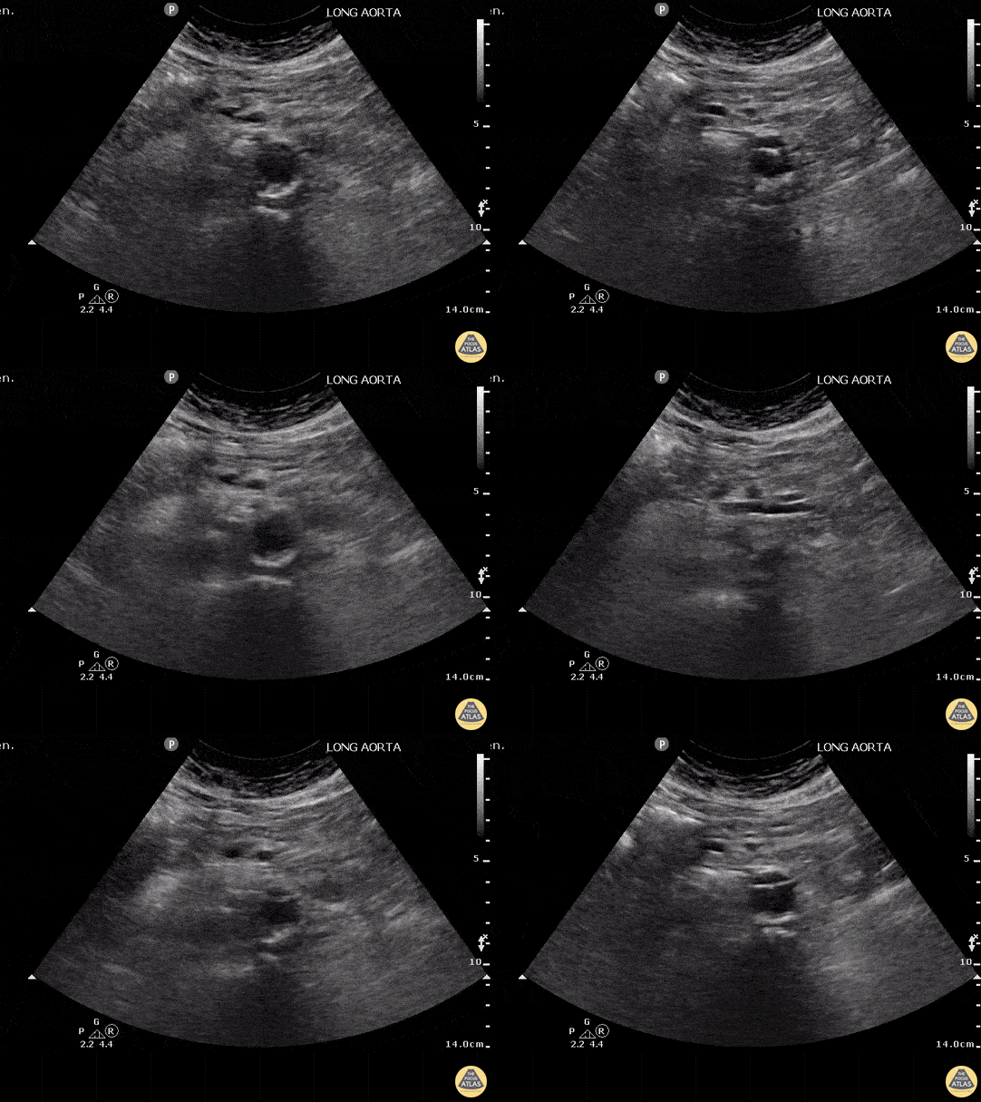
Abdominal Aortic Dissection (Transverse)
Aortic dissection carries an incredibly high mortality that increases 1%/hour. POCUS can be used as a rule-in test to quickly identify this life threatening diagnosis. If a dissection is not seen on POCUS, CT angiography should still be performed because the sensitivity of POCUS is not as high as for other indications.
The spine can be used as a landmark - the echogenic stripe with shadowing in the midline. The aorta is the large vessel anterior and slightly to the right of the spine. In this image an intimal flap can be seen in the anterior third of the aorta consistent with an aortic dissection. The IVC cannot be clearly visualized in this image but would normally be left, less pulsatile, with a less echogenic vessel wall. Non-visualization of the IVC is most often due to bowel gas or compression of the abdomen with the probe.
Justin Bowra MBBS, FACEM, CCPU Emergency Physician, RNSH et al.
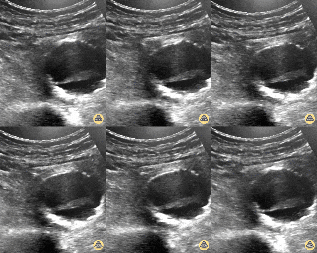
Pulsating Aortic Aneurysm
You may think this a dissection but it's actually an aortic aneurysm filled with thrombus, "with holes" making it a very "happy flappy." CT Confirmed.
Dr. Vincent Rietveld - Amsterdam, The Netherlands

Abdominal Aortic Aneurysm
AAA is defined as a localized balloon-like dilatation of the abdominal aorta greater than 3cm. Risk factors include male sex, increased age, and tobacco use. AAAs should be closely monitored for changes in size. Due to the risk of rupture, elective surgery is recommended when the dilatation is greater than 5-5.5cm, or it is growing in size by greater than 1cm/year. The classic triad of a ruptured AAA include pulsatile abdominal mass, hypotension and pain.
This AAA has an intramural thrombus. Some studies have claimed that POCUS has a ~100% sensitive for increased diameter. 3cm from outer wall to outer wall defines an aneurysm. Slow, graded compression is key to move the bowel out of the way in any abdominal study.
Sukh Singh, MD
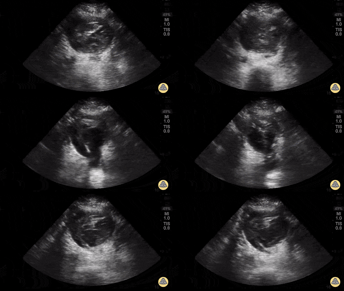
Aorta Posterior Wall Rupture
63yoM witnessed collapsed. Arrived VSA with PEA rhythm. A fast look abdominal aorta POCUS showed a large abdominal aortic aneurysm with internal thrombus and rupture through the posterior wall. Note the AAA is measured from echogenic outer wall to outer wall, not the hypoechoic internal lumen that is seen pulsating. The patient was resuscitated aggressively with blood and initially survived operative repair. Unfortunately he died in the ICU several days later from multiorgan failure.
Dr. Joey Newbigging

Fetal Aorta
Pulsating fetal aorta
Marco Garrone
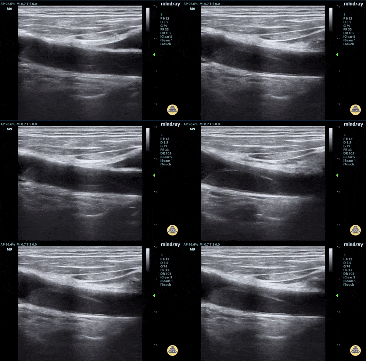
Aortic dissection flap tamponade
Elderly fellow who had a headache while bike riding, with some leg weakness. No chest or back pain. Stable for hours then came to hospital, suddenly hypotension and drowsy in ER
POCUS RUSH Exam performed lead to rapid diagnosis of Aortic Dissection with tamponade.
A dissection flap can clearly be visualized.
Claire Heslop - Pediatric Emergency Medicine - University of Toronto Hospital for Sick Children
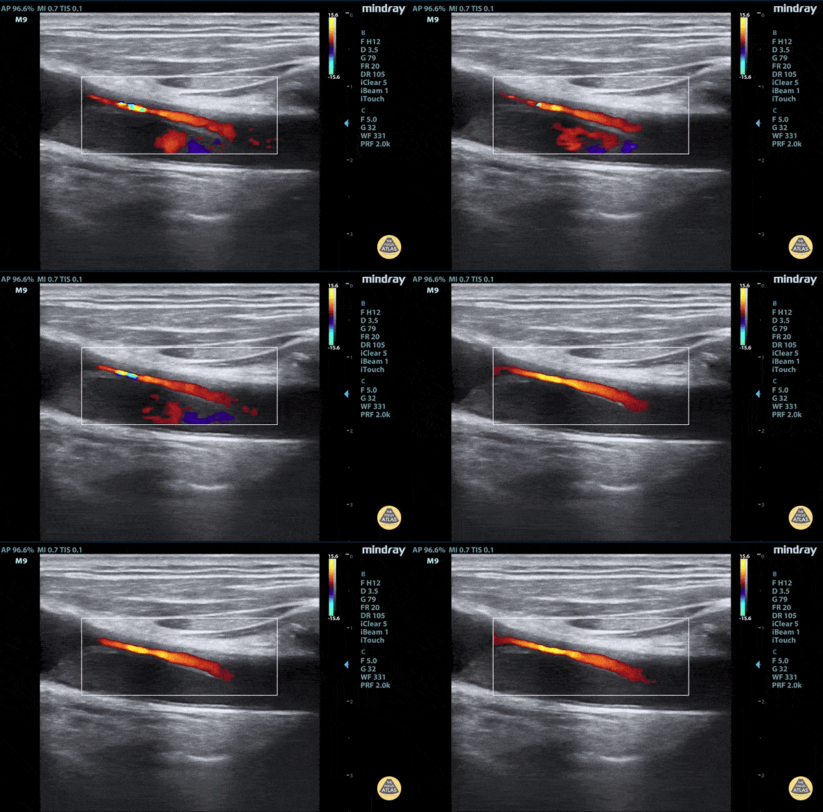
Aortic dissection flap tamponade color flow
Elderly fellow who had a headache while bike riding, with some leg weakness. No chest or back pain. Stable for hours then came to hospital, suddenly hypotension and drowsy in ER
POCUS RUSH Exam performed lead to rapid diagnosis of Aortic Dissection with tamponade.
A dissection flap can clearly be visualized with color flow around it.
Claire Heslop - Pediatric Emergency Medicine - University of Toronto Hospital for Sick Children
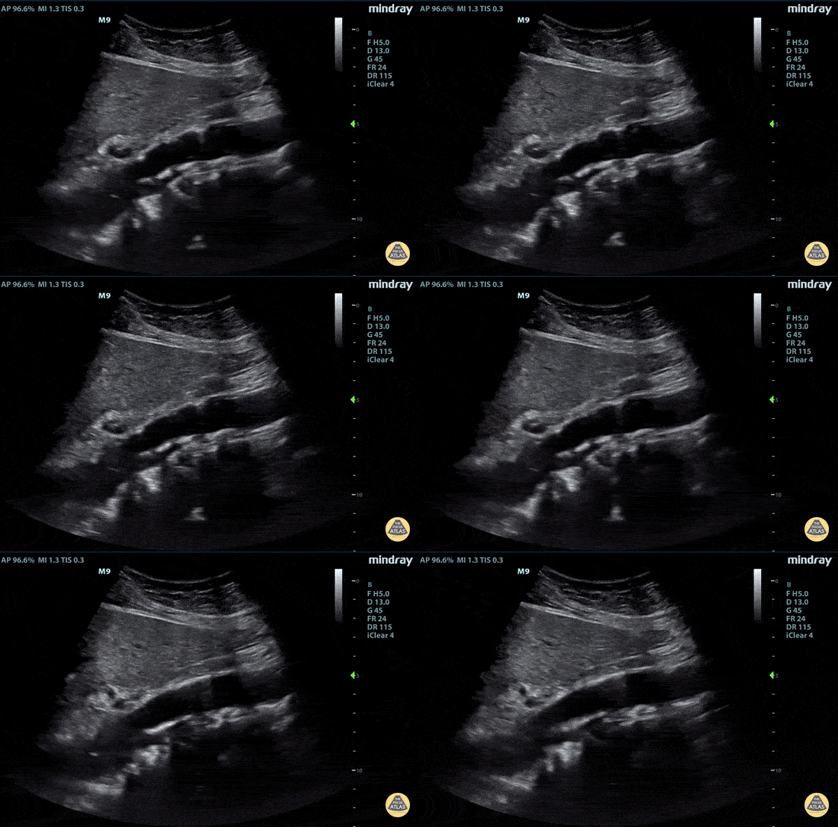
Aortic Dissection Flap
Elderly fellow who had a headache while bike riding, with some leg weakness. No chest or back pain. Stable for hours then came to hospital, suddenly hypotension and drowsy in ER
POCUS RUSH Exam performed lead to rapid diagnosis of Aortic Dissection with tamponade.
A pulsating dissection flap can clearly be visualized.
Claire Heslop - Pediatric Emergency Medicine - University of Toronto Hospital for Sick Children
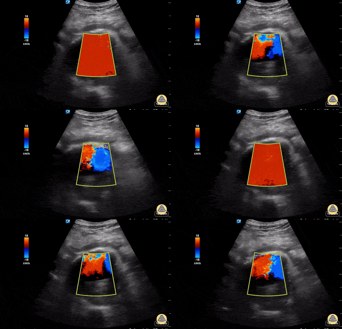
POCUS Utility in Suspected AAA Rupture
A 76 year old male presented to the ED for abdominal pain and syncope. He was tachycardic but normotensive. US performed with Sonosite 5-2MHz C60 probe with patient in supine position and probe held in transverse orientation revealed a AAA measuring ~9cm in AP diameter at its largest point. The AAA had an anechoic center with echogenic material in the posterior lumen, representing cholesterol deposit. Free fluid was seen anterior to the AAA suggestive of rupture which was confirmed by STAT CTA Aortagram. Patient was admitted to the OR for surgery. In this scenario, POCUS was a safe, non-invasive study that played a critical role in diagnosis of a ruptured AAA and accelerated surgical intervention.
Susan Dhamala, MS4, Drexel University College of Medicine
Brenton Elliot, MD, Crozer Chester Medical Center
Matthew Cully, DO, Nemours/Alfred I. duPont Hospital for Children
Kevin Conor Welch, DO, Crozer Chester Medical Center
Max Cooper, MD, RDMS, Director of Emergency Ultrasonography at Crozer Chester Medical Center
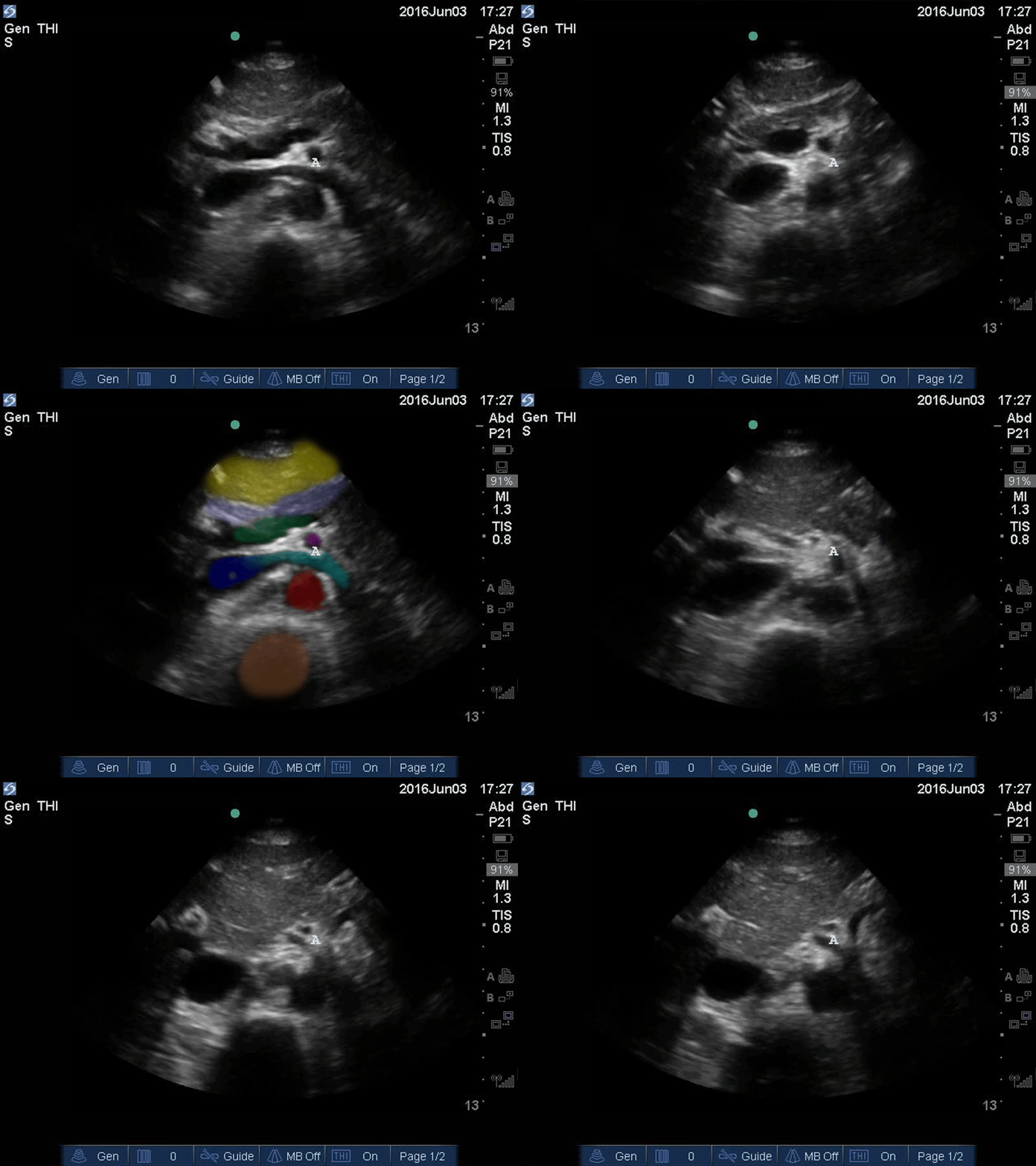
Upper Aorta - Colorized - The POCUS Atlas
Orange: Yellow: Liver, Light blue: Pancreas, Aqua: splenic vein/portal confluence, Blue: IVC with left renal vein, Purple: SMA, Red: Aorta, Orange: Spine
Images: Dr. Lindsay Davis, Dr. Hannah Kopinski. Image Editing: Michael Amador and Dr. Matthew Riscinti
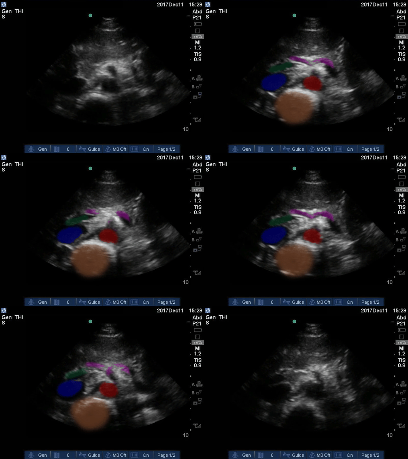
Seagull Sign - Colorized
Good Seagull Sign
Orange: Spine, Red: Aorta, Blue: IVC, Green: Portal venous confluence, Pink: “Seagull sign” aka celiac trunk
Images: Dr. Lindsay Davis, Dr. Hannah Kopinski. Image Editing: Michael Amador and Dr. Matthew Riscinti
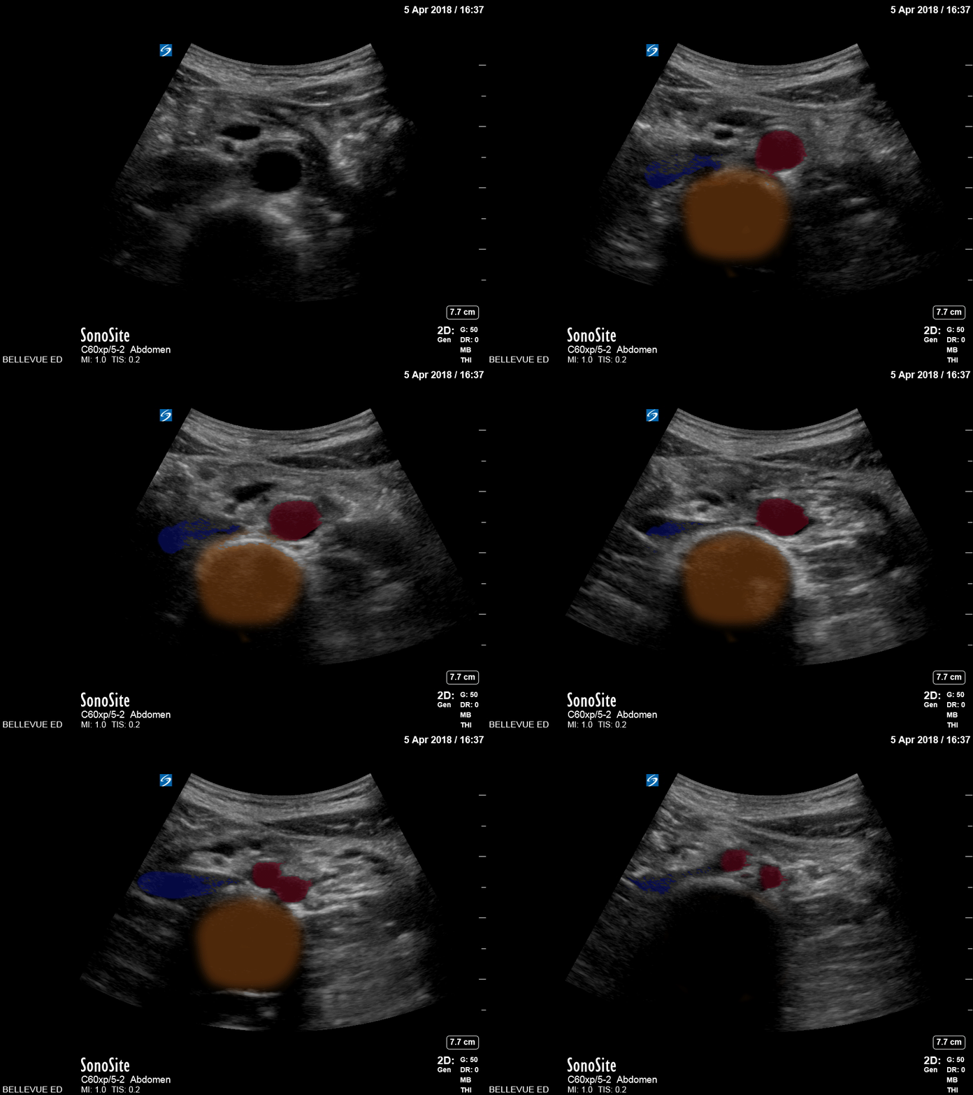
Distal Aorta - Colorized
Distal Aorta
Orange: Spine, Red: Aorta and Iliacs, Blue: IVC, Green: Portal venous confluence, Pink: “Seagull sign” aka celiac trunk
Images: Dr. Lindsay Davis, Dr. Hannah Kopinski. Image Editing: Michael Amador and Dr. Matthew Riscinti
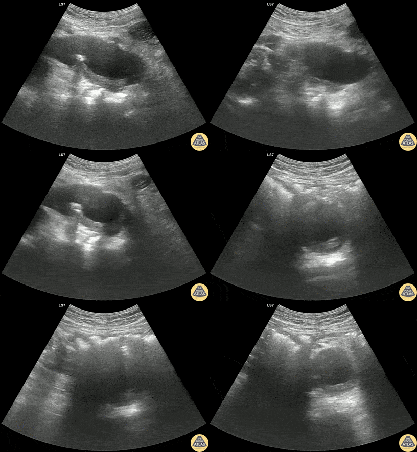
Thrombosed Type B Aortic Dissection
60s M PMH HTN, CKD stage 3 presented with bilateral flank pain x2 days. CT of the abdomen/pelvis with IV contrast showed a 3.8 cm x 4.6 cm infrarenal AAA with a 3.1 cm L internal iliac aneurysm. POCUS of the aorta was obtained re-demonstrating the AAA. Dedicated CTA showed that there actually demonstrating aneurysmal dilation of the descending thoracic aorta with a thrombosed type B dissection. The patient was managed with aggressive blood pressure and heart rate control with esmolol and was admitted to the ICU for medical management.
Dr. Henrik Galust, PGY4, Denver Health Residency in Emergency Medicine
Dr. Nimish Bhatt, Fellow, Denver Health Ultrasound Fellowship


