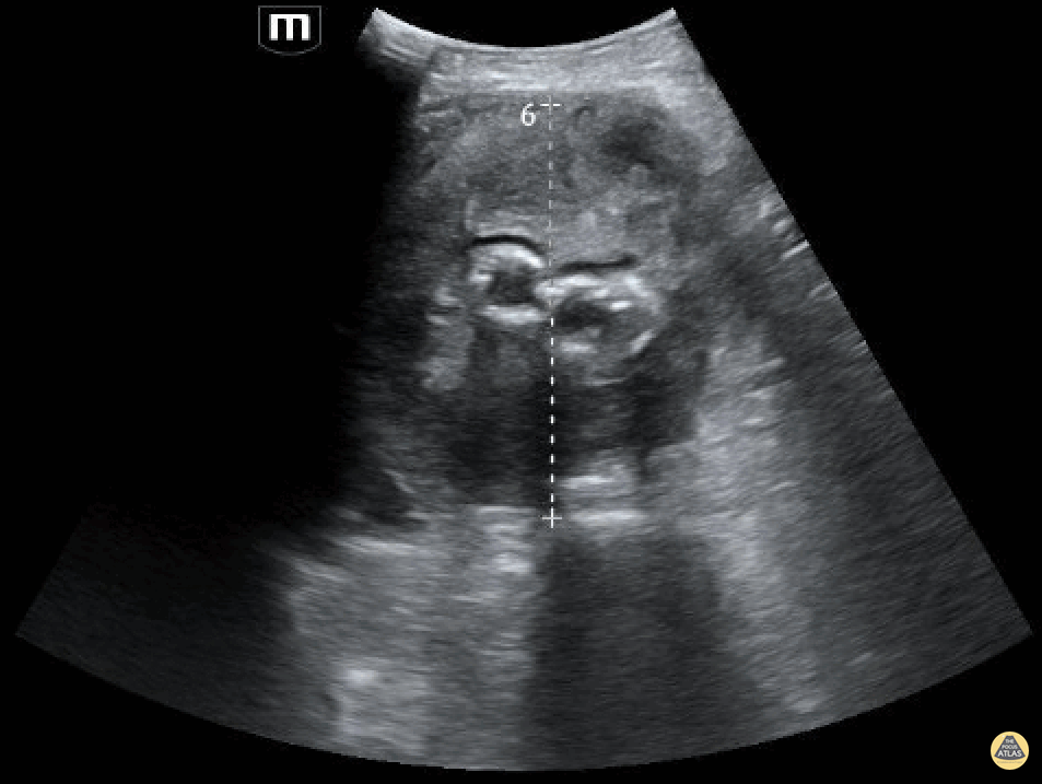
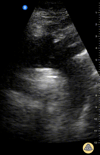
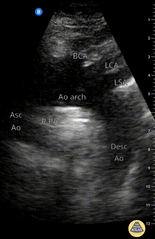
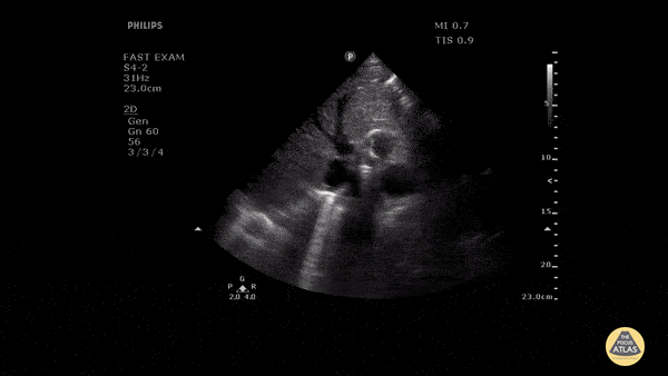
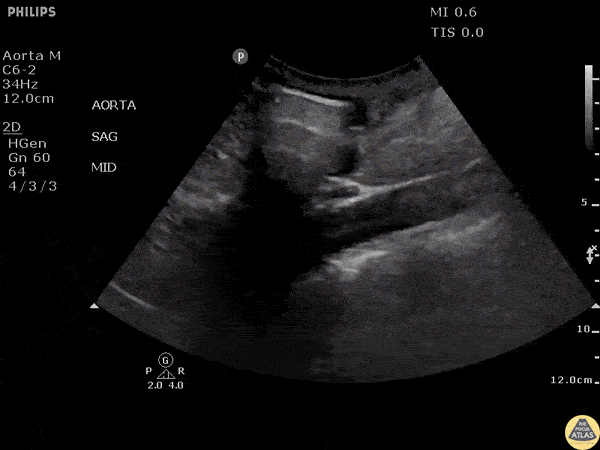
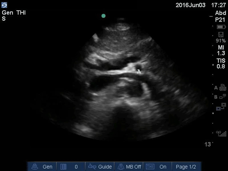
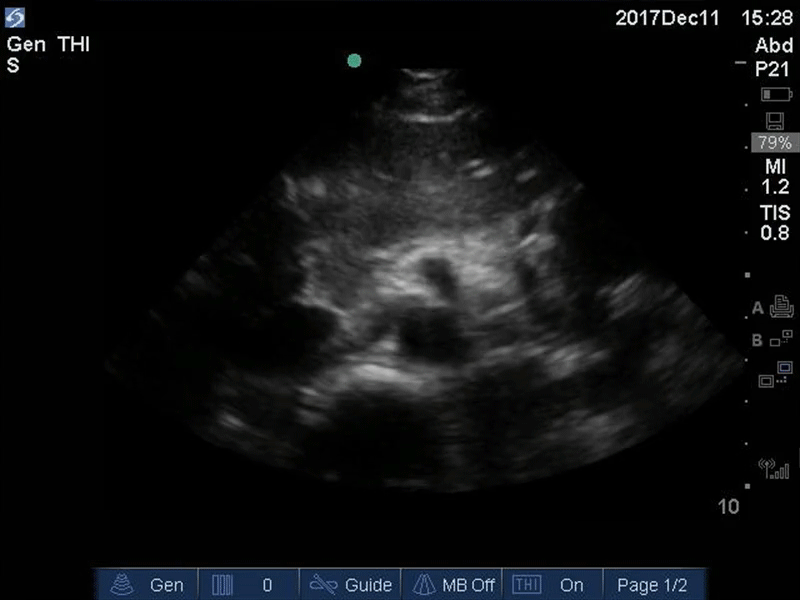
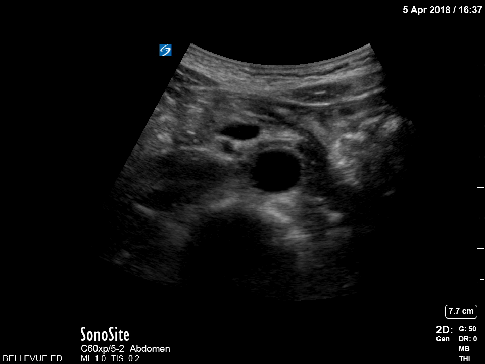
Aorta

Suprasternal view of the aorta
Suprasternal notch view of the aortic arch with brachiocephalic artery (BCA), left common carotid artery (LCA), and left subclavian artery (LSA) branching off. Right pulmonary artery is also visible adjacent to the ascending aorta.
Charles Jang, EM PGY-3

Suprasternal view of the aorta with labels
Suprasternal notch view of the aortic arch with brachiocephalic artery (BCA), left common carotid artery (LCA), and left subclavian artery (LSA) branching off. Right pulmonary artery is also visible adjacent to the ascending aorta.
Charles Jang, EM PGY-3

Faint Dissection Flap in Descending Aorta
Patient presented with severe chest pain and concerning ECG without STEMI. A faint dissection flap can be seen in the aorta on this transverse view in the subcostal region.
Image courtesy of Robert Jones DO, FACEP @RJonesSonoEM
Director, Emergency Ultrasound; MetroHealth Medical Center; Professor, Case Western Reserve Medical School, Cleveland, OH
View his original post here

Sagittal View of Abdominal Aortic Aneurysm
Sagittal view of large abdominal aortic aneurysm incidentally found after accidental fall.
Image courtesy of Robert Jones DO, FACEP @RJonesSonoEM
Director, Emergency Ultrasound; MetroHealth Medical Center; Professor, Case Western Reserve Medical School, Cleveland, OH
View his original post here

Abdominal Aortic Aneurysm Presented As Cough
Here is another example of an abdominal aortic aneurysm that measured approximately 10.6 cm. The patient originally presented to the clinic complaining of a cough and was eventually admitted for surgical operation.
Image courtesy of Robert Jones DO, FACEP @RJonesSonoEM
Director, Emergency Ultrasound; MetroHealth Medical Center; Professor, Case Western Reserve Medical School, Cleveland, OH
View his original post here

Ruptured AAA
Sagittal view of an abdominal aortic aneurysm with a hemorrhage anterior to the aorta. Be sure to distinguish a AAA from a dissection by assessing the abnormal wall motion seen at the site of rupture.
Image courtesy of Robert Jones DO, FACEP @RJonesSonoEM
Director, Emergency Ultrasound; MetroHealth Medical Center; Professor, Case Western Reserve Medical School, Cleveland, OH
View his original post here

Aortic Dissection
A chest pain received an echo and showed evidence of a dissection in the PLAX, PSAX, and AP4C views within the descending aorta.
Image courtesy of Robert Jones DO, FACEP @RJonesSonoEM
Director, Emergency Ultrasound; MetroHealth Medical Center; Professor, Case Western Reserve Medical School, Cleveland, OH
View his original post here

Aortic Dissection
Patient presented with symptoms of stroke. Echo revealed pericardial effusion and aortic US showed a dissection flap emphasizing the importance of POCUS prior to TPA.
Image courtesy of Robert Jones DO, FACEP @RJonesSonoEM
Director, Emergency Ultrasound; MetroHealth Medical Center; Professor, Case Western Reserve Medical School, Cleveland, OH
View his original post here

Upper Aorta - Colorized - The POCUS Atlas
Orange: Yellow: Liver, Light blue: Pancreas, Aqua: splenic vein/portal confluence, Blue: IVC with left renal vein, Purple: SMA, Red: Aorta, Orange: Spine
Images: Dr. Lindsay Davis, Dr. Hannah Kopinski. Image Editing: Michael Amador and Dr. Matthew Riscinti

Seagull Sign - Colorized
Good Seagull Sign
Orange: Spine, Red: Aorta, Blue: IVC, Green: Portal venous confluence, Pink: “Seagull sign” aka celiac trunk
Images: Dr. Lindsay Davis, Dr. Hannah Kopinski. Image Editing: Michael Amador and Dr. Matthew Riscinti

Distal Aorta - Colorized
Distal Aorta
Orange: Spine, Red: Aorta and Iliacs, Blue: IVC, Green: Portal venous confluence, Pink: “Seagull sign” aka celiac trunk
Images: Dr. Lindsay Davis, Dr. Hannah Kopinski. Image Editing: Michael Amador and Dr. Matthew Riscinti


