
Vascular
DVT, veins, arteries, aneurysms, color flow doppler, doppler, all POCUS point of care ultrasound.

Femoral Artery Stent
Femoral artery stent can be appreciated in this short axis view as a bright, hyperechoic structure within the femoral artery borders.
Contributed by: Brittany Garza, DO; Saleem Nasseh, MD; Sadie Ellerson, MS4

Occlusive DVT of the Left Popliteal Vein
This clip demonstrates a non-compressible left popliteal vein consistent with deep venous thrombosis.
Contributed by: Brittany Garza, DO; Saleem Nasseh, MD; Sadie Ellerson, MS4

Popliteal DVT
This is of a 52 year old male who presented to the emergency department complaining of leg pain and erythema while at work. He was evaluated prior to his arrival to the emergency department and was noted to have an elevated d-dimer level. Point of care ultrasound was performed starting from the great saphenous vein and common femoral vein, descending along his femoral vein. Initial compression did not reveal any thrombus until arriving at his popliteal vein which revealed a non-compressible vein due to a large thrombus.
Dr. Christopher Paulo, DO, PGY-1
Riverside Regional Medical Center Emergency Medicine Program (Newport News, VA)

PICC Line in Subclavian Vein
55 y/o man with septic shock and multiorgan failure due to SBP and liver failure. Picc line visualized within the R subclavian vein. longitudinal view.
Alex Steinberg

Dual Radial Artery
Very interesting find while performing a wrist ultrasound where we see the presence of two radial arteries
Image courtesy of Robert Jones DO, FACEP @RJonesSonoEM
Director, Emergency Ultrasound; MetroHealth Medical Center; Professor, Case Western Reserve Medical School, Cleveland, OH
View his original post here

DVT
Elderly patient with a history of dyspnea and desaturation. POCUS of the common femoral vein in its transverse axes with color Doppler and longitudinal axes, showing partially mobile hyperechoic content in its interior associated with the absence of venous compressibility.
Paula Martins, @paula_smrtns
Medical Student at The Faculdade de Medicina de Marília

Great Saphenous Thrombosis
POCUS of a patient with unilateral leg swelling and redness revealed thrombosis of the great saphenous vein seen as an isoechoic structure within the lumen. No DVT was noted in this patient.
Image courtesy of Robert Jones DO, FACEP @RJonesSonoEM
Director, Emergency Ultrasound; MetroHealth Medical Center; Professor, Case Western Reserve Medical School, Cleveland, OH
View his original post here

IVC Thrombus
This patient presented with bilateral LE swelling and negative DVT scans. Doppler waveform showed results concerning for proximal obstruction. Transverse abdominal scan shows a thrombus within the inferior vena cava.
Image courtesy of Robert Jones DO, FACEP @RJonesSonoEM
Director, Emergency Ultrasound; MetroHealth Medical Center; Professor, Case Western Reserve Medical School, Cleveland, OH
View his original post here

Spontaneous Echo Contrast
This clip shows spontaneous echo contrast (SEC) in the IJV. This finding is indicative of blood stasis and may be a precursor to thrombus formation.
Image courtesy of Robert Jones DO, FACEP @RJonesSonoEM
Director, Emergency Ultrasound; MetroHealth Medical Center; Professor, Case Western Reserve Medical School, Cleveland, OH
View his original post here

Venous Valves
Seen here is healthy appearing venous system with effectively functioning valves. Important distinction between the arterial and venous systems.
Image courtesy of Robert Jones DO, FACEP @RJonesSonoEM
Director, Emergency Ultrasound; MetroHealth Medical Center; Professor, Case Western Reserve Medical School, Cleveland, OH
View his original post here

DVT
POCUS revealed an acute DVT becoming easily mobile when the patient coughs.
Image courtesy of Robert Jones DO, FACEP @RJonesSonoEM
Director, Emergency Ultrasound; MetroHealth Medical Center; Professor, Case Western Reserve Medical School, Cleveland, OH
View his original post here

Common Femoral DVT
A 65 yo male presented with 2 week hx of proximal left LE pain. Pt denied trauma or risk factors for VTE. Also did not have visible abnormalities on clinical exam. Seen here is POCUS of his common femoral vein; note the hyperechoic thrombus and absent blood flow (when compared to adjacent femoral artery). This vein was also non compressible. This case highlights that even with low pre-test probability, POCUS can help drive ones differential diagnosis!
Mandy Peach, MD @mandy_peach
Saint John Regional Hospital. NB, Canada

Floating Venous Thrombus
A 60-year-old woman seen in the emergency department with pain and edema in her right lower limb. Bedside vascular US with a linear probe in the femoral territory showed a large floating venous thrombus . This condition has great potential for pulmonary embolization.
Renato Tambelli
@JediPocus // @R_Tambelli

Superficial Thrombophlebitis
A patient presented reporting a tender, erythematous, and edematous portion of his left forearm; notably at the site of an IV that had been in place at the time of a recent surgery. POCUS as seen here revealed a hyperechoic area within a vessel concerning for thrombus, consistent with the diagnosis of superficial thrombophlebitis. Point of care ultrasound can be useful in differentiating thrombophlebitis from other differential diagnoses including muscle strain or sprain.
Rupinder Sekhon, MD
Central Michigan University, Emergency Medicine

Pseudoaneurysm
POCUS of a suspected abscess revealed a pseudoaneurysm of the femoral artery. Note the “Pepsi sign” on color doppler made by the turbulence of the blood entering the pseudoaneurym.
Image courtesy of Robert Jones DO, FACEP @RJonesSonoEM
Director, Emergency Ultrasound; MetroHealth Medical Center; Professor, Case Western Reserve Medical School, Cleveland, OH
View his original post here

Carotid Dissection
A middle aged patient presented with severe chest pain without associated neurologic deficits. CTA revealed a Type A aortic dissection with equivocal carotid extension. Subsequent POCUS seen here confirmed presence of a mobile echogenic intimal flap consistent with carotid artery dissection.
Michael Cover, MD
@michaelc0ver

AV Fistula
A male presented with thigh pain and swelling with a history of an untreated stab wound 2 months prior. A sagittal view over the femoral artery revealed bidirectional flow indicative of an AV fistula.
Image courtesy of Robert Jones DO, FACEP @RJonesSonoEM
Director, Emergency Ultrasound; MetroHealth Medical Center; Professor, Case Western Reserve Medical School, Cleveland, OH
View his original post here

Right Femoral DVT
69-year-old male patient with no relevant chronic medical history presents to the ER complaining of two-day right inguinal pain and swollen lower extremity. Directed interrogation revealed one-month subacute dyspnea upon physical effort.
Femoral POCUS showed this image. The contractile femoral artery lies superficially and to the left of the screen. The common femoral vein is not fully compressible in this study, and an echogenic thrombus can clearly be identified in its interior. Subsequently, angio CT confirmed a massive bilateral PE, although the patient remained stable and did not require invasive interventions.
Dr. Felipe Urriola P. Emergency Unit, Puerto Aysen Hospital. Chilean Patagonia.

Normal IVC to RA - Longitudinal view
The IVC drains into the RA. A normal IVC frequently demonstrates transmitted pulsations from the RA. This clip is taken over the subxiphoid region, longitudinally to the body’s axis, and with the probe marker oriented to the patient’s head and RA. The hepatic vein can also be seen draining into the IVC.
Dr. Felipe Urriola P., Emergency Unit, Puerto Aysen Hospital, Chilean Patagonia.

Subclavian Vein
This POCUS revealss a supraclavicular long axis view of the subclavian vein. It is an ideal view for US-guided central venous access.
Renato Tambelli, mergency Physician Hospital das Clínicas de Marília, Brazil.
@R_Tambelli // @JediPocus

Internal Jugular Thrombus
This POCUS was obtained using a linear probe and reveals a short axis view of an internal jugular vein containing a large thrombus. This highlights the importance of evaluating vascular structures with ultrasound prior to placing central lines.
Renato Tambelli, Emergency Physician Hospital das Clínicas de Marília, Brazil
@R_Tambelli // @JediPocus

Left femoral DVT
A 45-year-old female presented to the ED with chest pain, dyspnea, and evidence of shock. Our FOCUS cardiac exam was notable for acute pulmonary hypertension. Subsequently using our multi-organ approach we identified evidence of a DVT (linear probe on the proximal left leg reveals hyperechoic material within the non-compressible femoral vein). Diagnosis of pulmonary thromboembolism secondary to VTE causing obstructive shock was immediately confirmed at the bedside.
Images from: Emergency Department of Marilia Clinic Hospital, Sao Paulo, Brazil. POCUSJEDI Team.
@JediPocus
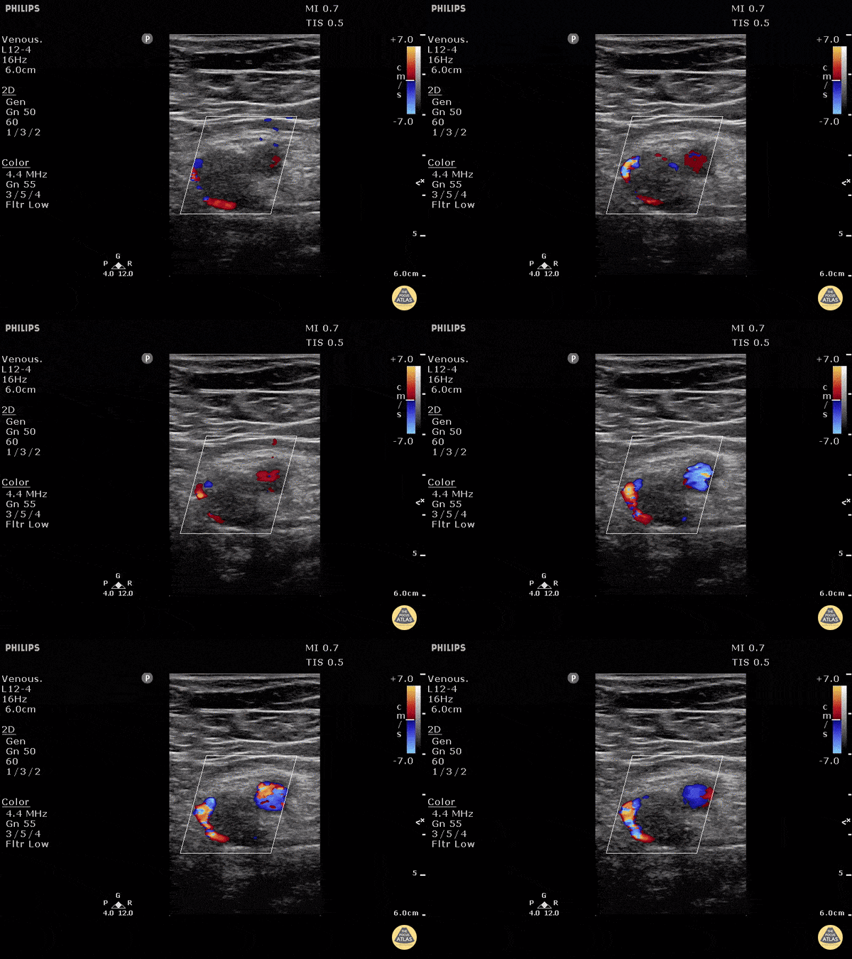
DVT of Left Common Femoral Vein
Patient with profound rapid onset lower extremity pain and swelling. POCUS can be used to rapidly assess for DVT with as high as 95% sensitivity and 96% specificity amongst ED providers.
In this image the artery can be seen as the pulsating thick walled vessel with strong pulsations under color doppler. The vein is the large vessel with echogenic material with flow only around the edge of the clot. Inability to compress the vessel would confirm the diagnosis of DVT (not pictured).
Dr. Justin Bowra et al.
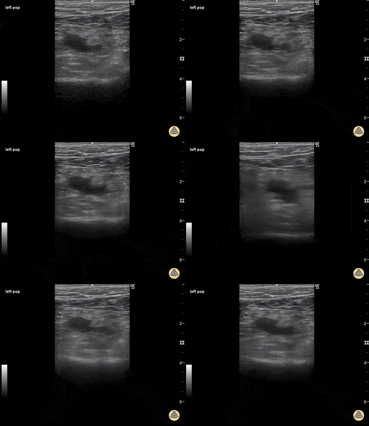
DVT - Popliteal
When pressure is applied to the ultrasound probe over blood vessels, veins usually collapse before arteries because of their much thinner walls. In this clip, enough pressure was applied to the ultrasound probe to partially collapse the popliteal artery, which is the pulsatile vessel visualized. Despite such high pressure, the popliteal vein remains fully patent, indicating a deep vein thrombosis within its walls.
Sukh Singh, MD

DVT Left Common Femoral Vein
Patient with pain and swelling of the entire left lower extremity. Three point POCUS carries a sensitivity of 95% and specificity of 96% for detecting DVT when performed by emergency physicians.
Mild external rotation of the hip can help open up the inguinal crease to find the femoral vein. The POCUS should be performed 1-2cm distal to the bifurcation of the common femoral for a complete study.
Dr. Justin Bowra et al.
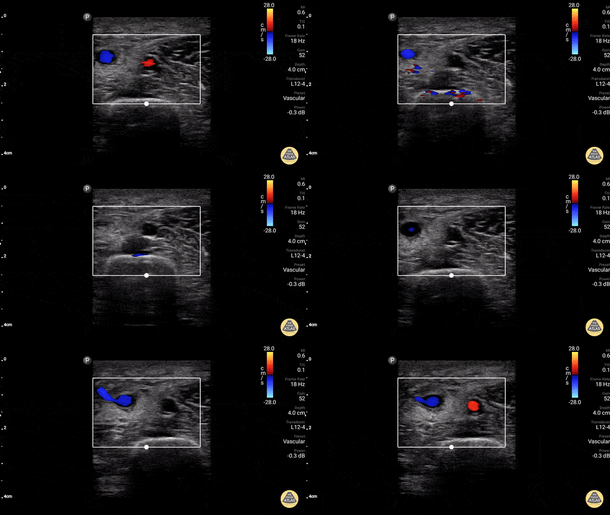
Axillary Vein (Normal Compressible)
Axillary vein thrombus ruled OUT in a patient with arm swelling. Doppler used to highlight vein and artery.
Dr. Gordon Johnson
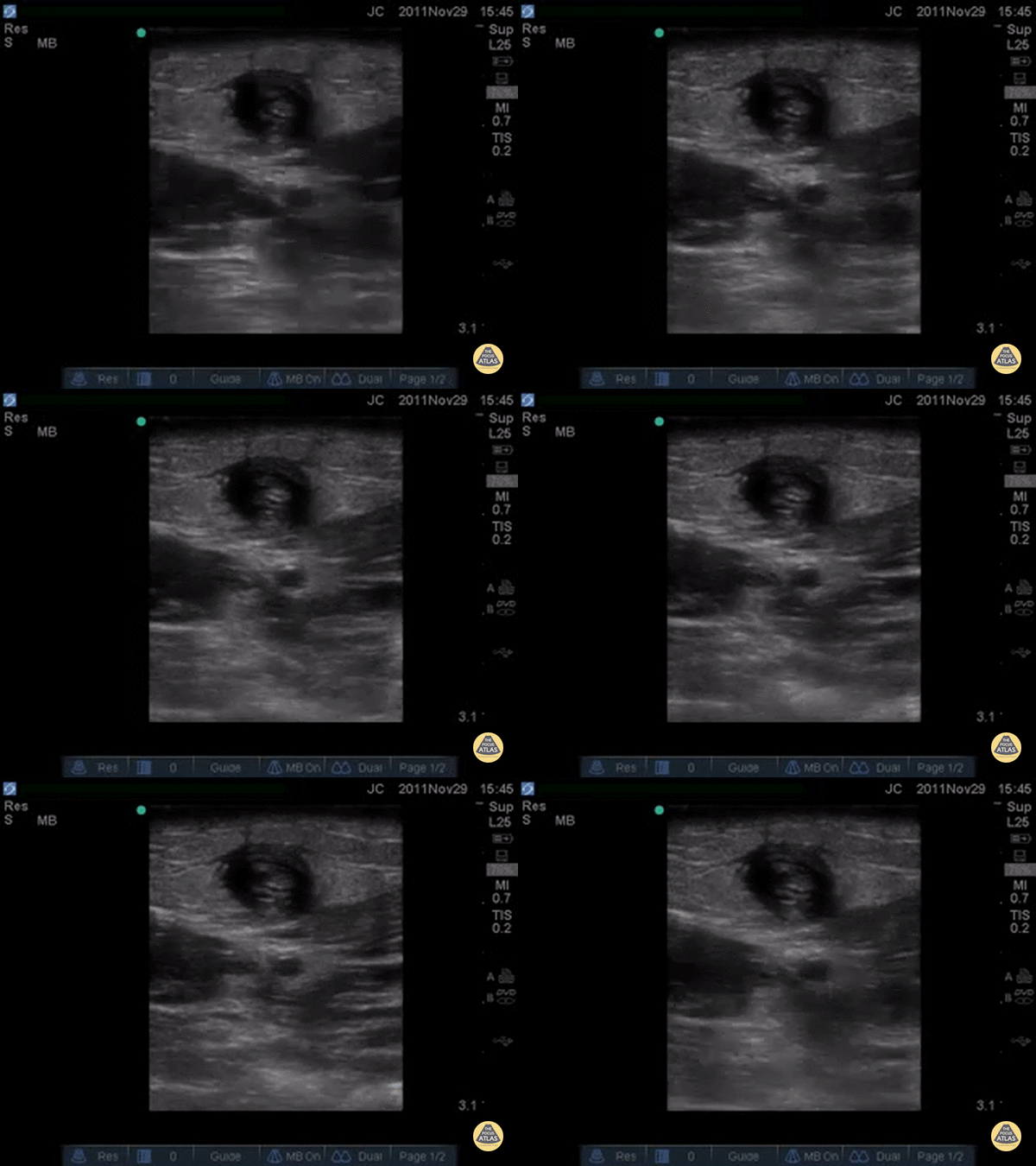
Cephalic Vein Thrombophlebitis
Patient with pain and swelling to area above the antecubital fossa where an IV had been the week before, a palpable cord was appreciated proximal to the former IV site.
POCUS demonstrated a clot, likely septic thrombophlebitis secondary to IV site infection. This is a similar techinque as what is done for DVT by finding the vessel, looking for a clot, and seeing if the vessel can be compressed.
Dr. Bowra et al. (Dr. Chiang)

Swirling Blood in the Internal Jugular Vein after Aortic Dissection
Patient presented with near syncope and unilateral neck pain. Ipsilateral neck ultrasound revealed swirling blood within the IJ; subsequent evaluations identified etiology to be a thoracic aortic dissection contributing to pericardial effusion and cardiac tamponade!
Peter Weimersheimer, MD. Director EM POCUS at ULA
@VTEMSONO

VTE in PE
A 27-year-old women presented to ED with acute onset of dyspnea and chest pain. She was hypoxic with an O2 saturation of 91% on room air and tachycardic. POCUS lung ultrasound revealed an A-line pattern bilaterally and was notably absent for pleural effusions. Venous scan of the proximal right femoral region revealed a non-compressible femoral vein with hypoechoic material within the lumen. High resolution chest CT confirmed diagnosis of PE.
Dr. Victor Bang. Emergency Physician at Hospital das Clínicas de Marília. Co-founder of Pocus Jedi.
@vmjbang
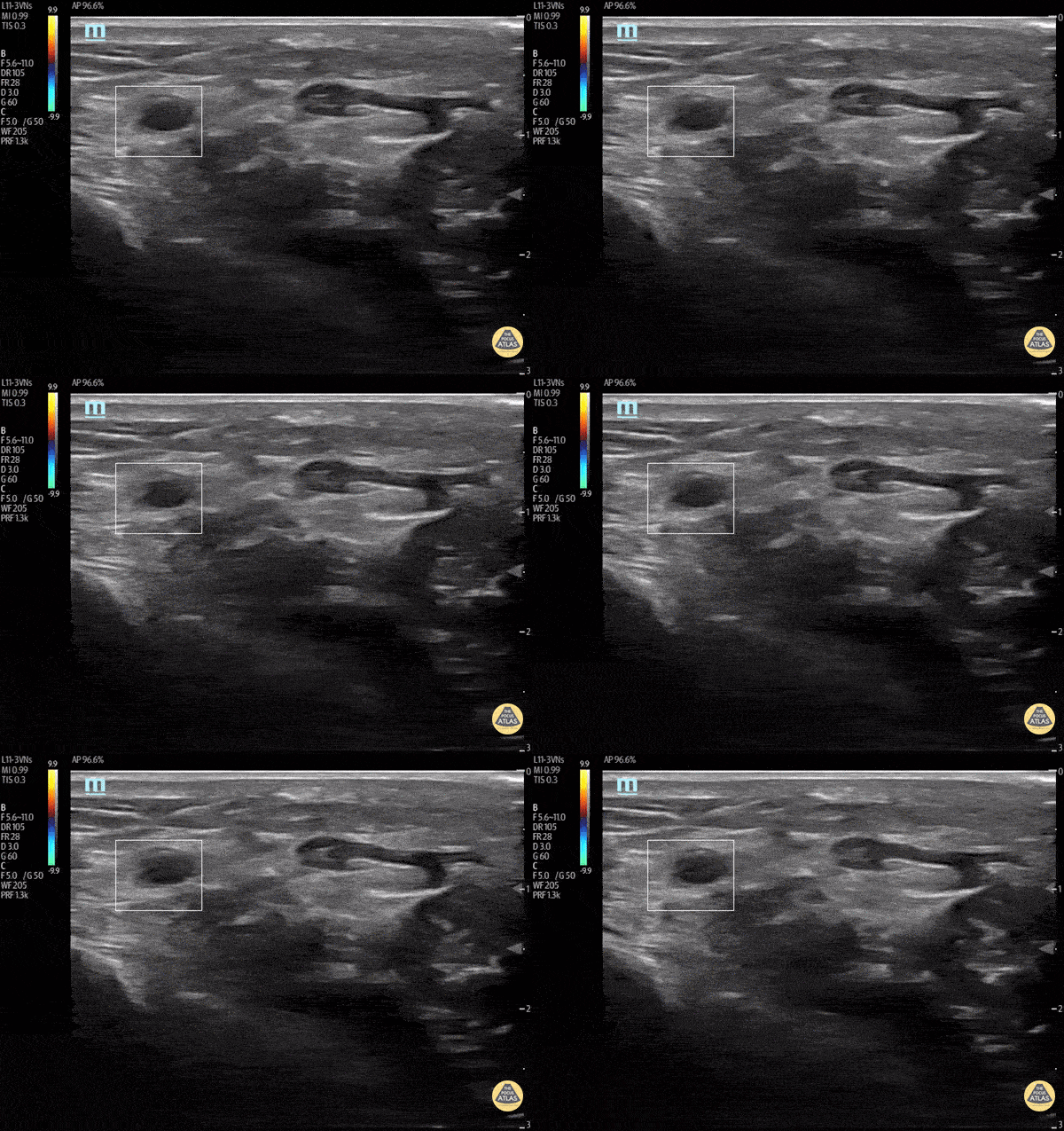
Post Arterial Catheterization Complication
A middle aged male with history of hypercoagulability and recent cardiac catheterization utilizing radial artery access, presented with right arm pain, numbness and tingling. POCUS revealed a complete occlusion of the distal right radial artery. The patient was started on anticoagulation therapy initially, with plan for potential thrombectomy with interventional radiology
Kathrin Parisi, MS-IV; Courtney Hollingsworth, MD; Therese Mead, DO, RDMS, FACEP; Matt French, DO - Central Michigan University
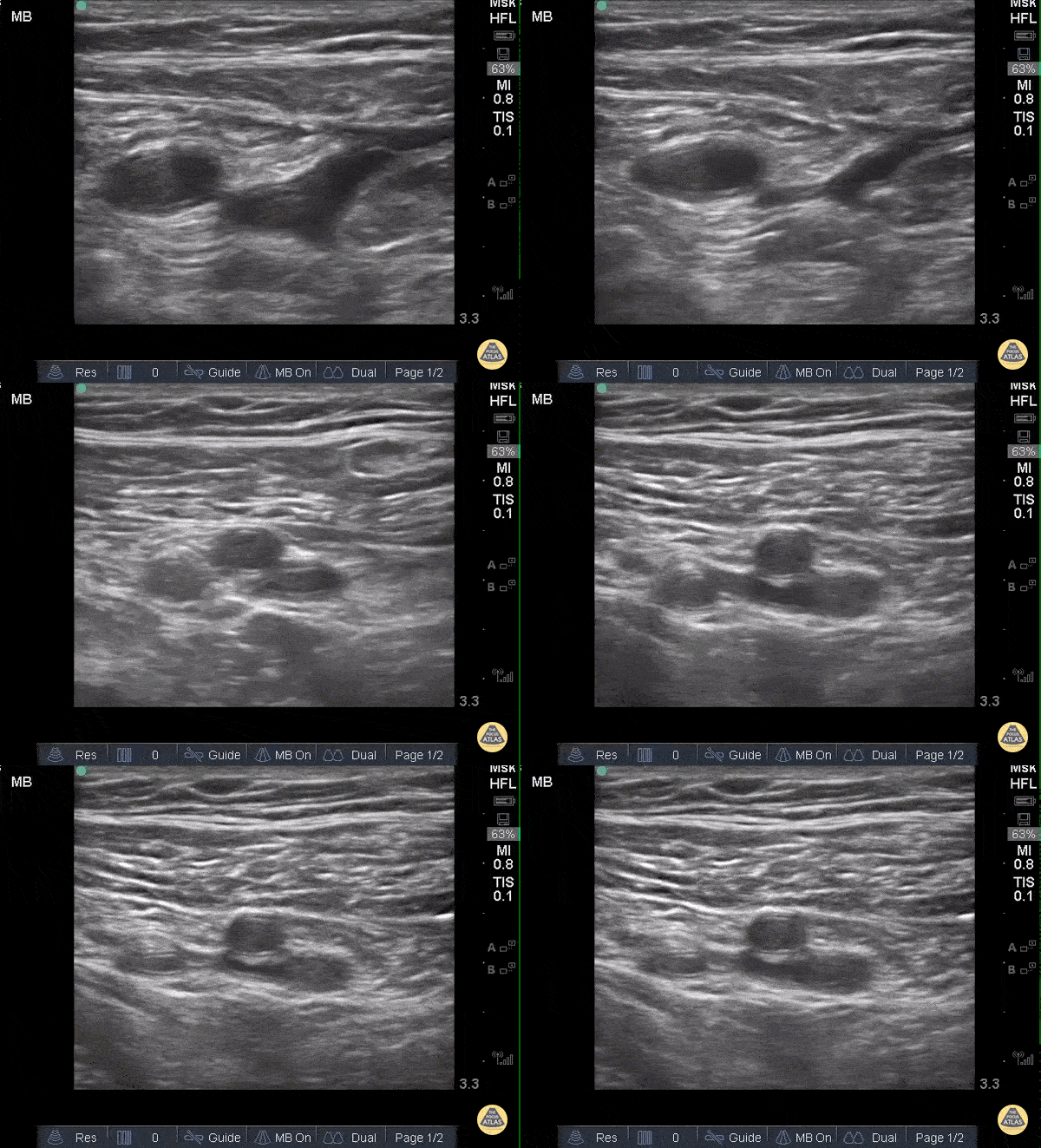
DVT - Common Femoral
When applying pressure to the ultrasound probe, veins collapse first while arteries remain patent and pulsatile. In this clip one can see that proximally, the large pulsatile vessel (which represents the common femoral artery) sits adjacent to a mostly compressible common femoral vein. As the probe moves distally and both the artery and vein split into deep and superficial branches, the superficial femoral vein is not entirely compressible along its medial aspect, representing a thrombus.
Sukh Singh, MD
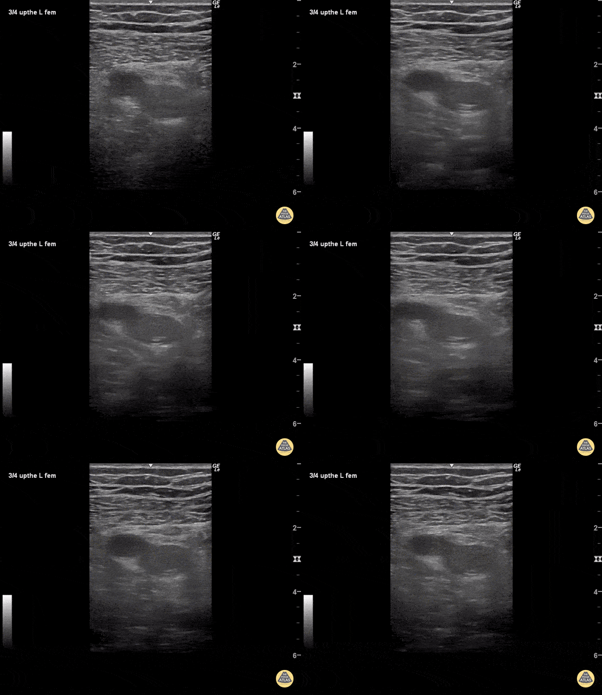
DVT - Superficial Femoral Vein
The superficial femoral artery (pulsatile, to the left) and superficial femoral vein are visualized. As pressure is applied to the probe, the artery becomes oblong but the vein does not collapse as it should, indicated a deep vein thrombosis within its walls. Veins should collapse before arteries under pressure as they have much thinner walls.
Sukh Singh, MD
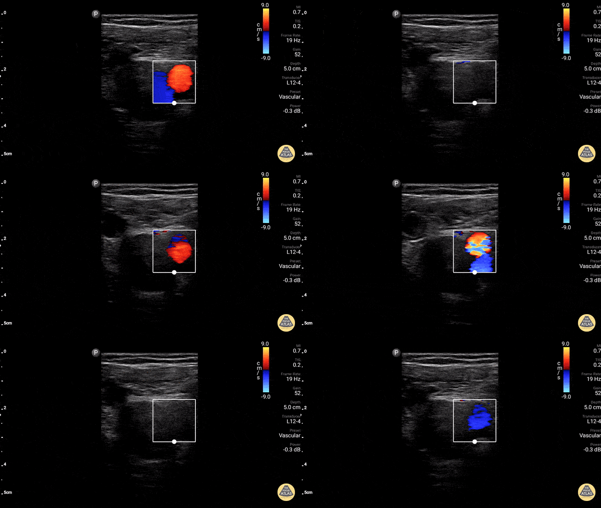
Great Saphenous Vein
The greater saphenous vein, as it takes off from the femoral vein. Make sure to scan up this high and start compressions here to rule out DVT in the lower extremity.
Dr. Gordon Johnson
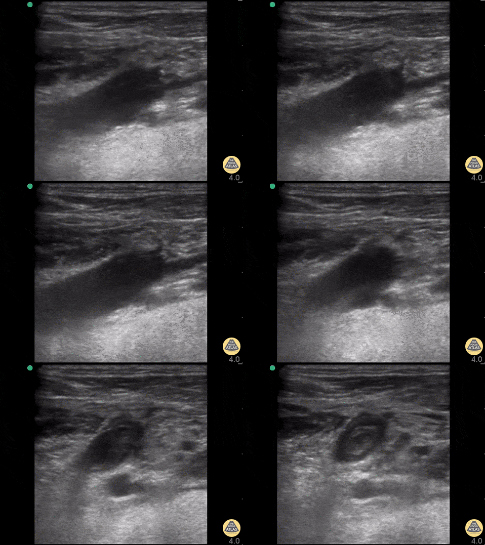
Lemierre Syndrome
25yo w/ no PMH had left molar extracted 10days prior, presents with four days of subjective fever, malaise, and increasing pain to L neck, tachypneic and hypoxic and was intubated w/ central line placed.
POCUS scans through the jugular vein to reveal a clot. CT angiogram showed left internal jugular vein thrombosis, low attenuation L temporal lobe concerning for parenchymal abscess.
Dr. John F. Kilpatrick - Kings County/SUNY Downstate Emergency Medicine
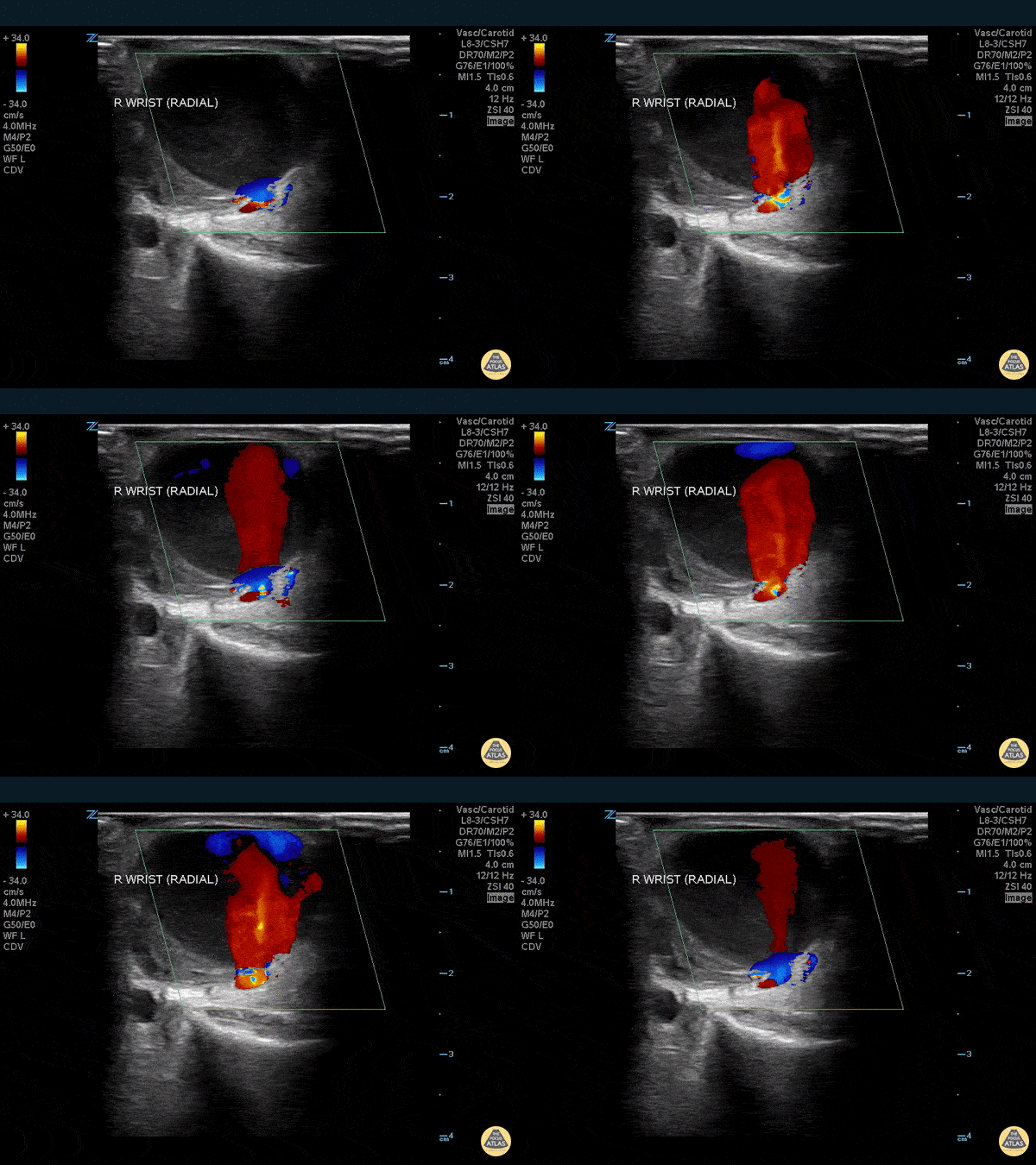
Pseudoaneurysm - Radial Artery
WCUME 2017 Submission and WINNER of "Best POCUS" Category
Radial artery pseudoaneurysm 5 days following transradial coronary angiography. Thrill. Instant diagnosis with POCUS.
Stephen Alerhand, MD - Mount Sinai Hospital
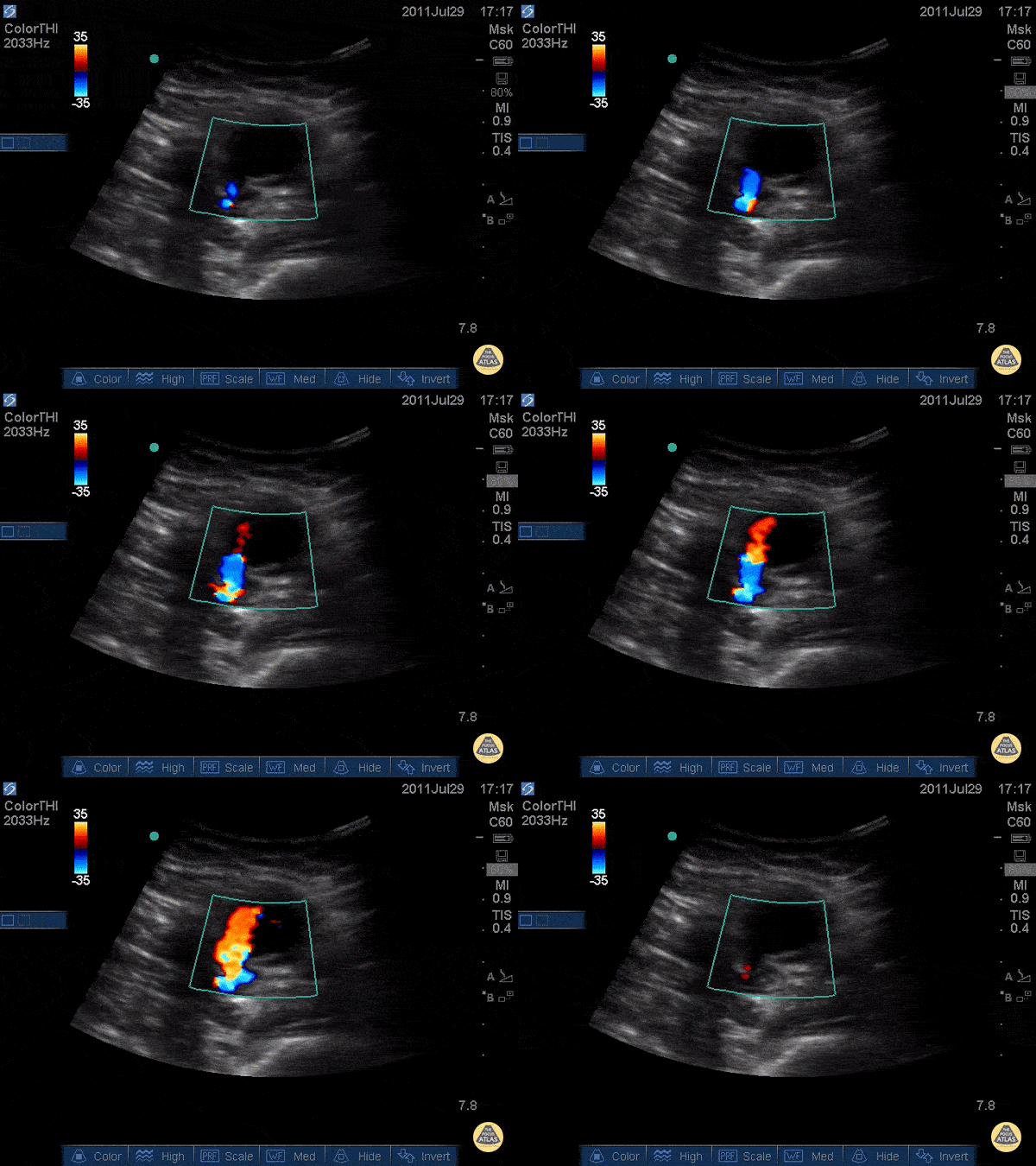
Pseudoaneurysm - "Thar She Blows"
WCUME 2017 Submission for "Best POCUS"
Thought this was an abscess and almost cut into it.
POCUS saved me.
Russell Horowitz, MD - Northwestern
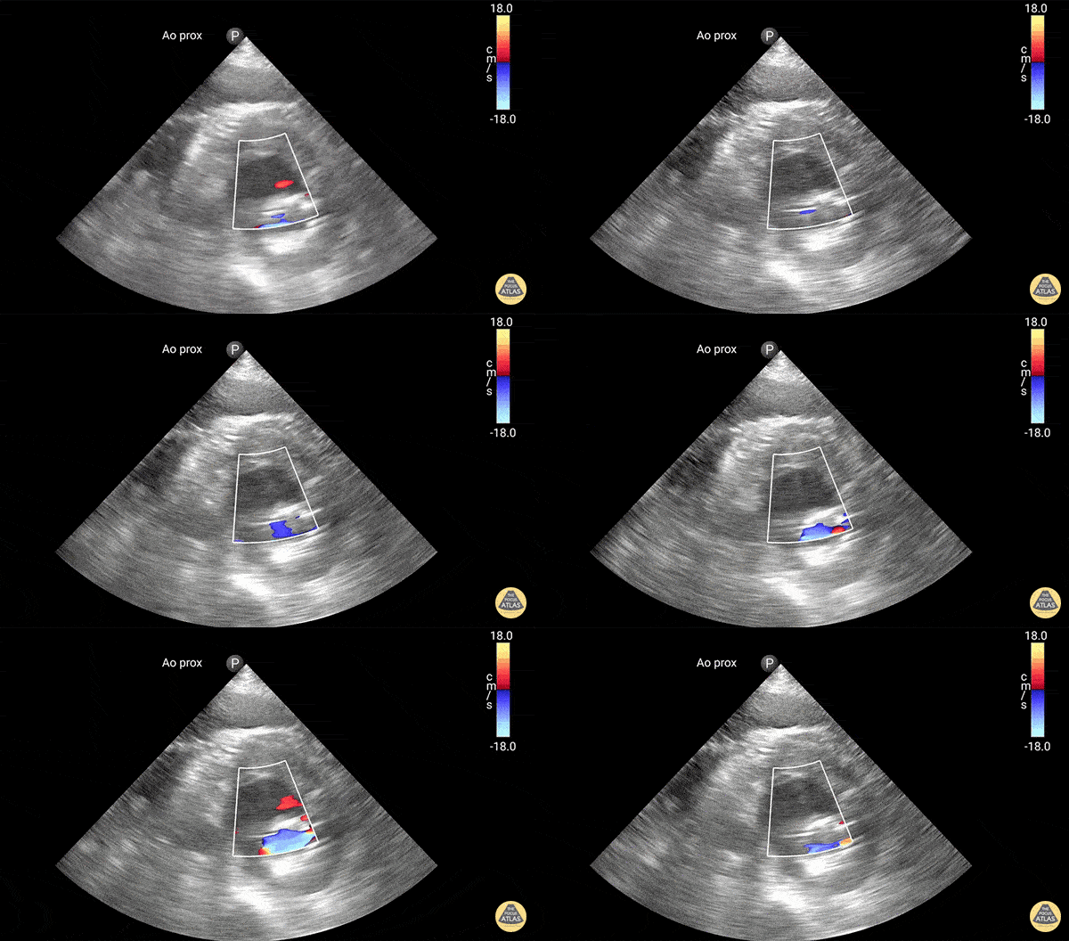
Color Extravasation
Active extravasation through endograft into false lumen. Known chronic endoleak, but CT 30 minutes prior to this study showed no active extravasation or impending rupture. Keep an open mind when reassessing your patients, and try not to anchor too much on prior results or others' opinions!
Dr. Elias Jaffa
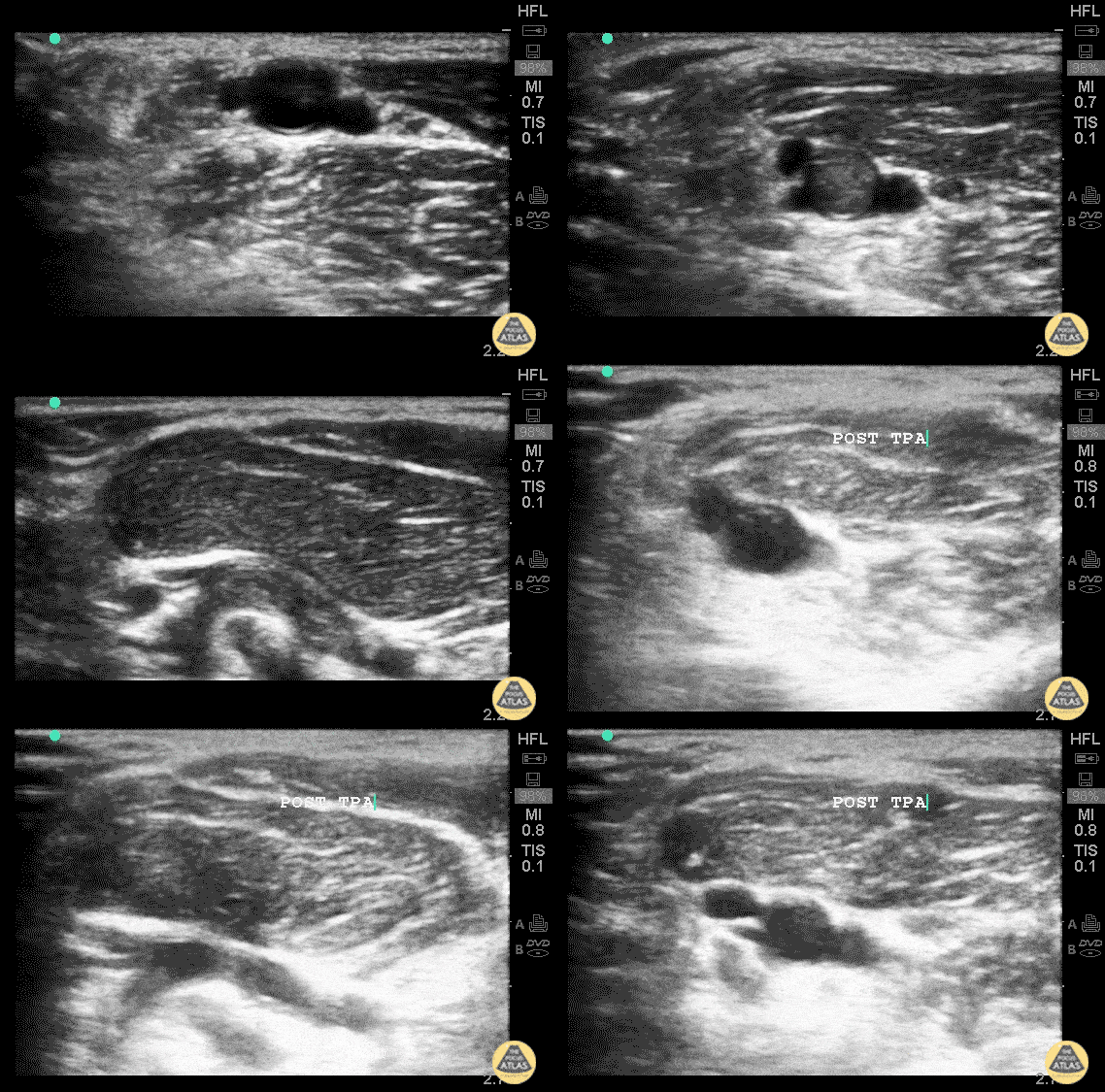
Radial Artery Thrombus (Pre/Post TPA)
The images above are from a middle-aged woman who presented to the emergency room as a stroke code. She complained of paresthesias in her right arm and had decreased sensation to
light touch. It was noted that this arm was colder than the left, so after she returned from head CT, POCUS was performed of her brachial artery.
There is a non-compressible hyperechoic structure inside the brachialartery, consistent with the clinical picture of an arterial embolus. Given the urgency of stroke treatment, these images were obtained after the stroke team had administered tenecteplase.
Vascular surgery consult was made aware of the findings and repeat images taken an hour after presentation and administration of tenecteplase were obtained at bedside with the vascular
surgery consult. These images show near resolution of the embolus.
Dr. Ben Kaufman
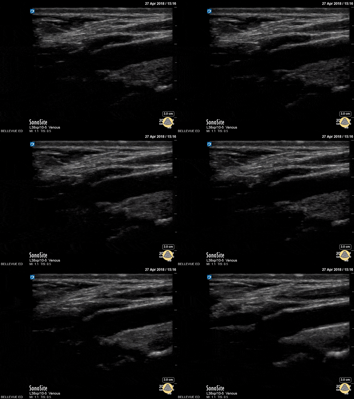
Carotid Bulb Long Axis
The pulsatile carotid artery in long axis at the level of the carotid bulb (widened segment in center of the screen). The intima is visible as a distinct layer of the carotid wall. The bifurcation of the internal and external carotid is not visible here. A portion of the internal jugular vein is seen superficial to the carotid.
Hannah Kopinksi and Dr. Lindsay Davis - NYU Emergency Medicine
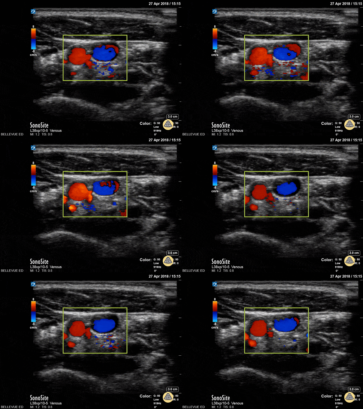
Carotid and IJ - Doppler
This clip shows the common carotid artery (red) and internal jugular vein (blue) in cross-section side by side using color doppler overlay. The sternocleidomastoid muscle is seen in cross-section at the top of the screen.
Hannah Kopinksi and Dr. Lindsay Davis - NYU Emergency Medicine
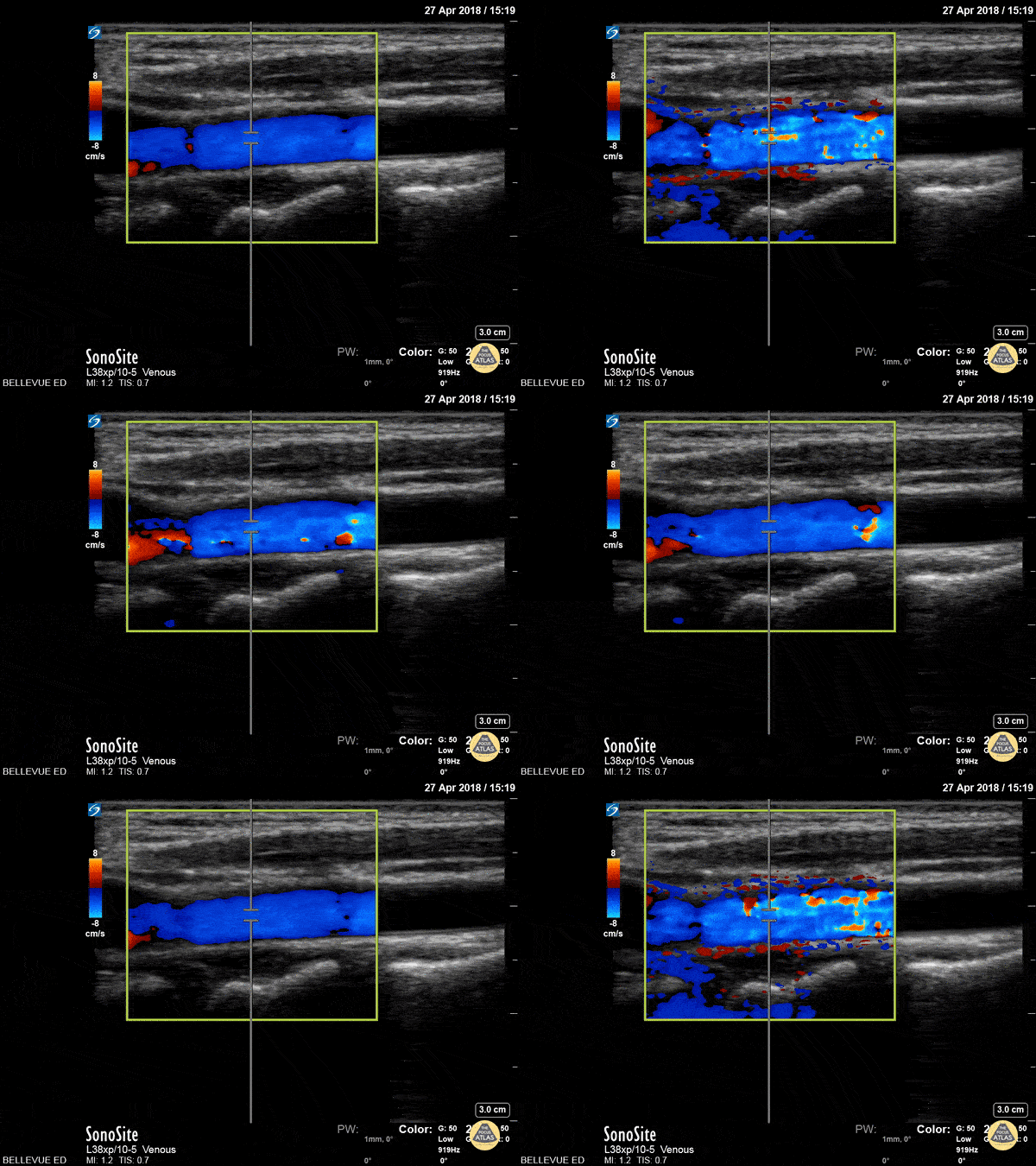
Carotid Long - Doppler
The common carotid artery in long axis with color doppler overlay. On the far left of the screen the wide carotid bulb is visible.
Hannah Kopinksi and Dr. Lindsay Davis - NYU Emergency Medicine
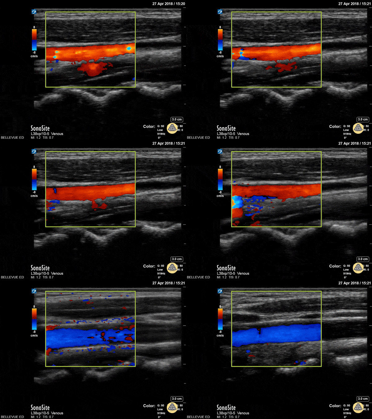
Carotid vs IJ - Doppler
In this clip the probe sweeps between the internal jugular vein (red, demonstrating respiratory variation) and the common carotid (blue, pulsatile) in long axis with color doppler overlay.
Hannah Kopinksi and Dr. Lindsay Davis - NYU Emergency Medicine
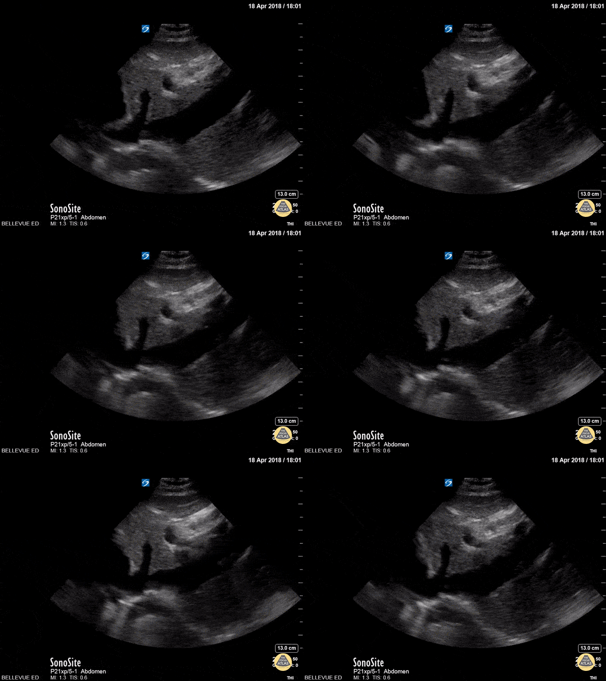
IVC and Hepatic Vein
In this longitudinal subxiphoid view, we see the liver in the center and beating heart to the left of the screen. The IVC in long axis empties into the right atrium. A hepatic vein is seen draining into the IVC just prior to its confluence with the heart.
Hannah Kopinksi and Dr. Lindsay Davis - NYU Emergency Medicine
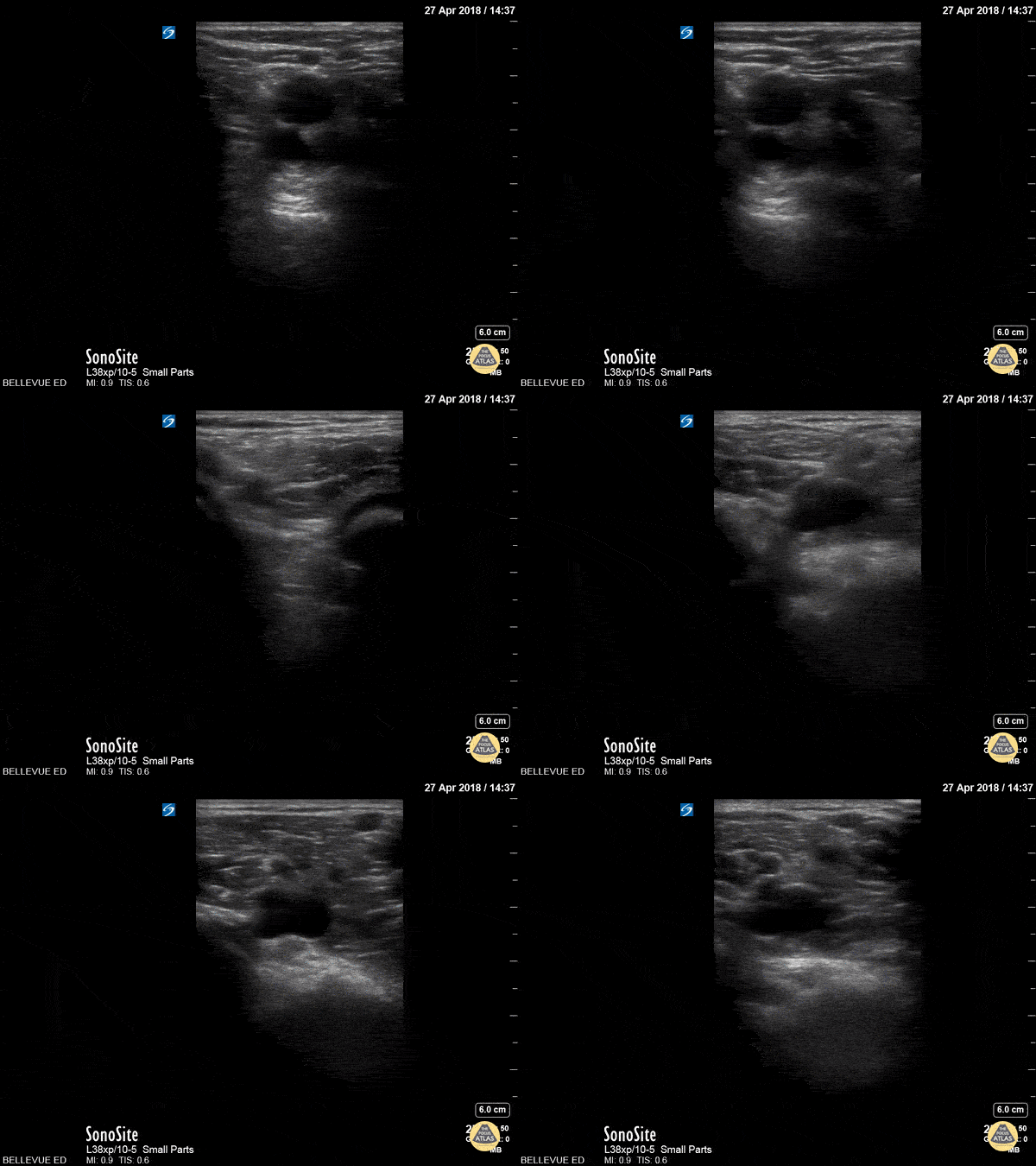
Popliteal Compression Test
This clip demonstrates compression of the popliteal vein moving distally from the popliteal fossa down the calf as is often done to assess for DVT. The non-compressible vessel deep and medial to the popliteal vein is the popliteal artery.
Hannah Kopinksi and Dr. Lindsay Davis - NYU Emergency Medicine
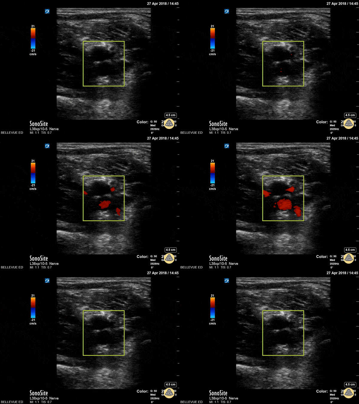
Popliteal Vasculature - Doppler
In this clip the color doppler helps to differentiate the popliteal artery (deep, red, pulsatile) from the popliteal vein (no color, superficial).
Hannah Kopinksi and Dr. Lindsay Davis - NYU Emergency Medicine

Lemierre Syndrome
A young male presented to the ED with high fever and right lateral neck pain with swelling and associated chest pain. He has a recent history of a sore throat. Transverse view of the neck reveals IJ thrombus with enlarged surround lymph nodes. Chest CT revealed a septic pulmonary emboli.
Image courtesy of Robert Jones DO, FACEP @RJonesSonoEM
Director, Emergency Ultrasound; MetroHealth Medical Center; Professor, Case Western Reserve Medical School, Cleveland, OH
View his original post here
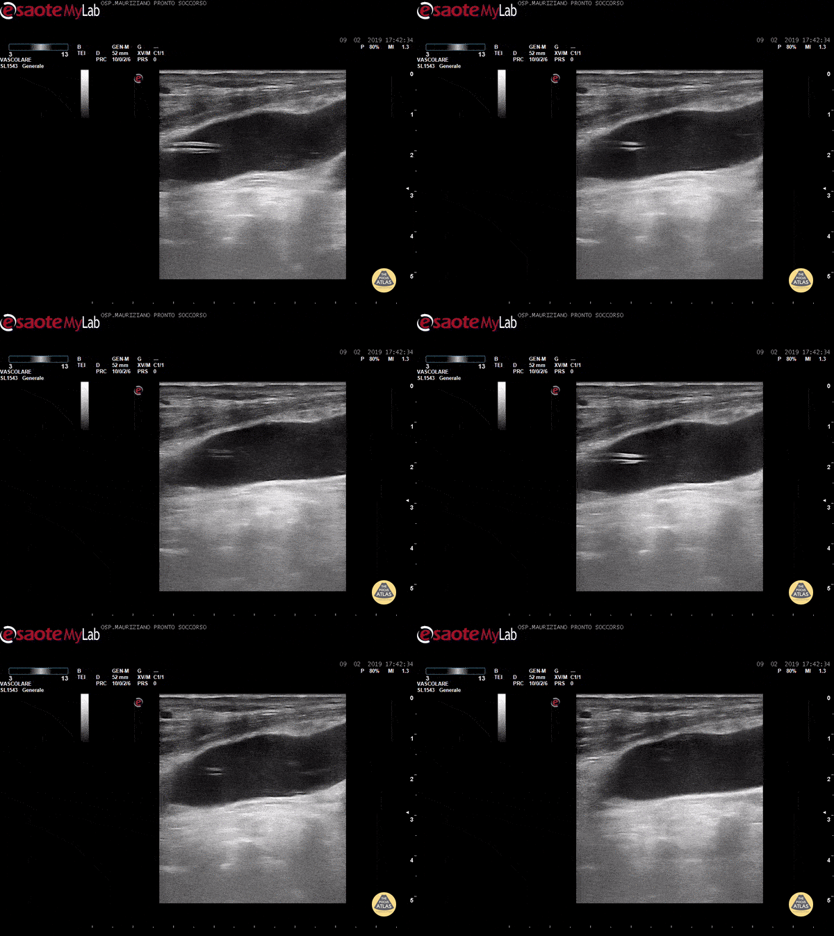
CVC kinking in IJV
After a troubled implant (Seldinger wire got somehow stuck) there was normal IV dripping and normal blood reflux after positioning IV bottle below the patient. However, no flush in AD was visible. We scanned to find a 180° degrees kinking in IJV just below insertion site .
Dr. Garrone
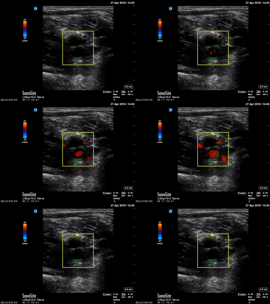
Popliteal Vasculature - Colorized
Popliteal vessels
Green: Popliteal artery, Yellow: Popliteal vein
Images: Dr. Lindsay Davis, Dr. Hannah Kopinski. Image Editing: Michael Amador and Dr. Matthew Riscinti
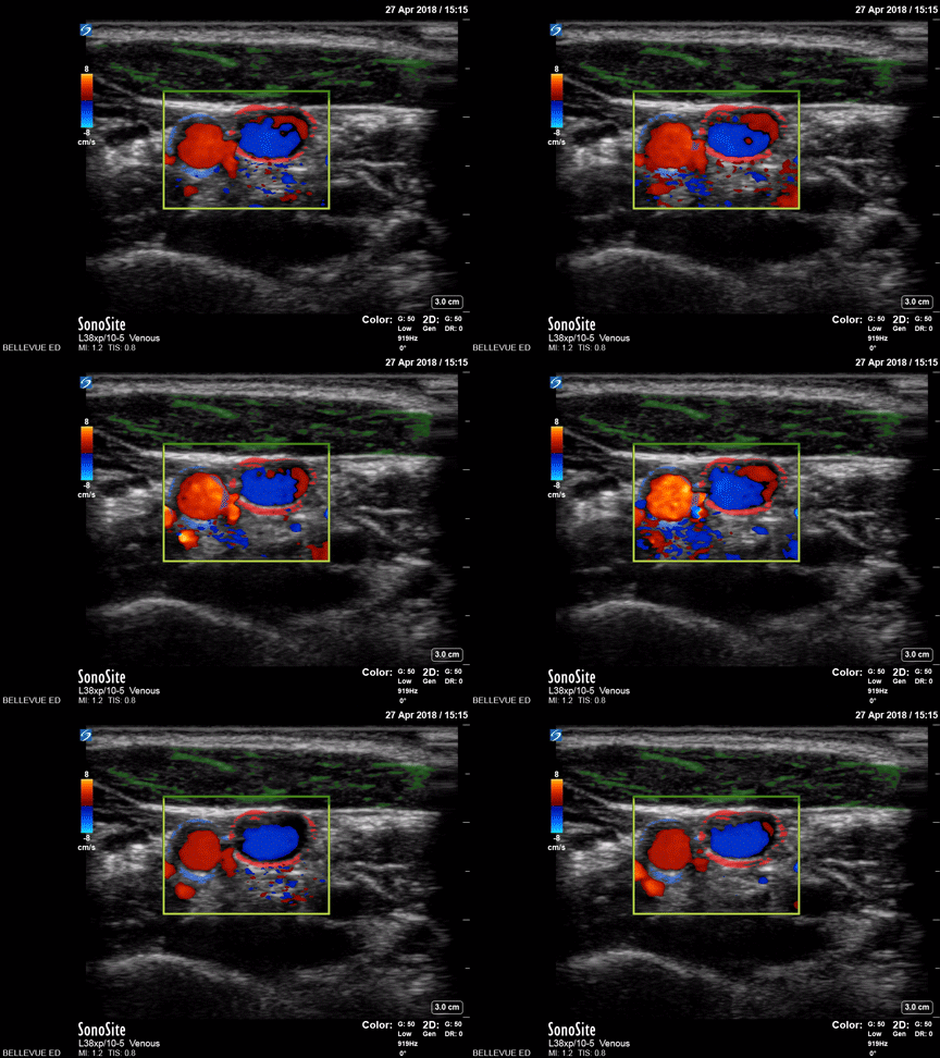
Carotid and Internal Jugular Doppler - Colorized
Carotid Artery and Internal Jugular Doppler
Green: Sternocleidomastoid, Blue outline: Common carotid artery, Red outline: Internal jugular vein
Images: Dr. Lindsay Davis, Dr. Hannah Kopinski. Image Editing: Michael Amador and Dr. Matthew Riscinti
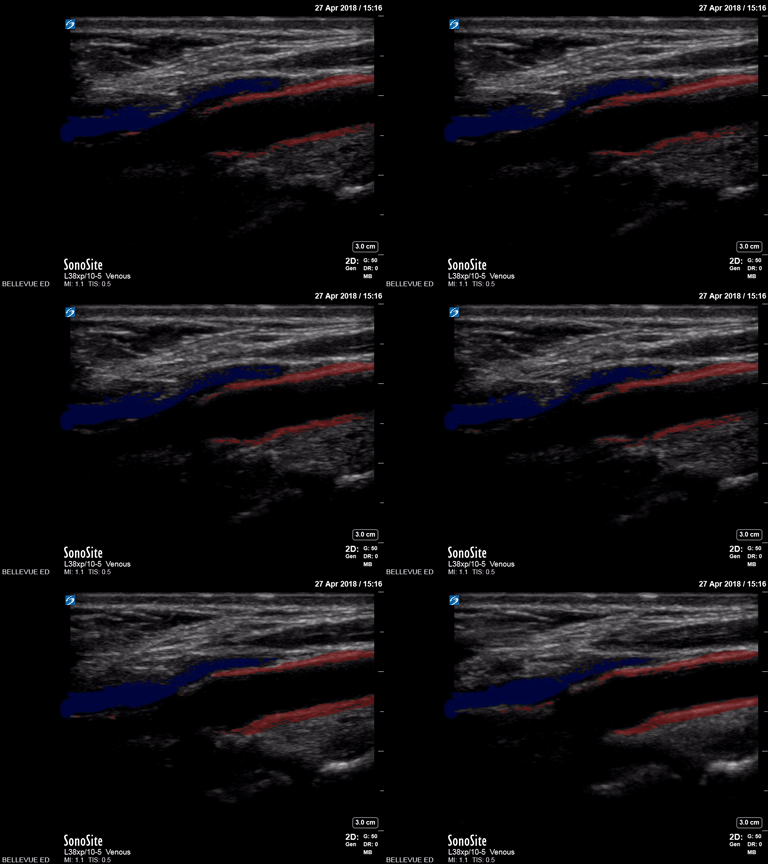
Carotid Bulb Long Axis - Colorized
Carotid Bulb (long axis)
Red: Common carotid artery, Blue: Lumen of internal jugular vein
Images: Dr. Lindsay Davis, Dr. Hannah Kopinski. Image Editing: Michael Amador and Dr. Matthew Riscinti
Common Carotid Long Axis - Colorized
Long Axis of Common Carotid Artery
Red: Common carotid artery, Green: Carotid bulb
Images: Dr. Lindsay Davis, Dr. Hannah Kopinski. Image Editing: Michael Amador and Dr. Matthew Riscinti

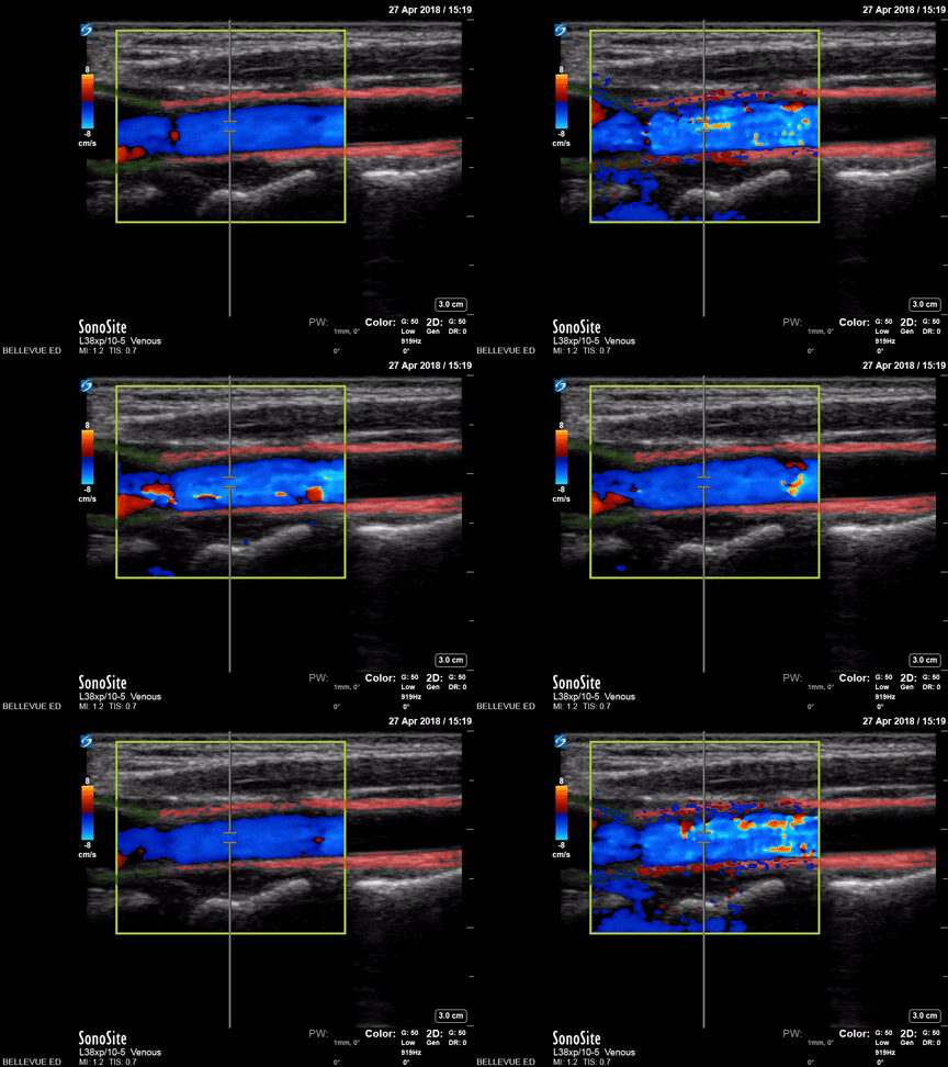
Internal Jugular and Common Carotid Doppler (long axis) - Colorized
Long Axis Doppler of Common Carotid Artery and Internal Jugular Vein
Yellow outline: Internal jugular vein, Green outline: Common carotid artery
Images: Dr. Lindsay Davis, Dr. Hannah Kopinski. Image Editing: Michael Amador and Dr. Matthew Riscinti


