EA Echo Examples

Abnormal End Point Septal Separation (EPSS)
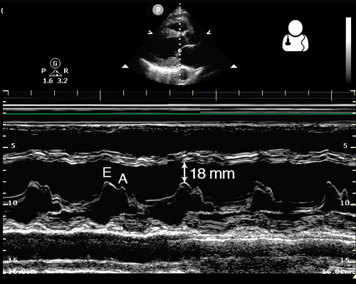
One way to evaluate cardiac function is to measure the distance from the anterior leaflet of the mitral valve to the septum during diastole. This is known as end point septal separation (EPSS).
An EPSS < 7 mm is considered normal, while EPSS > 10 mm suggests decreased cardiac function.
This measurement should be obtained in a parasternal long axis view, using M-mode with the cursor placed through the tip of the mitral valve.
Image credit: Ultrasound of the Week

Reduced Ejection Fraction
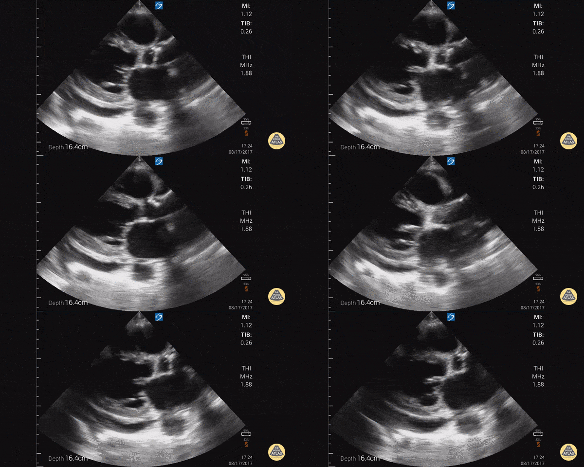
Parasternal long axis with cardiomyopathy, pericardial effusion, and decreased EF.
Dr. Gordon Johnson MD
Internist
Portland Oregon & Uganda

Cardiac Standstill
A patient presented with out-of-hospital cardiac arrest. POCUS was used to confirm presence of cardiac standstill. Note the absence of movement of the left ventricular free wall.
Melissa Myers, MD. Emergency Medicine in Texas. Ultrasound Fellowship Program Director.
@melissamyersmd

RV Thrombus & McConnell's Sign
60 year-old smoker presents with dyspnea and chest pain. Apical 4 chamber view is notable for a thrombus within the RV and associated evidence of RV strain including increased RV size and impaired systolic function with sparing of the apex (known as McConnell’s sign).
Renato Tambelli, @R_Tambelli
Emergency Physician Hospital das Clínicas de Marília, Sao Paulo/Brazil

B-Lines - Pulmonary Edema
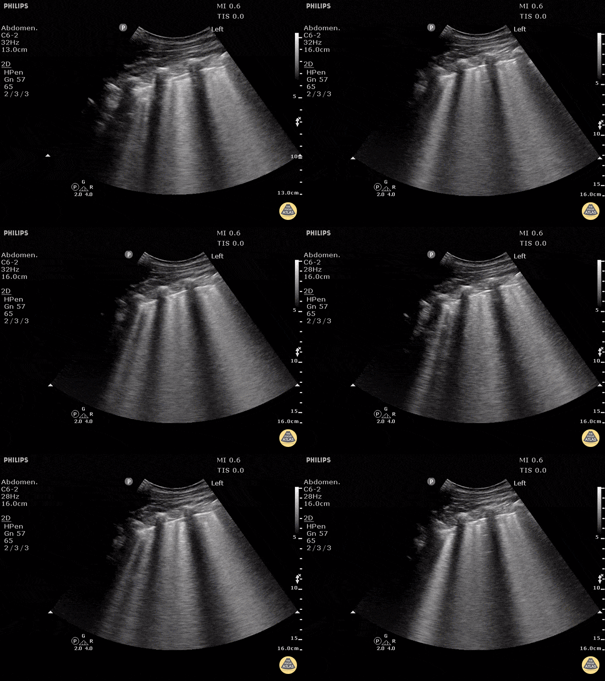
B-lines obtained with curved probe.
B-lines are vertical artifacts that move with respiration from the pleural surface. They represent increased water in an area of the lung. In the right clinical context this could represent pulmonary edema. An increase in B-lines correlates with the degree of pulmonary edema.
3 B-lines in an intercostal space represent a "positive" region of the lung, and if there are two regions of the lung that are positive, you can diagnose pulmonary edema.
Dr. Justin Bowra et al. (Dr. D Browne and Dr. J Knights)


