
Biliary

Pyogenic Liver Abscess with Pleural Effusion
A elderly female with severe sepsis with E. coli bacteremia initially thought to be from urinary source developed worsening hypoxia overnight.
Using a Butterfly US, a moderate-sized right pleural effusion with associated ~6cm liver abscess is seen. These images helped mobilize pulmonology and IR to place a chest tube and hepatic drain, which led to resolution of hypoxemia and sepsis. Hepatic drain cultures grew E coli. Although one may appreciate a diaphragmatic defect on the image, the pleural fluid remained sterile.
POCUS in this case led to successful identification of the true sepsis source and associated pleural effusion as a complication of pyogenic liver abscess.
Contributors: Eric Reid, MD

Gallbladder cancer
Patient presented with right upper quadrant pain. Clip shown demonstrated what appeared to be sludge however it was non-mobile and color doppler demonstrated internal flow. Further work up revealed gallbladder malignancy.
Image courtesy of Robert Jones DO, FACEP @RJonesSonoEM
Director, Emergency Ultrasound; MetroHealth Medical Center; Professor, Case Western Reserve Medical School, Cleveland, OH
View his original post here

Dilated cystic duct and spiral valves of Heister
Patient originally presented with right upper quadrant to mid epigastric pain with jaundice. An obstructing mass can be seen at the head of the pancreas along with intra-heaptic and extra-hepatic duct dilation. The “candy cane” appearing structure is the dilated cystic duct with spiral valves of Heister visible.
Image courtesy of Robert Jones DO, FACEP @RJonesSonoEM
Director, Emergency Ultrasound; MetroHealth Medical Center; Professor, Case Western Reserve Medical School, Cleveland, OH
View his original post here

Incidental Hepatic Arterial Calcification
An elderly patient presented with abdominal pain and hypotension. On eval of liver a linear intrahepatic hyperechoic area was seem consistent with hepatic artery calcification.
Pneumobilia and portal venous gas would be include in differentials however pneumobilia would present with air more centrally while portal venous gas would be more peripherally.
Here is an example of pneumobilia.
Image courtesy of Robert Jones DO, FACEP @RJonesSonoEM
Director, Emergency Ultrasound; MetroHealth Medical Center; Professor, Case Western Reserve Medical School, Cleveland, OH
View his original post here

Gangrenous Cholecystitis
Stone trapped in cystic duct resulted in gallbladder distention as well as sloughing of intraluminal membranes.
Image courtesy of Robert Jones DO, FACEP @RJonesSonoEM
Director, Emergency Ultrasound; MetroHealth Medical Center; Professor, Case Western Reserve Medical School, Cleveland, OH
View his original post here

Gallbladder Polyp
51 year old male with a chief complaint of RUQ abdominal pain for 3 days with associated nausea and vomiting.
The curvilinear probe was used to evaluate the gallbladder for stones or other pathology. A gallbladder polyp at the neck was discovered (also subsequently confirmed on abdominal CT).
Lindsay Davis, DO, MPH, @Lindsadavis18
Lydia Mansour, DO
Emily Nagourney, MS4
Central Michigan University

RUQ Color Doppler
51 year old male with a chief complaint of RUQ abdominal pain for 3 days with nausea and vomiting. When looking to evaluate the aorta, IVC or gallbladder and you find multiple anechoic structures you can you use color doppler to help determine which structures have pulsatile blood flow, non-pulsatile or no flow in a effort to distinguish these anatomic structures.
Lindsay Davis, DO, MPH, PGY1; @Lindsadavis18
Lydia Mansour, DO, PGY3
Emily Nagourney, MS4
Central Michigan University Emergency Medicine Residency

Hepatic Abscess
A 50-year-old male presented with right upper quadrant pain, vomiting, and fever. A point-of-care ultrasound demonstrated a large, approximately 20cm heterogenous, well-demarcated mass in the liver. CT scan confirmed the presence of an abscess. IR-guided drainage yielded "anchovy paste" purulent aspirate, which later grew out Entamoeba histolytica. Amebic liver abscesses are the most common form of extra-intestinal amebiasis, but abscesses can also form in the lung and brain. He was treated with metronidazole without complications.
Mark Zhang

Cholelithiasis - Impacted Stone
It’s fundamental to study the gallbladder in its whole extension. In this image, there is an impacted stone at the neck.
Dr. Felipe Urriola P.
www.thepocus.com

Cholelithiasis - Multiple Stones
The gallbladder is studied in both planes, revealing multiple small stones generating acoustic shadowing.
Dr. Felipe Urriola P

Distended Gallbladder
Elderly male with history HTN, DM presented with jaundice and elevated LFTs, bilirubin, and alkaline phosphatase. POCUS showed distended gallbladder with normal wall thickness, and some areas of increased echogenicity in the gallbladder which may represent debris. Patient was later found to have a proximal pancreatic mass likely compressing bile ducts and causing distention of gallbladder and elevated lab findings.
Hannah Gadway MS4
Therese Mead, DO, RDMS, FACEP
Central Michigan University College of Medicine

Intrahepatic Ductal Dilation
Transverse scan of the liver shows diffuse dilation of intrahepatic biliary ducts.
Image courtesy of Robert Jones DO, FACEP @RJonesSonoEM
Director, Emergency Ultrasound; MetroHealth Medical Center; Professor, Case Western Reserve Medical School, Cleveland, OH
View his original post here

Cholelithiasis with Adenomyomatosis
Longitudinal view of the gallbladder revealing two unique findings. A hyperechoic structure with posterior acoustic shadowing indicative of a stone is located in the neck. Two smaller hyperechoic structures can be seen attached to the wall of the GB body, likely adenomyomatosis.
Image courtesy of Robert Jones DO, FACEP @RJonesSonoEM
Director, Emergency Ultrasound; MetroHealth Medical Center; Professor, Case Western Reserve Medical School, Cleveland, OH
View his original post here

Liver Hematoma
66 y/o male with a history of a recent CBD stent placement complicated by a ruptured hepatic artery resulting in a subcapsular liver hematoma that required IR drainage. He now presents to the ED with progressively worsening right upper quadrant abdominal pain.
POCUS was instrumental in monitoring for continued bleeding following the initial drainage. In the clip, you can see the large hematoma, measuring 15 x 13 cm, obscuring most of the liver. Patient was admitted for surgical deroofing.
Jennifer Kaminsky, MD PGY-2; @jen_kaminskyMD
Pamela Santivanez, MD PGY-1
Sean Beckman, Rocky Vista University COM OMS-4
Joshua Greenstein, MD, Director of ED Ultrasound
Northwell Health – Staten Island University Hospital

Emphysematous Cholecystitis
RUQ US of a patient revealed air within the gallbladder consistent with emphysematous cholecystitis, which can easily be confused with bowel gas.
Image courtesy of Robert Jones DO, FACEP @RJonesSonoEM
Director, Emergency Ultrasound; MetroHealth Medical Center; Professor, Case Western Reserve Medical School, Cleveland, OH
View his original post here

Pneumobilia
Air can be noted within the biliary tract in this ultrasound of the RUQ. This patient was s/p Whipple procedure.
Image courtesy of Robert Jones DO, FACEP @RJonesSonoEM
Director, Emergency Ultrasound; MetroHealth Medical Center; Professor, Case Western Reserve Medical School, Cleveland, OH
View his original post here

Portal Venous Gas
RUQ ultrasound reveals air within the portal venous system in a patient with ischemic colitis. Something that can be difficult to distinguish from pneumobilia.
Image courtesy of Robert Jones DO, FACEP @RJonesSonoEM
Director, Emergency Ultrasound; MetroHealth Medical Center; Professor, Case Western Reserve Medical School, Cleveland, OH
View his original post here

Gallstone in Neck Causing Mirizzi's Syndrome
Note the hyperechoic structure with posterior acoustic shadowing indicative of a gallstone. Patient was moved and the stone remained lodged in the neck. Additionally, gallbladder sludge can be seen. The patient was found to have elevated LFTs. MRCP performed demonstrating external compression of biliary tree this large stone in GB neck consistent with Mirizzi’s syndrome.
Image courtesy of Robert Jones DO, FACEP @RJonesSonoEM
Director, Emergency Ultrasound; MetroHealth Medical Center; Professor, Case Western Reserve Medical School, Cleveland, OH
View his original post here

Choledocholithiasis
A 65-year-old female with no PMH presented to ED with 6 hour hx RUQ abdominal pain. She reported no associated vomiting or fever.
POCUS seen here reveals the cystic duct emerging from the GB neck to then reach a severely dilated CBD. A hyperechoic round structure with posterior shadowing lies inside the CBD, consistent with a diagnosis of choledocholithiasis.
Dr. Felipe Urriola
Emergency Unit. Puerto Aysen Hospital, Chilean Patagonia.

Cholelithiasis
An elderly female presented to the emergency department reporting abdominal pain. POCUS seen here revealed the presence of a gallstone within her gallbladder with shadowing; and a notable absence of findings consistent with cholecystitis including gallbladder sludge, wall thickening, and/or ductal dilatation. This enabled appropriate triage of this patient to outpatient follow-up rather than considering immediate surgical intervention.
Rupinder Sekhon, MD & Peter Biggane, MD
Central Michigan University, Emergency Medicine

Adenomyomatosis
A middle aged patient presented with epigastric pain and vomiting. Ultrasound identified anechoic cystic thickening of the gallbladder wall associated with comet-tail artifact and twinkling artifact. Findings were consistent with adenomyomatosis, a generally benign condition caused by hyperplastic growth of the gallbladder mucosa often associated with chronic inflammation.
Michael Cover, MD
@michaelc0ver

Choledocolithiasis
A patient presented to the ED with RUQ pain. Sagittal view of the common bile duct revealed dilation and stones indicative of choledocolithiasis.
Image courtesy of Robert Jones DO, FACEP @RJonesSonoEM
Director, Emergency Ultrasound; MetroHealth Medical Center; Professor, Case Western Reserve Medical School, Cleveland, OH
View his original post here

Cholecystitis with Gallbladder Rupture
Patient presented with right upper quadrant abdominal pain and fever. RUQ ultrasound showed an impacted stone in the gallbladder neck with gallbladder wall perforation. Note the flow through the perforation and the associated pericholecystic abscess. Labelled image can be found on the original post with the link below.
Image courtesy of Robert Jones DO, FACEP @RJonesSonoEM
Director, Emergency Ultrasound; MetroHealth Medical Center; Professor, Case Western Reserve Medical School, Cleveland, OH
View his original post here

Normal Common Bile Duct
Often, a left lateral position delivers a clear image of the common bile duct (CBD) on its entire length, as is the case in this clip. Notice the normal hyperechoic walls and its small diameter. The CBD inner wall diameter should be < 6 mm in healthy adults, although it can be enlarged in post-cholecystectomy patients.
Dr. Felipe Urriola P. Emergency Unit, Puerto Aysen Hospital. Chilean Patagonia.

Normal Portal Triad without Doppler
The large, pulsatile inferior vena cava lies at the bottom of the screen. Anterior to it and from left to right we see the large portal vein and small, round hepatic artery. Just above the hepatic artery lies another tubular structure which presents hyperechoic walls and an anechoic lumen; this is the common bile duct.
Dr. Felipe Urriola P. Emergency Unit, Puerto Aysen Hospital. Chilean Patagonia.

Normal Portal Triad - Doppler
Both the adjustment of depth and color-doppler allow for better identification of structures. The large, pulsatile inferior vena cava lies at the bottom of the screen. Color-doppler helps to discern blood vessels from other anatomical structures. Anterior to the IVC and from left to right of the screen, we can see the portal vein and hepatic artery with color-flow. Just above the hepatic artery lies another tubular structure which presents hyperechoic walls and an anechoic lumen; this is the common bile duct.
Dr. Felipe Urriola P. Emergency Unit, Puerto Aysen Hospital. Chilean Patagonia.

Twinkling Artifact in Choledocholithiasis
A patient presented with RUQ abdominal pain.
Ultrasound showed cholelithiasis and choledocholithiasis. Left image shows a portion of the common bile duct containing a small hyperechoic shadowing mobile stone. Right image shows the same with color Doppler added demonstrating twinkling artifact.
Learning point: Twinkling artifact is most often associated with urinary stones but also can be caused by gallstones (depending on composition), bowel gas, foley balloons, metallic objects, and many others. Twinkling artifact may help identify small stones that do not cause prominent acoustic shadowing.
Michael Cover, MD
@michaelc0ver

Extreme GB distention in setting of cholangiocarcinoma
A 40-year-old male presented to the ED with several week hx unintentional weight loss and new onset jaundice. POCUS of his RUQ demonstrated an overtly distended gallbladder with plethoric intra-hepatic circulation causing compression of the right kidney. As expected, he had laboratory evidence of an elevated direct bili and acute kidney injury. Subsequent MRI confirmed the diagnosis of cholangiocarcinoma.
Dr. Victor Bang. Emergency Physician at Hospital das Clínicas de Marília. Co-founder of Pocus Jedi.
@vmjbang

Angry gallbladder
A patient presented with altered mental status and anorexia; clinically he had findings consistent with sepsis. POCUS revealed one angry gallbladder! You can appreciate gallbladder wall thickening (measuring 6mm), trace pericholecystic fluid, and shadowing from gallbladder sludge.
Garrett Ghent, Resuscitationst and diagnostician; Norfolk, VA
@garrettghentMD

Biliary Colic
A 33-year-old female presented to the ED reporting acute onset abdominal pain immediately after eating a meal. She was well-appearing and afebrile; physical exam notable for reproducible tenderness to palpation of her upper abdomen. POCUS long axis view obtained to right of epigastrium notable for hyperechoic material within the lumen of the gallbladder (anechoic) producing posterior shadowing. This confirmed our clinical suspicion of biliary colic, and the patient was able to be discharged with scheduled outpatient elective surgery.
Victor Bang, Medical Student Brazil
@vmjbang
Gallstone Stuck in GB Neck with Shadowing
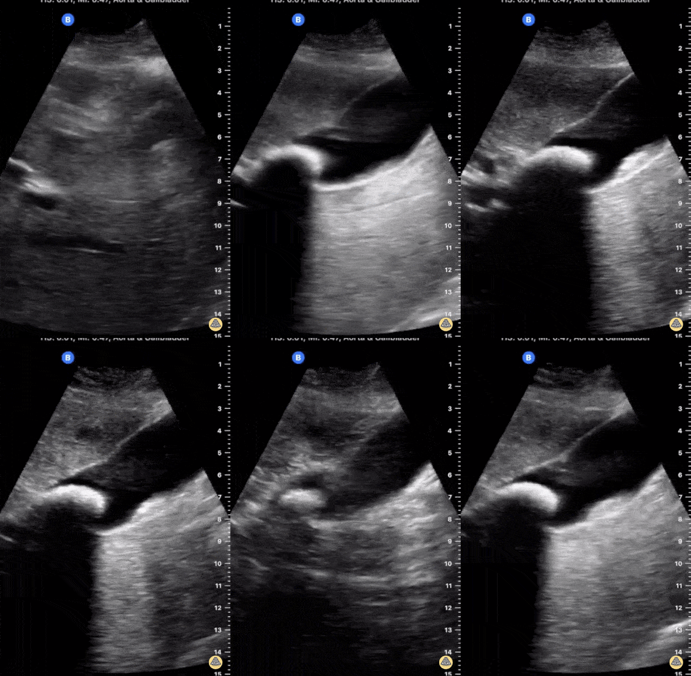
This image demonstrates a large gallstone lodged in the neck of a distended gallbladder with posterior shadowing. There is no evidence of gallbladder wall thickening seen here.
Jonny Wilkinson, Consultant in ICU and Anesthesia
Pneumobilia
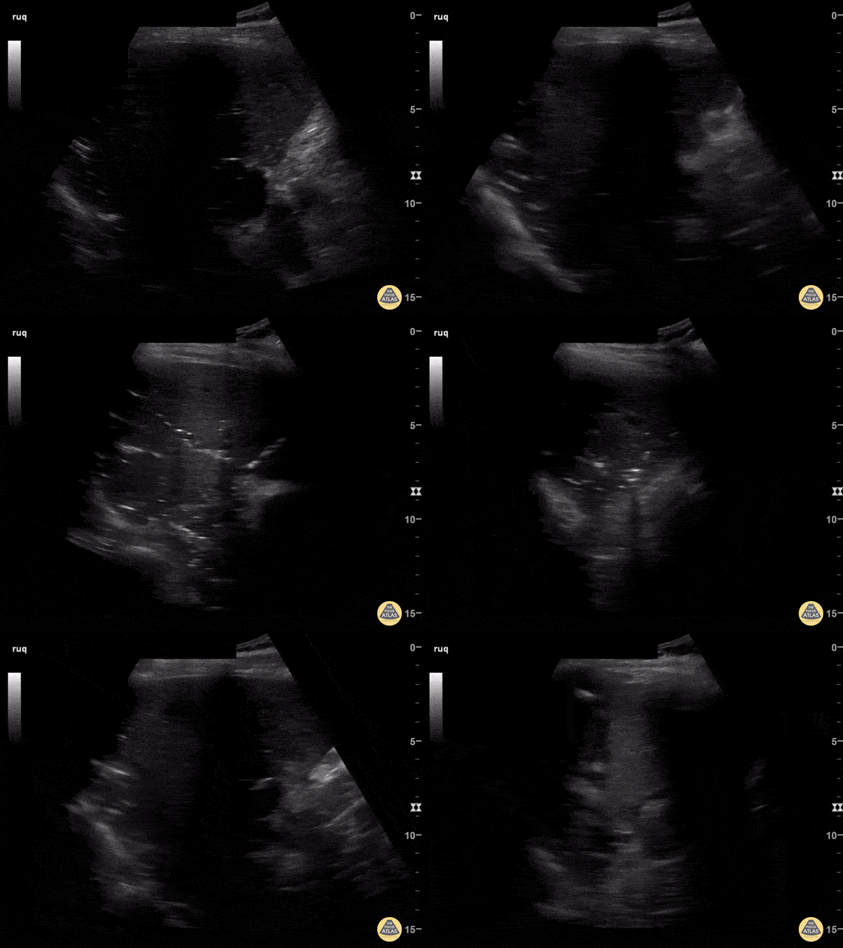
82-year-old male with history of recurrent cholangitis presenting to the ED with abdominal pain. US imaging of the RUQ reveals presence of pneumobilia. Click here to read the full caption.
Andrew Morris, M.D. Gaurav Patel, M.D. Vu Huy Tran, M.D. - Aventura Hospital & Medical Center, Emergency Medicine Program
Adenomyomatosis
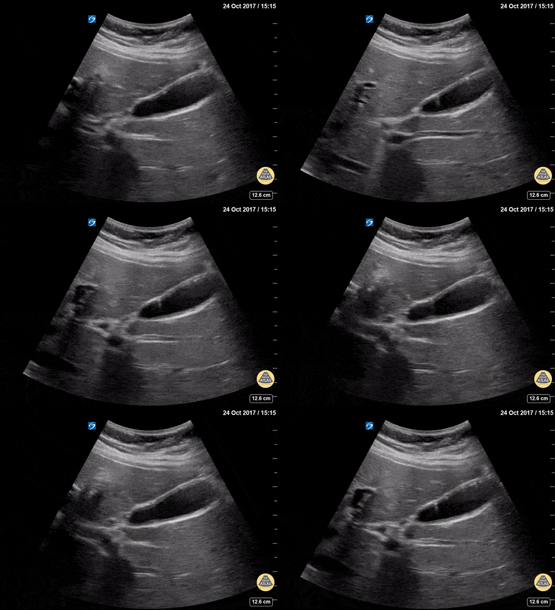
Adenomyomatosis is a generally benign condition characterized by diffuse thickening of the gallbladder wall and intramural diverticula. On ultrasound it creates comet tail artifacts (reverberation artifacts that appear as tiny echogenic beams) originating from distinct locations on the gallbladder wall.
This is often seen in asymptomatic patients; however it can be associated with chronic inflammation from gallstones and pancreatitis.
Dr. Lindsay Davis - NYU/Bellevue Department of Emergency Medicine
Cholelithiasis, Not Impacted
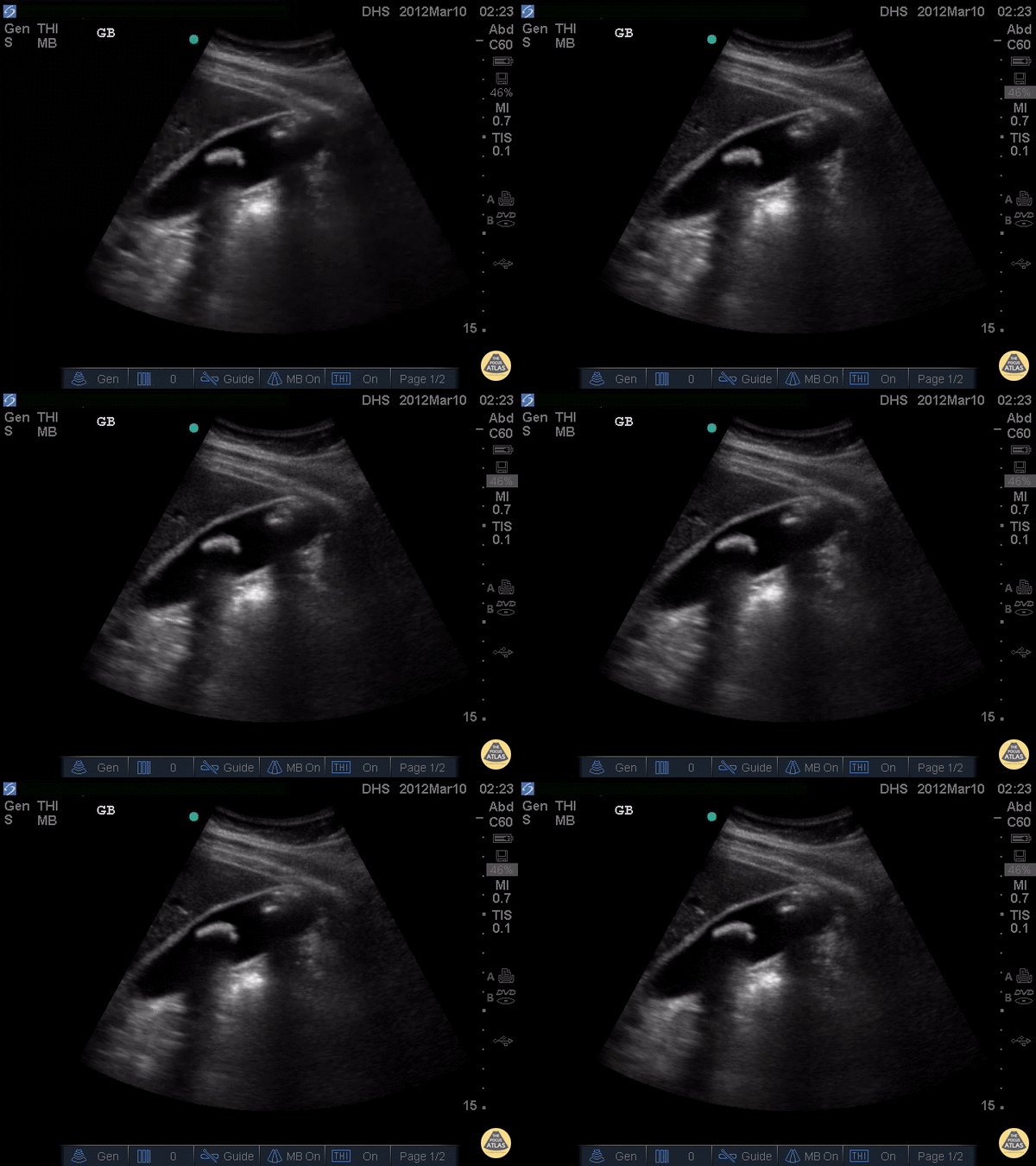
Two large, hyperechoic stones can be appreciated in this gallbladder. These are likely two stones because of the degree of echogenicity and posterior shadowing. If you have one or two objects in the gallbladder and you're not sure if they're stones, it can be useful to have the patient lay on their side. If the objects moves when the patient moves, they are stones. If not, they are likely polyps or other masses growing from the gallbladder wall.
Justin Bowra MBBS, FACEM, CCPU Emergency Physician, RNSH et al. (Dr. Darmas)
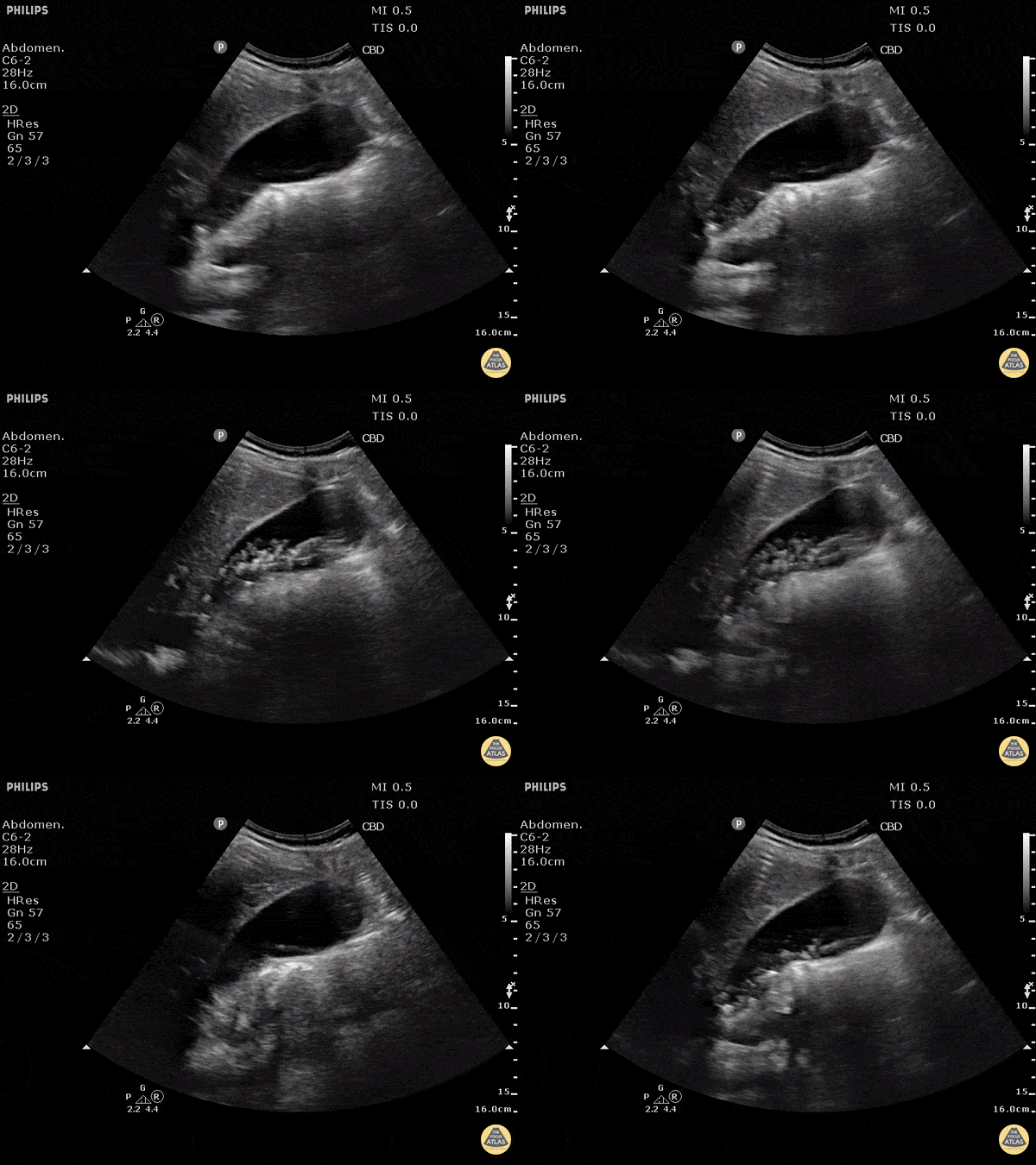
Cholelithiasis with Many Stones
This non-inflamed gallbladder contains many stones. The stones are hyperechoic with posterior shadowing. The gallbladder wall is not thickened, there is no pericholecystic fluid, and no sonographic Murphy's sign, thus cholecystitis is unlikely.
Justin Bowra MBBS, FACEM, CCPU Emergency Physician, RNSH et al. (Dr. Ken Lee)
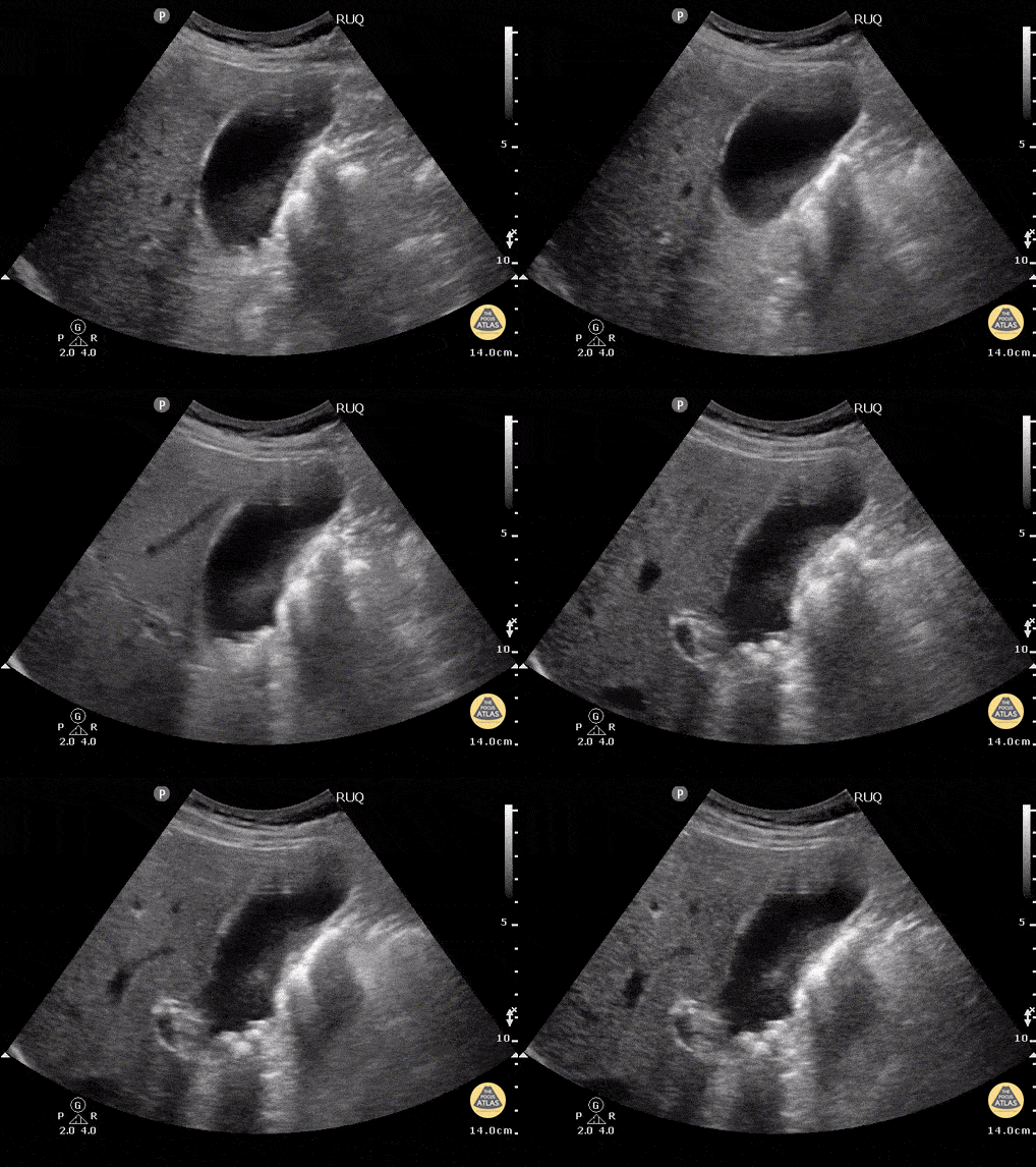
Cholelithiasis - Neck Stones
This patient w/o cholecystitis clearly demonstrates cholelithiasis. A 2002 study showed that ER doctors can diagnose gallstones with a sensitivity of 88% and specificity 96% with POCUS.
Gallstones can be identified by hypoechoic "shadowing" behind hyperechoic stones. If there isn't shadowing, hyperechoic structures could represent polyps or sludge. In this image the stones are resting mainly in the neck of the gallbladder.
Kendall JL, Shimp RJ. Performance and interpretation of focused right upper quadrant ultrasound by emergency physicians. J Emerg. Med. 2001; 21(1):7-13
Submitted by Justin Bowra MBBS, FACEM, CCPU Emergency Physician, RNSH et al.
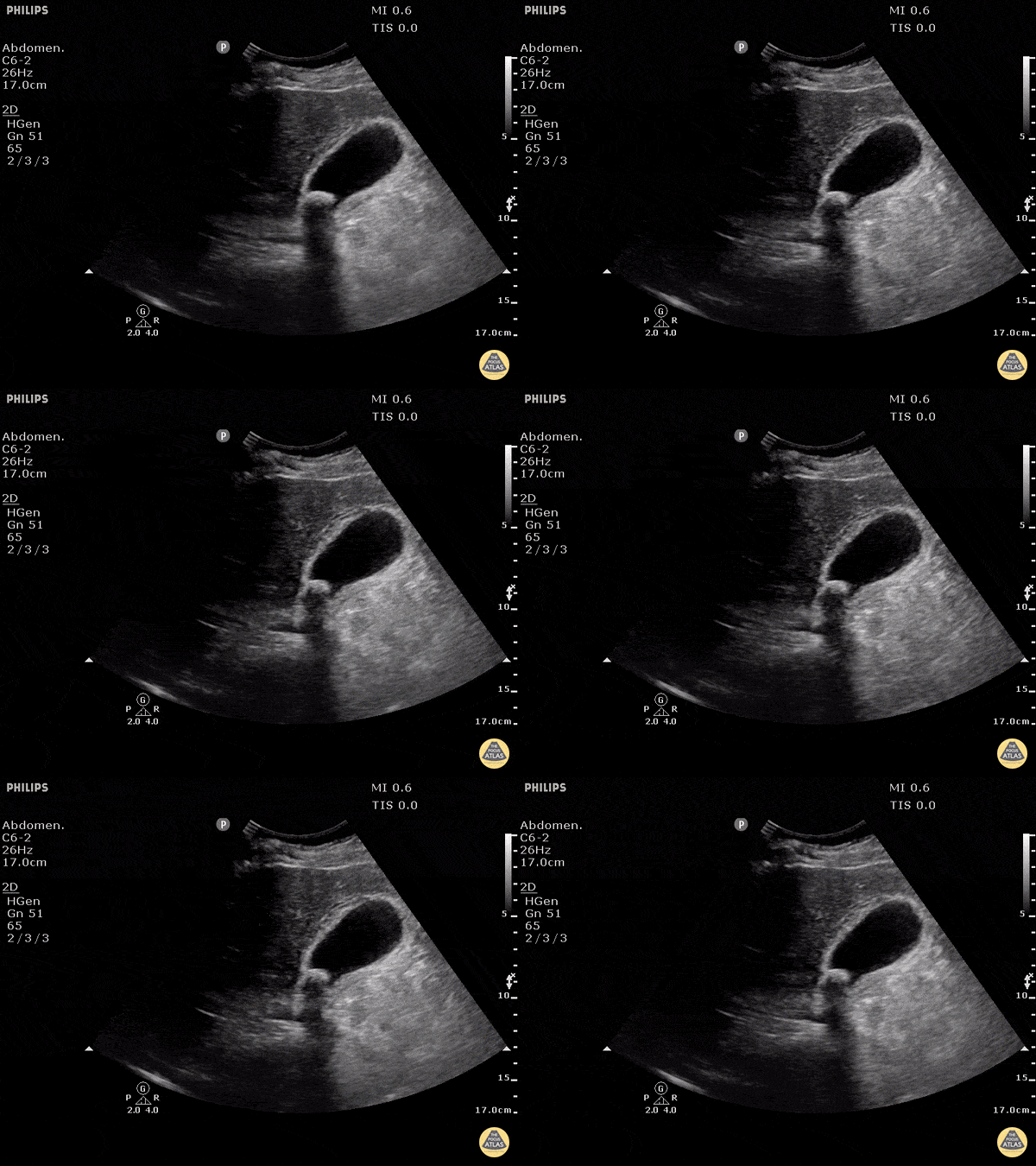
Cholecystitis with Obstructing Stone
This clearly demonstrates a stone in the gallbladder neck as a hyperechoic structure with posterior shadowing. The neck is not always clearly visualized on first glance so it is important to scan through the gallbladder in two planes to exclude stones in the neck. Other signs of acute cholecystitis include pericholecystic fluid, gallbladder wall thickening (>3mm) common bile duct dilatation, and positive sonographic Murphy's sign.
Justin Bowra MBBS, FACEM, CCPU Emergency Physician, RNSH et al. (Mo)
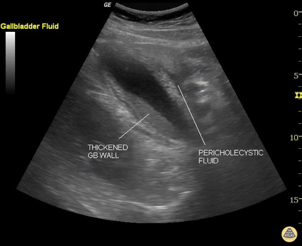
Cholecystitis
This still image shows two clear signs of acute cholecystitis. The gallbladder wall is thickened and there is pericholecystic fluid. Of note, when measuring the gallbladder wall it's better to measure the anterior wall as the posterior wall can appear enlarged due to artifact. The cut off for wall thickening is 3mm. The anterior wall is thickened in this case but is not labeled. Other signs to look for are stones, sludge, common bile duct dilatation, and overall gallbladder enlargement.
Of note, a positive sonographic Murphy's carries a sensitivity of 63%, specificity of 93.6% and positive predictive value of 72.5%.
Justin Bowra MBBS, FACEM, CCPU Emergency Physician, RNSH et al. (Sharon)

Cholecystitis - Emphysematous
Emphysematous cholecystitis is the result of gas formation within the wall or lumen of the gallbladder as a result of infection by a gas forming organism. It is a surgical emergency. The air in the gallbladder appears highly echogenic with mild posterior shadowing, as air next to tissue is highly reflective.
Sukh Singh, MD

Choledocholithiasis - Dilated Common Bile Duct with Stone
A normal common bile duct should be < 4mm plus 1mm per decade after 40 years of age. A stone can clearly be seen in the area of color flow doppler. The bile duct is obviously dilated here (although not measured).
When searching for the CBD, it can be useful to turn color flow doppler on to ensure you're visualizing a biliary structure and not vessels. This technique was employed here.
POCUS is highly sensitive for acute cholecystitis but as many people already know, it can be hard to find the CBD, which is reflected in the lower sensitivity and specificity for this indication in many studies.
Justin Bowra MBBS, FACEM, CCPU Emergency Physician, RNSH et al. (Dr. Dop Ahilan)
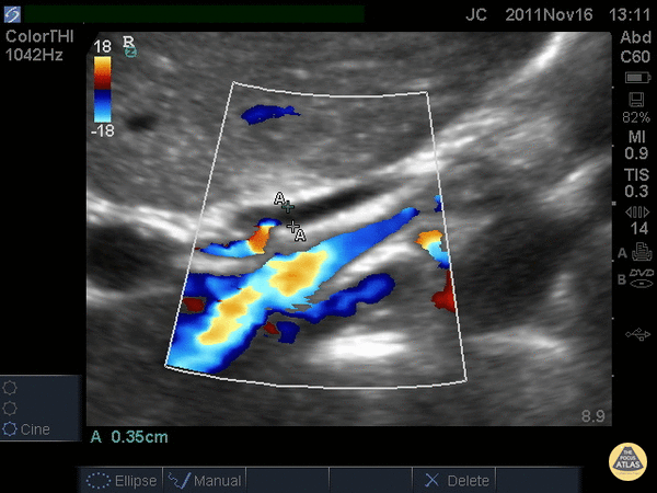
Common Bile Duct (Normal)
When searching for the common bile duct (CBD), color doppler can be useful to differentiate vascular structures from non-vascular structures. In this case the CBD is being measured anteriorly and does not have any pulsations on color doppler. The portal vein and artery can be seen and with the CBD, make the portal triad.
A normal CBD should be less than 4mm plus 1mm per decade after 40 years old. It should be measured inner wall to inner wall.
Justin Bowra MBBS, FACEM, CCPU Emergency Physician, RNSH et al. (Dr. Chiang)

Common Bile Duct (Normal Labeled)
A normal common bile duct should be <4mm plus 1mm per decade after 40 years of age. It should be measured from inner wall to inner wall.
You should attempt to find the portal triad: the CBD, the portal vein, and the hepatic artery. You may need color flow to find all three. Here the CBD and portal vein are labeled (no labeled hepatic artery).
To get better images, especially if you're dealing with a lot of rib shadow, it can be helpful to have your patient take a deep breath and/or roll onto their left side.
Justin Bowra MBBS, FACEM, CCPU Emergency Physician, RNSH et al.

Dilated Common Bile Duct
The CBD measures at 1.89cm indicating it is grossly dilated, as a normal common bile duct should be <4mm plus 1mm per decade after 40 years of age. It should be measured from inner wall to inner wall. The color doppler is used to ensure that the dilated structure is indeed a bile duct rather than a vascular abnormality.
Sukh Singh, MD

Dilated Common Bile Duct
This patient has a dilated common bile duct and obstructive pattern LFTs, highly suggestive of choledocholithiasis or other biliary obstruction. No stone is visualized on this study.
A normal common bile duct should be <4mm plus 1mm per decade after 40 years of age. Here it is 9.78 mm.
POCUS is highly sensitive for acute cholecystitis but as many people already know, it can be hard to find the CBD, which is reflected in the lower sensitivity and specificity for this indication in many studies.
Justin Bowra MBBS, FACEM, CCPU Emergency Physician, RNSH et al. (Dr. Ken Lee)

Gallbladder Wall Measurement
This normal gallbladder image shows how to measure the anterior wall of the gallbladder. A normal gallbladder wall should be less than 3mm. The posterior wall should not be measured as it can appear enlarged due to artifact.
Justin Bowra MBBS, FACEM, CCPU Emergency Physician, RNSH et al.
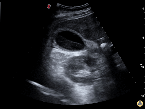
Gallbladder with Biliary Sludge
The image above is from a patient presenting to the emergency department with recurrent abdominal pain. The image demonstrates a large amount of biliary sludge within a normal sized gallbladder. Biliary colic from intermittent obstruction was suspected. While the gallbladder wall is noted to be mildly thick here, cholecystitis was ruled out based on other clinical factors.
Marco Garrone, MD - Torino, Italy

Gallbladder Sludge
Echogenic material can be seen within this gallbladder but there are no stones. It is not bright white (it is not highly echogenic) and furthermore there isn't any shadowing. When there is no shadowing consider polyps or in this case biliary sludge.
Justin Bowra MBBS, FACEM, CCPU Emergency Physician, RNSH et al. (Sharon)
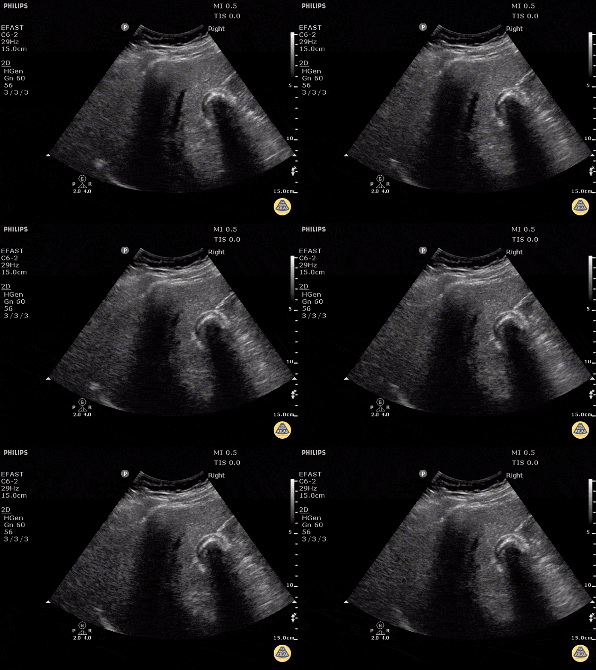
Gallbladder Wall Echo Shadow (WES Sign)
Gallstones can be seen on the right side of the image with a hyperechoic front edge and posterior shadowing.
Justin Bowra MBBS, FACEM, CCPU Emergency Physician, RNSH et al.
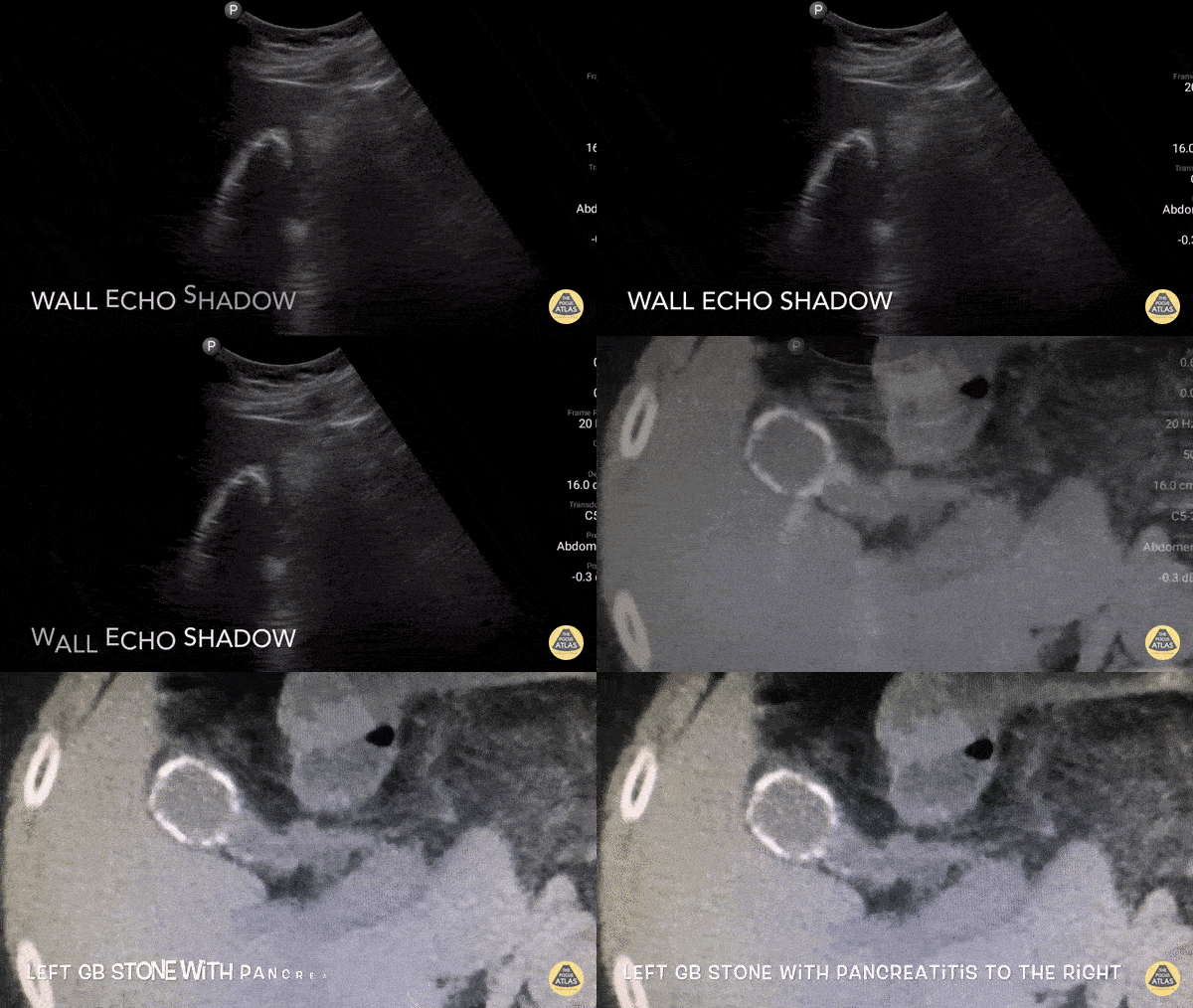
Wall Echo Shadow
Pt with epigastric pain, elevated LFTs and lipase. U/S shows the whole gallbladder shadowed by a large calcified stone or "wall echo shadow-WES." The novice may not be able to find the GB and can mistake WES for bowel (but bowel does not shadow like this).
Dr. Gordon Johnson

Portal Triad
Gallbladder with non-obstructing stone. Fanning down one sees the portal triad with bile duct (black) 7 mm, hepatic artery (pulsitile) and portal vein (red).
Dr. Gordon Johnson

Pericholecystic Fluid
Free fluid can be appreciated around this gallbladder. This is called pericholecystic fluid. However there are no stones, nor gallbladder wall thickening, and the sonographic Murphy's is negative. This free fluid is unlikely to be caused by cholecystitis. In this case, the free fluid came from a different source.
Justin Bowra MBBS, FACEM, CCPU Emergency Physician, RNSH et al.
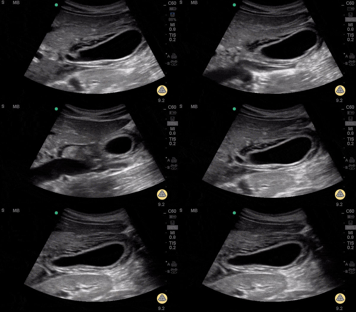
Pericholecystic Fluid - Long
26 y/o F PMH HIV presents with non-bloody, non-bilious vomiting for one day associated with upper abdominal pain. POCUS revealed gallbladder wall thickening and pericholecystic fluid, but no gallstones or sonographic Murphy’s sign. The patient received symptomatic treatment and a surgical evaluation. The patient ultimately improved and was determined to not have cholecystitis. The patient was discharged home after her symptoms resolved and she was able to tolerate PO.
Dr. Guru Shan, Dr. Guy Carmelli, Dr. Scott Kendal - Kings County Emergency Medicine
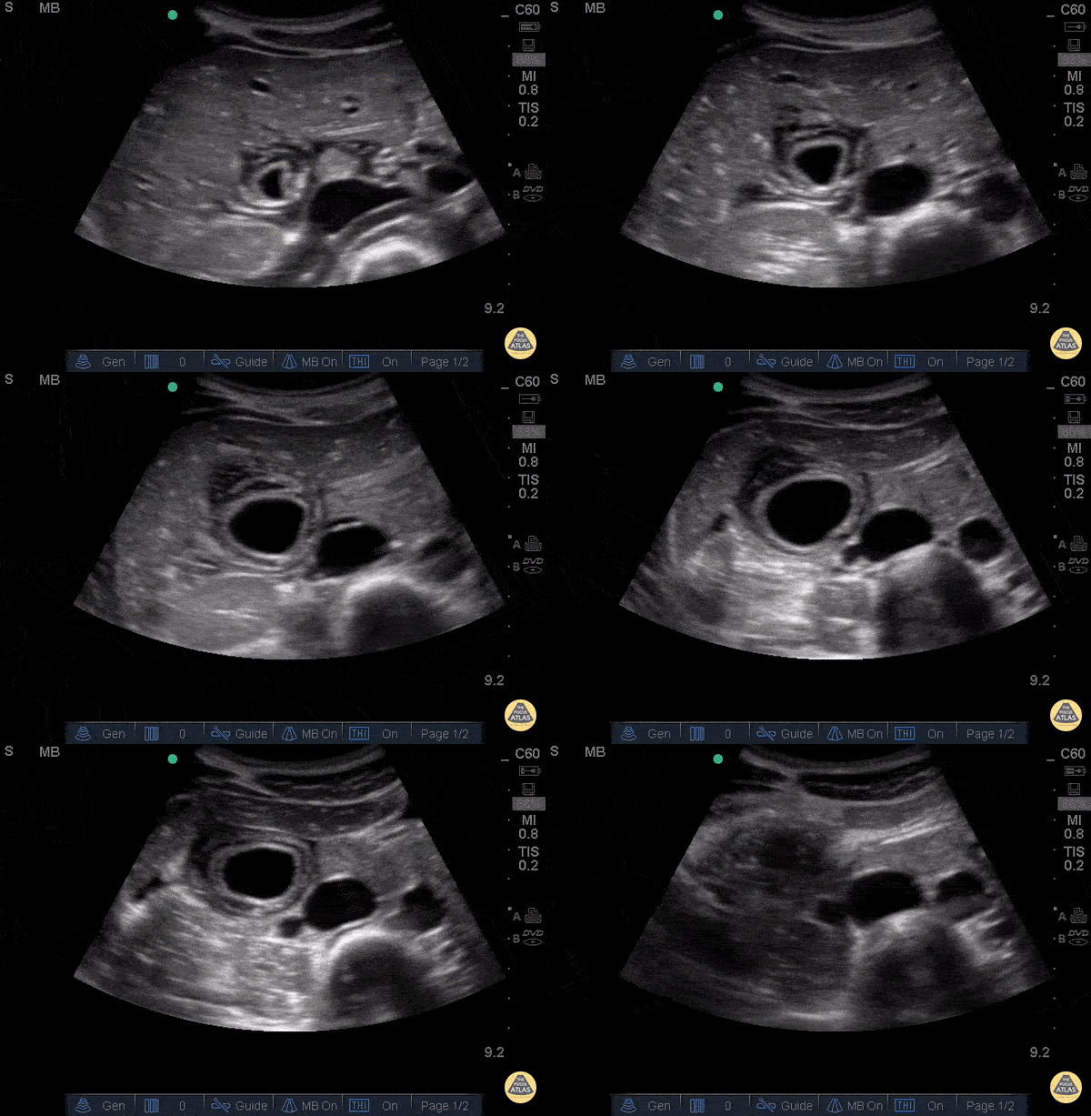
Pericholecystic Fluid - Transverse
26 y/o F PMH HIV presents with non-bloody, non-bilious vomiting for one day associated with upper abdominal pain. POCUS revealed gallbladder wall thickening and pericholecystic fluid, but no gallstones or sonographic Murphy’s sign. The patient received symptomatic treatment and a surgical evaluation. The patient ultimately improved and was determined to not have cholecystitis. The patient was discharged home after her symptoms resolved and she was able to tolerate PO.
Dr. Guru Shan, Dr. Guy Carmelli, Dr. Scott Kendal - Kings County Emergency Medicine
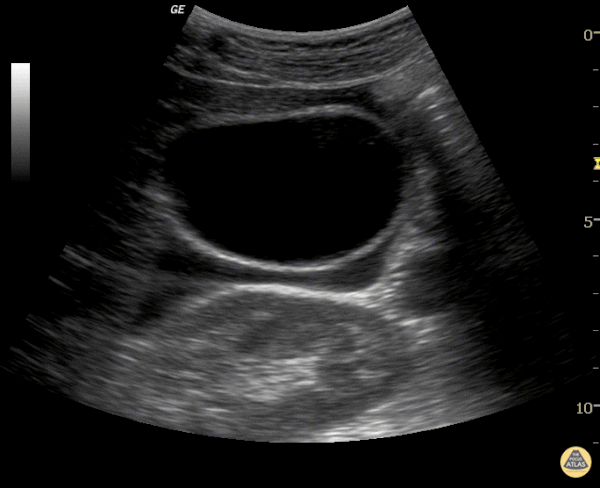
Free Fluid around Normal Gallbladder
There is a lot of pericholecystic fluid surrounding this gallbladder but no other signs of inflammation. The gallbladder wall is normal, there are no stones, and there is no sonographic Murphy's sign. The free fluid is likely from another source, not acute cholecystitis.
Justin Bowra MBBS, FACEM, CCPU Emergency Physician, RNSH et al.
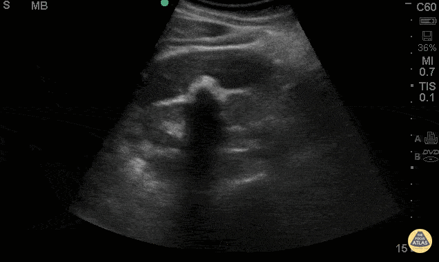
Wall Echo Shadow
38 y/o F presented with abdominal pain, nausea & vomiting. POCUS shows the WES (Wall-Echo-Shadow) sign. This shows a curvelinear hyperechoic line representing the gallbladder wall, followed by a thin hypoechoic line representing a small amount of bile, then a curvelinear hyperechoic line, followed by acoustic shadowing. This sign is seen when the gallbladder has either a large gallstone or multiple small gallstones in a contracted gallbladder taking up the extent of the lumen. Sometimes this sign is misinterpreted as an air filled loop of adjacent bowel.
Jaramillo, Juliana MD; Shah, Rushabh MD; Maurelus, Kelly MD - Kings County Emergency Medicine
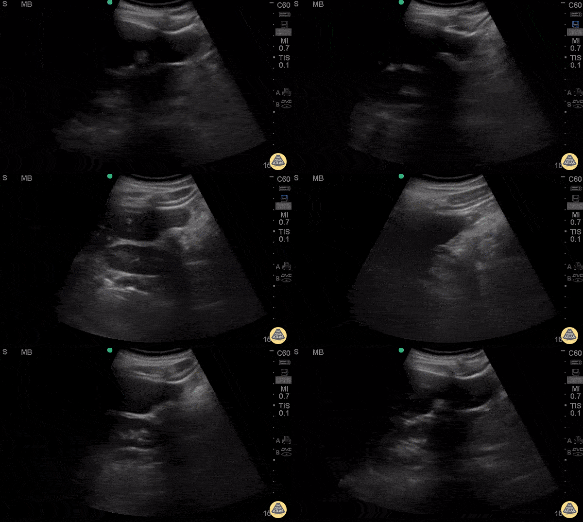
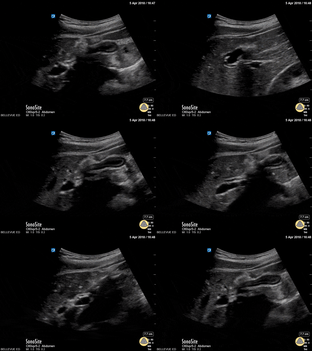
Contracted Gallbladder
In this clip, the gallbladder (center right of the screen) appears collapsed and the wall looks thickened. This is the normal appearance of a postprandial gallbladder which is contracted because it has just released its bile content into the duodenum for digestion of a meal.
Hannah Kopinksi and Dr. Lindsay Davis - NYU Emergency Medicine
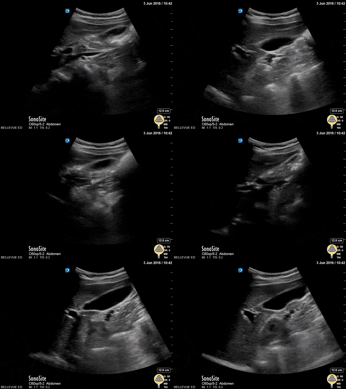
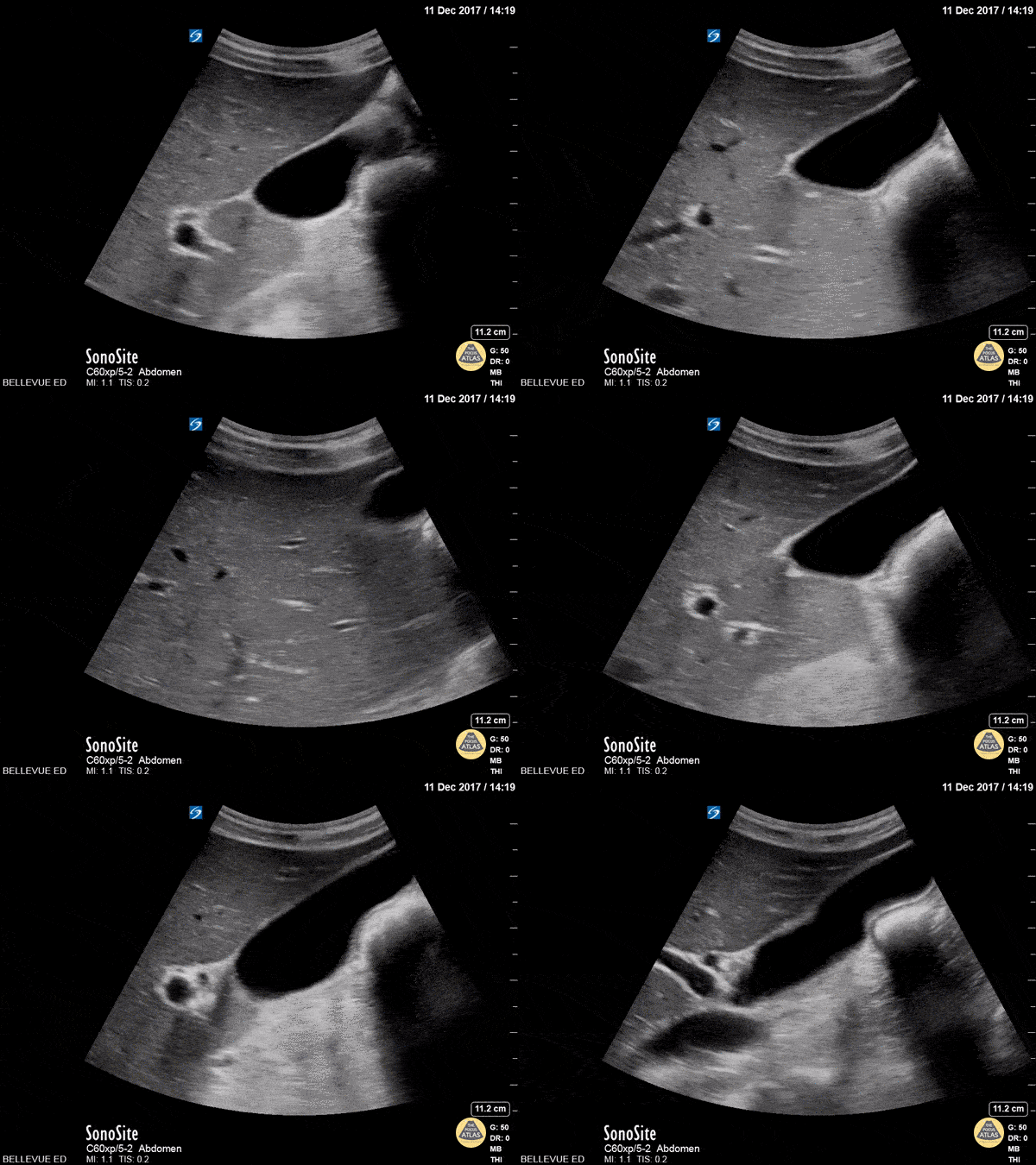
Gallbladder Long Axis
In this clip we see the liver on the left of the screen, and in the center right a large anechoic ovoid structure which is the gallbladder. The gallbladder in long axis together with the portal triad in cross-section form the “exclamation point sign”.
Hannah Kopinksi and Dr. Lindsay Davis - NYU Emergency Medicine
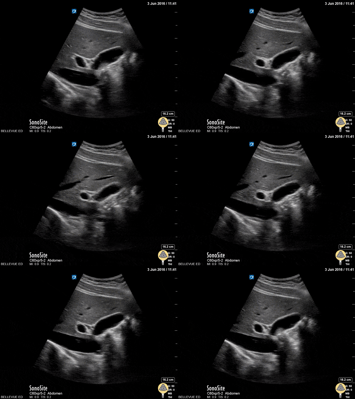
Gallbladder and CBD
In this clip we see the liver on the left, and the IVC inferior to the liver with a hepatic vein draining into it. The ovoid anechoic gallbladder is in long axis in the center of the screen. To the left of the gallbladder we see the portal vein in cross-section. Running just superior to the portal vein, in long axis, is the narrow common bile duct.
Hannah Kopinksi and Dr. Lindsay Davis - NYU Emergency Medicine
Gallbladder and Right Hepatic Artery
Here we see a normal anatomic variant: the right hepatic artery (small perfectly round vessel in cross section) runs superior to the CBD (seen in long axis towards the end of the clip).
Hannah Kopinksi and Dr. Lindsay Davis - NYU Emergency Medicine
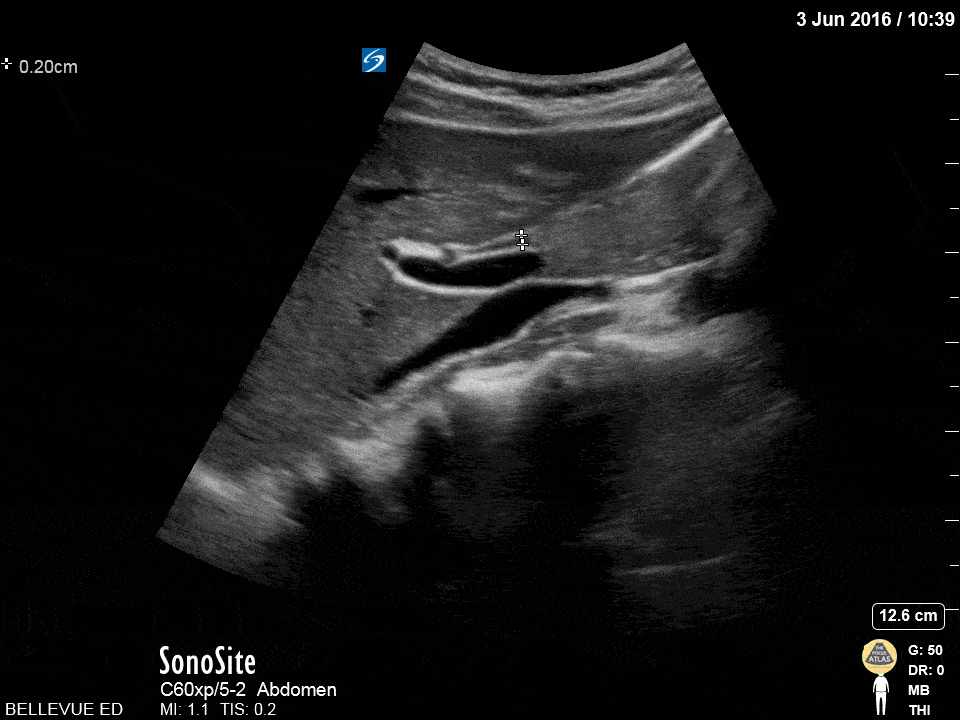
Olive Sandwich
The “olive sandwich” sign: the small round hepatic artery (in cross section) sandwiched between long axis views of the CBD superiorly and portal vein inferiorly. The IVC is seen in long axis deep to the portal vein.
Hannah Kopinksi and Dr. Lindsay Davis - NYU Emergency Medicine

Pancreatic Pseudocyst
40s F PMH severe ETOH use disorder and ETOH pancreatitis as well as recent COVID diagnosis presented with continued dyspnea and sore throat as well as abdominal bloating with a 15-20lb weight loss in 2 months. POCUS of the upper abdomen is shown here, demonstrating a large pseudocyst, which was confirmed on CT. The patient was admitted for her symptoms and was seen by GI but ultimately was planned for outpatient drainage rather than urgent/emergent.
Dr. Nimish Bhatt, Ultrasound Fellow
Denver Health Ultrasound Fellowship

Impacted Gallbladder Neck Stone
20s F presented with recurrent postprandial RUQ pain. POCUS demonstrated multiple hyperechoic stones in the gallbladder including one in the gallbladder neck. No pericholecystic fluid or wall thickening were noted, and the common bile duct was normal caliber. Formal RUQ US confirmed these findings, and the patient was taken to the OR by the surgery team for cholecystectomy.
Dr. Nhu-Nguyen Le, Ultrasound Fellow
Denver Health Ultrasound Fellowship
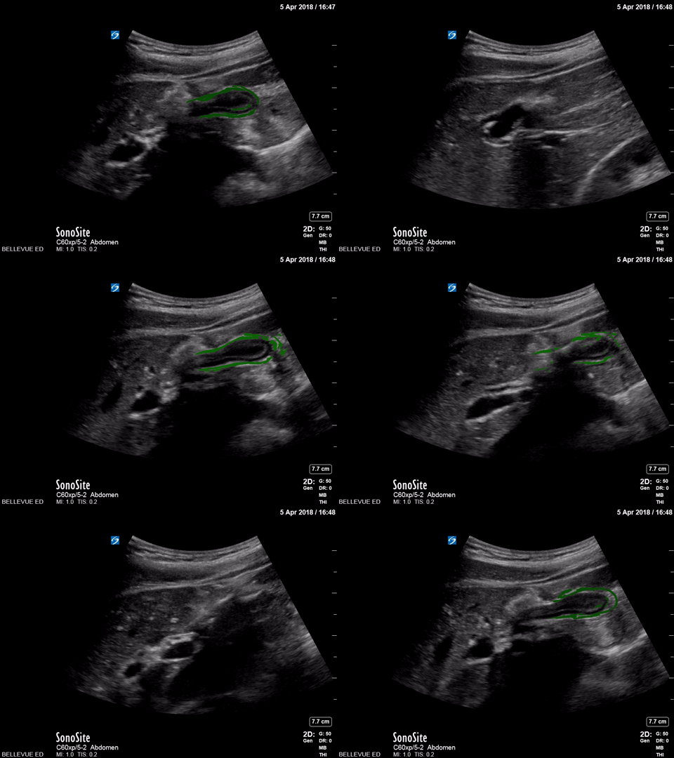
Contracted Gallbladder - Colorized
Contracted Gallbladder
Green- Gallbladder
Images: Dr. Lindsay Davis, Dr. Hannah Kopinski. Image Editing: Michael Amador and Dr. Matthew Riscinti
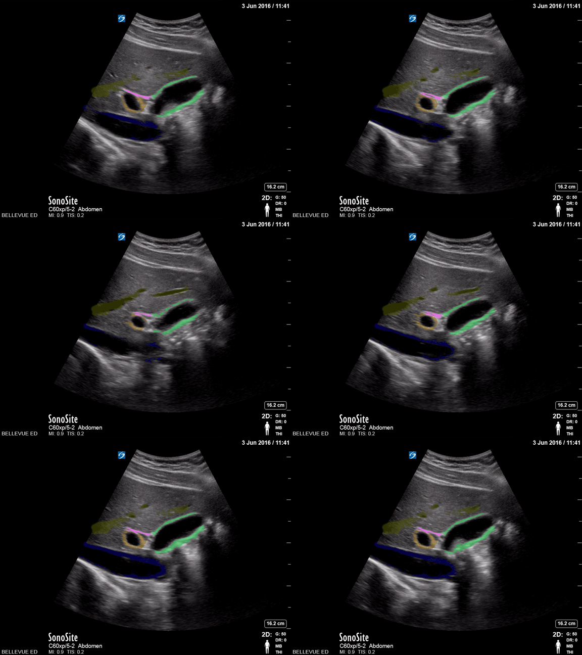
Gallbladder and Commmon Bile Duct - Colorized
Gallbladder and Common Bile Duct
Green: Gallbladder, Blue: IVC, Orange: Portal vein, Pink: Common bile duct, Yellow: Lumen of hepatic vein
Images: Dr. Lindsay Davis, Dr. Hannah Kopinski. Image Editing: Michael Amador and Dr. Matthew Riscinti
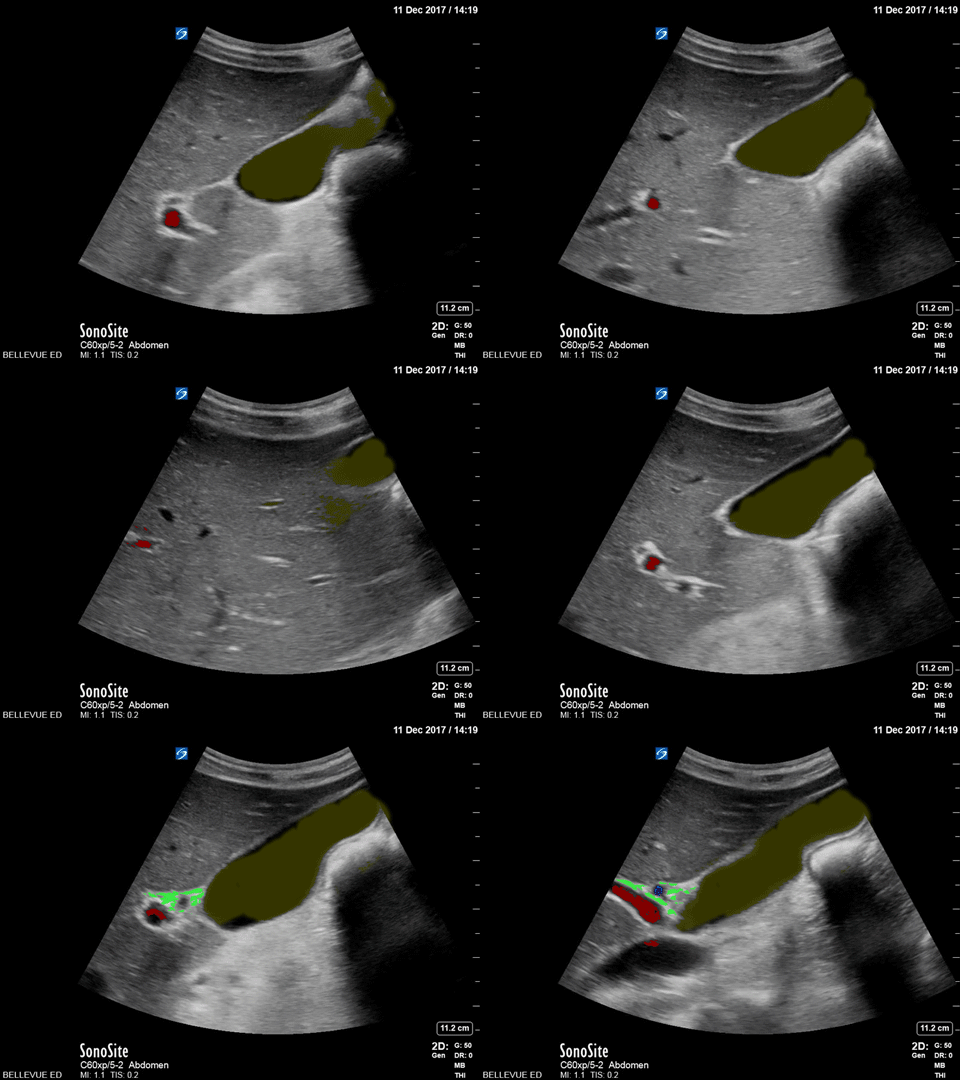
Normal Gallbladder - Colorized
Normal Gallbladder
Yellow: Lumen of gallbladder, Red: Portal vein, Green: Common bile duct
Images: Dr. Lindsay Davis, Dr. Hannah Kopinski. Image Editing: Michael Amador and Dr. Matthew Riscinti
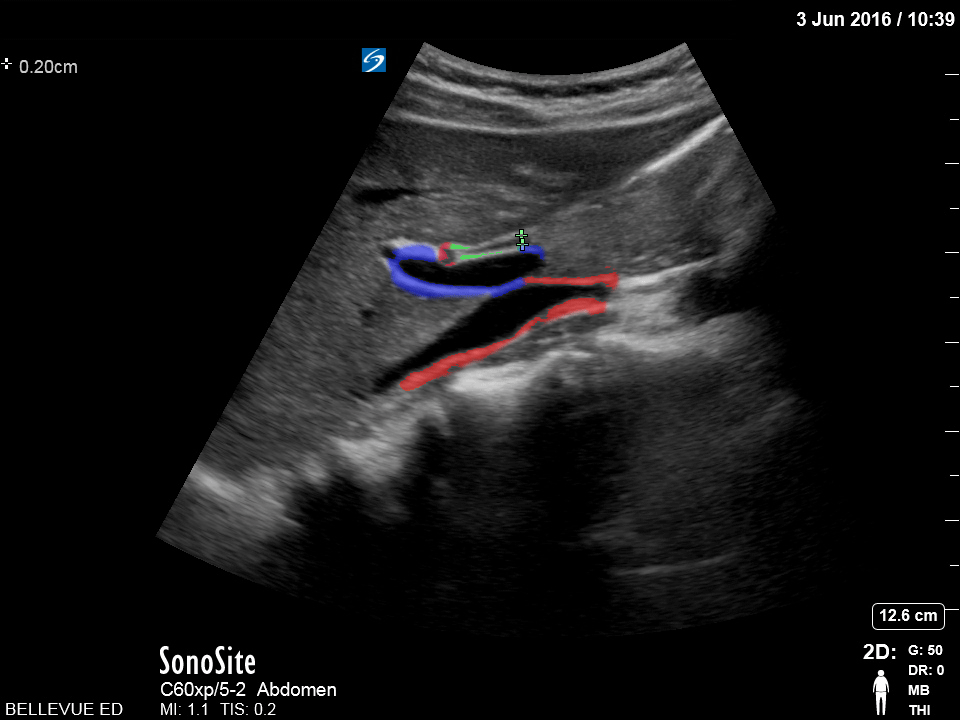
Olive Sandwich Sign - Colorized
Olive Sandwich Sign
Red (small): “Olive” or hepatic artery, Green: Common bile duct, Blue: Portal vein, Red (large): IVC
Images: Dr. Lindsay Davis, Dr. Hannah Kopinski. Image Editing: Michael Amador and Dr. Matthew Riscinti


