
Pulmonary

Lung Cavitary Lesion
HIV+ male patient presents with cough and shortness of breath. Thoracic US demonstrated a left upper lobe cavitary lesion with calcifications.
Differentials included TB, aspergilloma, lung abscess.
Contributors: Sara Schambach, MD; Garrett Mason, MD

Snowstorm Sign in Endotracheal Intubation
This is a clip of a cervical airway view during intubation attempt.
Some artifact is visible in the trachea when passing the tube demonstrating a "Snowstorm Sign" which can be used to confirm the position of an endotracheal tube. In this view it is possible to see the esophagus next to the trachea. As demonstrated there is no motion detected in esophagus during intubation therefore confirming tracheal intubation.
Contributor: Renato Tambelli (@R_Tambelli @Jedipocus)

Lung Point Finding
Lung point is a pathognomonic finding on US for pneumothorax. It refers to the junction between healthy lung and collapsed lung.
This is represented in the US recording as lung sliding seen on the left of the pleural line but no lung sliding seen on the right of the pleural line.
Contributors: Dimitri Livshits, DO; Jane Belyavskaya, MD; Chris Hanuscin, MD
Kings County/SUNY Downstate

Large pleural effusion with hiatal hernia
72 year old with past medical history of hiatal hernia presenting to the ED with new onset shortness of breath requiring rescue BPAP. She was diagnosed and treated with CHF exacerbation. Lung ultrasound showed a large pleural effusion with uncertain mass-like object with heterogenous fluid contents on the right (shown here), along with dense B-lines. Correlation with prior CT suggested that the structure on the right chest was a large hiatal hernia.
Contributed by: William McGill, PA-C

Subpleural Consolidation with Shred Sign
Patient admitted to ICU with sepsis of unknown origin. Complaints of tachypnea led to ultrasound scanning, which showed a subpleural hypoechoic image in the posterolateral region of the left chest compatible with pneumonia.
Contributed by: @intensivanaveia
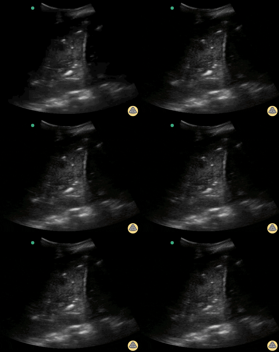
Massive dynamic air bronchogram
Massive lung consolidation in a patient diagnosed with COVID-19 with bacterial component.
Contributor: Rafael Intensivanaveia - Critical Care Physician at Hospital Israelita Albert Einstein

Pleural Effusion with Compressed Lung and Spine Sign
Pleural effusion with compressed lung and positive spine sign
Contributed by: Dimitri Livshits DO, Ultrasound Fellow; Jane Belyavskaya MD, Ultrasound Fellow; Chris Hanuscin MD, Ultrasound Division Director (Kings County/SUNY Downstate)

Subpleural Consolidation in COVID-19
Subpleural consolidation (shred sign) in a COPD patient with Covid 19, admitted to the ICU with acute respiratory failure progressing to intubation and mechanical ventilation.
Contributor: Bruno Souza @BrunoSo03038122

Lung Point in Iatrogenic Pneumothorax
Lung ultrasound in the right apical region after puncture attempt for central venous access. The image demonstrates a LUNG POINT, a specific sign of a pneumothorax.
Contributed by: Breno Moura

Color Doppler of Consolidation in Pneumonia
Massive lobar consolidation showing pulsatile flow on color doppler, befitting pneumonia.
Contributor: Rafael Intensivanaveia

Lung Re-expansion With Pigtail Catheter
Pictured here is an amazing return of observable lung sliding as a pigtail catheter is advanced through. Reestablishment of lung sliding indicates lungs have re-expanded.
Image courtesy of Robert Jones DO, FACEP @RJonesSonoEM
Director, Emergency Ultrasound; MetroHealth Medical Center; Professor, Case Western Reserve Medical School, Cleveland, OH
View his original post here

Lung Consolidation from Bronchial Obstruction
This is an image demonstrating dense lung consolidation with surrounding pleural effusion. Patient ultimately was diagnosed with bronchogenic CA with complete bronchial obstruction.
Image courtesy of Robert Jones DO, FACEP @RJonesSonoEM
Director, Emergency Ultrasound; MetroHealth Medical Center; Professor, Case Western Reserve Medical School, Cleveland, OH
View his original post here

Lung Slide M-mode
Lung Sliding with M mode; looking like a seashore.
Mary Fran King

Septated Pleural Effusion
76yo male with shortness of breath on exertion, w/o fever or other signs of infection. POCUS showed a large septated unilateral pleural effusion. Note the irregular and thickened pleura adjacent to the diaphragm (right).
Thoracentesis was only partially successful with a large residual effusion after drainage of 1000ml of exudative fluid with signs of lymphocytic inflammation w/o malignant cells on cytopathologic analysis. Thoracoscopic biopsy confirmed the diagnosis of a pleural mesothelioma 60 years after exposure to asbestos.
The presence of a septated complex effusion is 94% specific for an exudative effusion and warrants further investigation (Shkolnik, 2020). Cytopathology of pleural fluid is often negative in malignant mesothelioma and pleural biopsies are needed to confirm the diagnosis (Porcel, 2014).
Victor Speidel
Langenthal Regional Hospital, Switzerland

PLAPS consolidation
23 yo male, comes to Emergency Department with cough, fever and dyspnea. Lung Ultrasound in the right PLAPS point show this beautiful image: a large consolided lung with dynamic air bronchograms - numerous tortuous hyperechoic opacities that move with the respiratory cycle. This finding has a specificity of 94% and a positive predictive value of 97% for pneumonia as the cause of the consolidation.
Renato Tambelli
@JediPocus @R_Tambelli

Pneumonia
A 22-year-old patient without a medical background presents to the ED with a 2-day history of left costal stabbing pain. There was no shortness of breath nor fever and barely any respiratory symptoms. Lung ultrasound revealed a thick, interrupted pleural line and a mobile, hypoechoic structure with irregular edges compatible with a consolidation. Covid PCR was negative.
Dr. Felipe Urriola P.

Lung Point
18-year-old patient who presented following a motorcycle accident in which he sustained closed chest trauma with bilateral hemopneumothoraces. In this sequence taken with a linear transducer in left pulmonary zone 1, the "pulmonary point" indicative of pneumothorax can be seen.
Libardo Valencia Chicue

Pericardial & Pleural Effusions
Seen here is a subxiphoid view taken from a patient admitted with symptoms of acutely decompensated heart failure. You can appreciate both a small pericardial effusion as well as a more noticeable pleural effusion.
Reyna Huerta Sánchez, MD
@DraHuerta09

No Lung Sliding
This image demonstrates lack of lung sliding which is a finding that can be seen in a pneumothorax.
Francisco Norman

Parapneumonic Effusion
Right lower lobe pneumonia with lobar atelectasis and pleural fluid accumulation. These ultrasonographic findings are collectively referred to as jelly fish sign. This anechoic parapneumonic effusion measured 2.5 cm on ultrasound, and chest tube insertion resulted in drainage of 800 mL of clear fluid.
Gábor Csupor

Large Pleural Effusion
Patient presented to the emergency room reporting dyspnea. POCUS revealed presence of severe ascites and pleural effusion.
Reyna Huerta Sanchez, MD
@DraHuerta09

Air Bronchograms
Seen here are dynamic air bronchograms obtained via POCUS on a patient in whom we had clinical suspicion of a diagnosis of pneumonia.
Francisco Norman

Pneumothorax with Lung Point
A patient presented with spontaneous pneumothorax; lung point identified on POCUS.
ChunYi Tsai, @jerry1231213

Lung Re-expansion
POCUS reveals the return of lung sliding following insertion of a chest tube for pneumothorax.
Image courtesy of Robert Jones DO, FACEP @RJonesSonoEM
Director, Emergency Ultrasound; MetroHealth Medical Center; Professor, Case Western Reserve Medical School, Cleveland, OH
View his original post here

Malignant Pleural Effusion
A patient with a history of lung cancer presented to the ED with a fever and hypotension. Subcostal window revealed a septated large malignant pleural effusion.
Image courtesy of Robert Jones DO, FACEP @RJonesSonoEM
Director, Emergency Ultrasound; MetroHealth Medical Center; Professor, Case Western Reserve Medical School, Cleveland, OH
View his original post here

B Lines with Irregular Pleura
Irregular pleural line with patchy B-lines. Seen in viral (including COVID19) and bacterial pneumonia.
Image courtesy of Robert Jones DO, FACEP @RJonesSonoEM
Director, Emergency Ultrasound; MetroHealth Medical Center; Professor, Case Western Reserve Medical School, Cleveland, OH
View his original post here

Pneumonia-Subpleural Consolidation
Irregular pleural lining, B lines, and subpleural consolidation best seen with a linear probe.
Image courtesy of Robert Jones DO, FACEP @RJonesSonoEM
Director, Emergency Ultrasound; MetroHealth Medical Center; Professor, Case Western Reserve Medical School, Cleveland, OH
View his original post here

Normal Lung Sliding
Normal lung sliding with regular appearing pleural lining.
Image courtesy of Robert Jones DO, FACEP @RJonesSonoEM
Director, Emergency Ultrasound; MetroHealth Medical Center; Professor, Case Western Reserve Medical School, Cleveland, OH
View his original post here

Pneumonia-Parapneumonic Effusion
Large echogenic right parapneumonic effusion in patient with bacterial pneumonia/sepsis. Phased array probe used.
Image courtesy of Robert Jones DO, FACEP @RJonesSonoEM
Director, Emergency Ultrasound; MetroHealth Medical Center; Professor, Case Western Reserve Medical School, Cleveland, OH
View his original post here

Pneumonia-Parapneumonic Effusion
Small right anechoic parapneumonic effusion. Note B-lines seen at lung base. Curvilinear probe used. More commonly seen in bacterial pneumonia.
Image courtesy of Robert Jones DO, FACEP @RJonesSonoEM
Director, Emergency Ultrasound; MetroHealth Medical Center; Professor, Case Western Reserve Medical School, Cleveland, OH
View his original post here

Pneumonia-Shred Sign
The shred sign is a sign of consolidation in the lung that appears as subpleural hypoechoic areas with an irregular “shredded” border.
Image courtesy of Robert Jones DO, FACEP @RJonesSonoEM
Director, Emergency Ultrasound; MetroHealth Medical Center; Professor, Case Western Reserve Medical School, Cleveland, OH
View his original post here

Pnuemonia-Thickened Pleura
Increased thickness of the pleura with diffuse B-lines can be seen in this patient with pneumonia.
Image courtesy of Robert Jones DO, FACEP @RJonesSonoEM
Director, Emergency Ultrasound; MetroHealth Medical Center; Professor, Case Western Reserve Medical School, Cleveland, OH
View his original post here

Air Bronchograms in LLL Pneumonia
Dynamic air bronchograms present in a patient with left lower lobe pneumonia. LLL shows signs of hepatization. Find a labeled image at the original post here.
Image courtesy of Robert Jones DO, FACEP @RJonesSonoEM
Director, Emergency Ultrasound; MetroHealth Medical Center; Professor, Case Western Reserve Medical School, Cleveland, OH

COVID-19 Pneumonia
Seen here is an irregular and thickened pleural line with associated focal and confluent B lines in a patient with COVID-19 pneumonia.
Edgar Miranda

Unexpected Jellyfish
Seen here is an unexpected finding while acquiring an apical 4 chamber view with a phased array probe. The four chambers of the heart are difficult to bring into view due to the presence of large left pleural effusion with ipsilateral collapsed lung floating within the pleural fluid. This visual of atelectatic lung swimming within a pleural effusion is referred to as “jellyfish sign”.
Renato Tambelli, Emergency Physician Hospital das Clínicas de Marília, Brazil.
@R_Tambelli // @JediPocus

Dynamic Air Bronchograms
Pictured here are dynamic air bronchograms in a patient with bacterial pneumonia. Image was acquired using convex probe on the left PLAPS.
Renato Melo, PocusJedi co-founder, Emergency Physician HC Marília-SP, Brazil.
@Renato_Melo_

Pulmonary Contusion
A 23-year-old male was admitted to the ED following a motorcycle accident. He subjectively reported dyspnea; objectively was hypoxic. POCUS seen here (obtained from L3 zone using curvilinear probe) reveals multifocal B-lines consistent with pulmonary contusion. This case illustrates that bedside US is useful beyond diagnosing pneumothorax and hemothorax in trauma patients.
Renato Tambelli, Emergency Physician Hospital das Clínicas de Marília, Brazil.
@R_Tambelli / @JediPocus

Light Beam Sign
This image was taken from a 58-year-old man with cough and fever x 8 days who tested positive for SARS-CoV-2 Virus.
Pictured here is a POCUS view (R3 Zone) obtained in oblique position with a curvilinear probe. Appreciate the lung sliding, regular pleural line, and alternating multiple separated B-lines and A-lines; this constellation of findings is referred to as “Light Beam Sign”.
Renato Tambelli, Emergency Physician, Hospital das Clínicas de Marília, Brazil
@R_Tambelli / @JediPocus

Hepatization of Lung
Patient presented with septic shock. POCUS was used to assess for etiology in this hemodynamically unstable patient. Shown here is a lung US windows (patients LLL) revealing a consolidation pattern with echoginicity similar to liver, referred to as “hepatization” of the lung. These findings are consistent with a diagnosis of pneumonia.
Johannes Achenbach

Jellyfish Sign
A middle age man presented with a subacute dyspnea, orthopnea, non productive cough, and paroxysmal nocturnal dyspnea. Physical exam was notable for bilateral, fine, end-inspiratory crackles over the lung bases; an S3 on cardiac auscultation; and an SpO2 of 92% on 2L nasal cannula.
POCUS identified the presence of atelectatic lung in an anechoic large right-sided pleural effusion. The sonographic appearance of atelectatic lung "swimming" within a large pleural effusion is often referred to as “jellyfish sign”. Subsequent laboratory evaluation of the pleural fluid confirmed a transudative effusion secondary to decompensated left-sided heart failure.
Al Chalaby, Shahad. PGY3. Highland Hospital. Alameda Health System Internal Medicine Residency Program.
@shahad_Chalaby
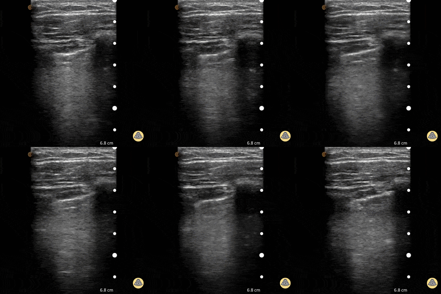
Normal Lung Slide
To assess for lung slide, look between two ribs. The two layers of pleura can be seen as the hyperechoic, shimmering line just under the subcutaneous tissue. This represents the sliding of the parietal and visceral pleura.
Normal lung slide should have: Shimmering aka "ants marching"
This may be augmented by m-mode as pictured in another post.
Dr. Matthew Riscinti - Kings County Emergency Medicine
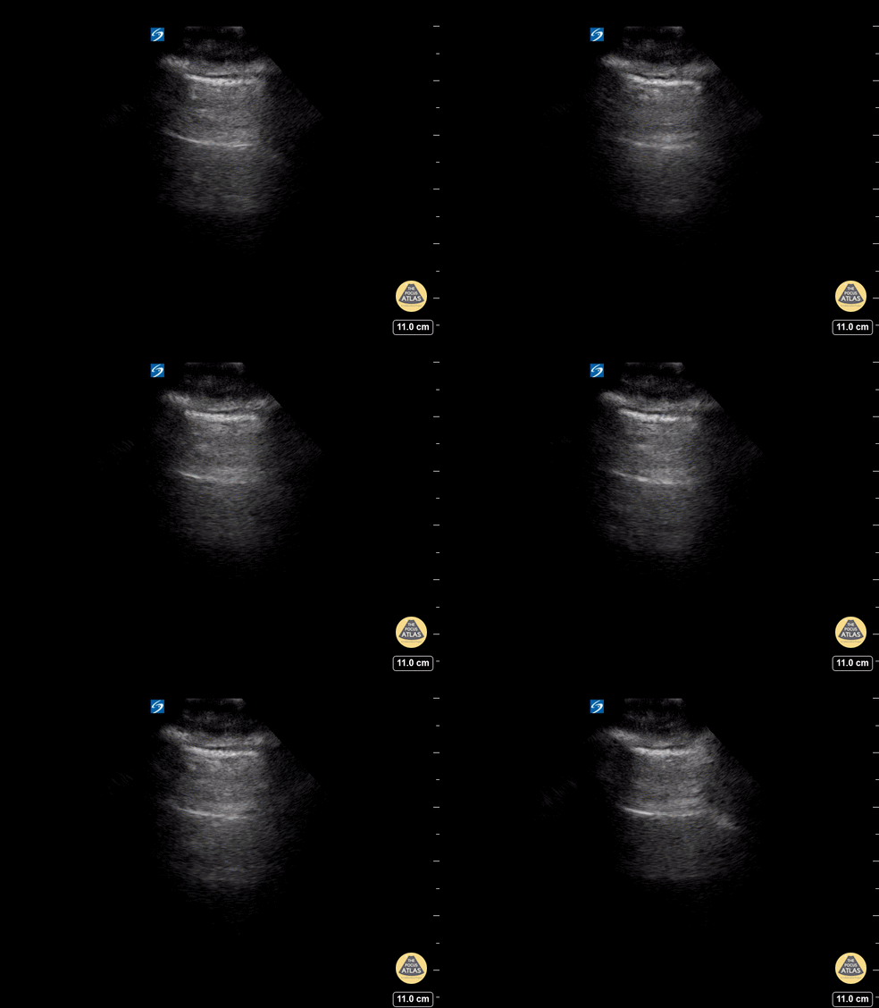
A - Lines - Normal Lung
A lines appear as horizontal lines that represent normal aerated lung (dry interlobular septa). They are a reverberation artifact caused by the sound waves bouncing off the highly echogenic pleura and back to the probe, and repeating.
Hannah Kopinski (MS4) and Dr. Lindsay Davis - NYU Emergency Medicine, Matthew Riscinti - Kings County Emergency Medicine

Normal lung
Lung ultrasound of a normal lung. Note both lung slide (shimmery, hyperechoic line on top of the screen, a result of parietal and visceral pleura sliding against each other) as well as multiple parallel A-lines (normal artifact from reverberation of the pleural line). The presence of lung sliding and no more than 3 B lines on lung ultrasound help exclude inersitital pulmonary edema and pneumothorax.
Shahad Al Chalaby, MD. PGY-2, Internal Medicine
Highland Hospital, Alameda Health System Internal Medicine Residency Program. CA, USA
@shahad_Chalaby

Lung Windows
This still-shot image captures the differences in view as obtained using a linear vs curvilinear to assess a lung window.
Submitted by Anibal Artero
@ECOPULMONAR
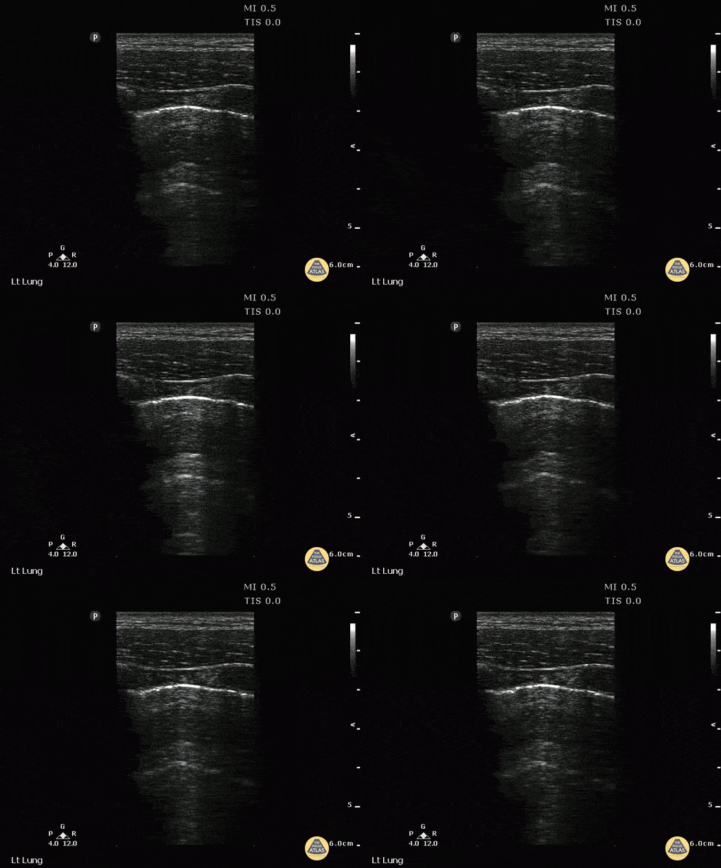
Double Lung Point Seen in Pneumothorax
A young male presented to ED with stab wound to chest. Upright CXR was normal. POCUS showed double lung point indicative of small PTX. This was monitored without evidence of progression. No intervention was required.
Image courtesy of Robert Jones DO, FACEP @RJonesSonoEM
Director, Emergency Ultrasound; MetroHealth Medical Center; Professor, Case Western Reserve Medical School, Cleveland, OH
View his original post here

Light Beam Sign (COVID-19)
Seen here is a "Light Beam Sign" in a patient with COVID-19 pneumonia. The on-and-off effect of the hyperecchyoic vertical artifact is believed to occur as a result of ground glass alterations in lung parenchyma. At times the light beams or B-lines cover A-lines; and at other times the A-lines remain visible in the background.
Renato Melo, Emergency Physician at Hospital das Clínicas de Marília-SP, Brazil. Co-founder of Pocus Jedi
@Renato_Melo_

Atypical presentation COVID-19
An elderly male presented from home with complaints of mild confusion, non-specific abdominal pain, and a 3-day history of dyspnea. He required no supplemental oxygen. 12-lung zone ultrasound was performed using the linear transducer. Zone R5 revealed the pictured confluent B-line pattern and small areas of consolidation. Patient subsequently tested positive for the SARS-CoV-2 virus.
Cian McDermott, Emergency Physician Dublin, Ireland
@cianmcdermott

COVID-19 Pneumonitis
Patient presented to the Emergency Department with 2-day history of worsening dyspnea and increased work of breathing. He was profoundly hypoxic upon arrival with EMS (O2 sat on 2L via nasal prongs was 42%; improved to 75% upon switching to high flow nasal cannula at 40L/min). A 12-zone lung ultrasound was performed using a linear probe and what is pictured is from L6 (left inferior posterior zone). You can appreciate the coarse irregular pleura, patchy B-lines, and and small areas of consolidation. Findings are typical for clinically-suspected COVID-19 pneumonitis.
Cian McDermott, Emergency Physician; Dublin, Ireland
@cianmcdermott

Pleural Shred Sign
Seen here is a pleural shred sign (discontinuity of a thickened pleura) and B-lines. Images were acquired from the LLL BLUE point on a patient who presented with fever and purulent discharge from tracheostomy site; all findings consistent with diagnosis of pneumonia.
Renato Melo, Emergency physician, H.C. de Marília-SP, Brazil
@Renato_Melo_

PLAPS pneumonia
This is a lung ultrasound image in PLAPS view in which we appreciate a consolidation pattern including dynamic air bronchograms as well as a small pleural effusion. The PLAPS view with these findings is a highly sensitivity and specificity for the diagnosis of pneumonia.
Renato Tambelli, Emergency Physician Hospital das Clínicas de Marília
@R_Tambelli

Jellyfish Sign
This a PLAPS-point view located slightly above the diaphragm. This window is a key component of lung ultrasound evaluation for pleural effusion. This image reveals what is know as "Jellyfish Sign,” or consolidated lung floating in anechoic fluid (pleural effusion). Also note the static air bronchograms within the lung parenchyma.
Renato Tambelli, Emergency Physician from Emergency Department of Marilia Clinics Hospital - Sao Paulo/ Brazil

Improve Lung Sliding Visualization
Normal lung sliding becoming more visible with decreased gain.
Aaron Inouye - @PAintheED
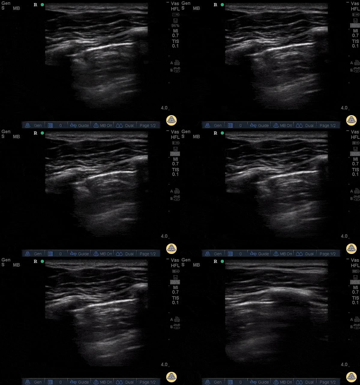
Decreased Lung Slide - Pneumothorax
26 yo male presents to ED stating he was kicked in the chest. He went home to “try to relax and smoke some weed” now short of breath and with pleuritic chest pain after smoking. POCUS demonstrating decreased lung slide on the left.
What are the signs of pneumothorax on ultrasound?
Decreased lung sliding - In normal lungs, lung sliding refers to the parietal pleura moving against the visceral pleura - described as “ants marching.”
Lack of B-lines or comet tails – These artifacts will not be present if there is a pneumothorax and the presence of B-lines or comet tails can rule out a pneumothorax.
No Lung pulse – the visceral pleura moving along a stationary parietal pleura due to cardiac motion when lung sliding is not present. These are so called “T lines” on M-mode. These signify that the parietal and visceral pleura are opposing one another and therefore that there is no pneumothorax
Lung point – 100% specific for pneumothorax, this is the cutoff point above which you can appreciate the lung sliding and below which there is no lung sliding. The lung point is the pneumothorax border.
Dr. Stacey Frisch, Dr. John F Kilpatrick - Kings County Emergency Medicine
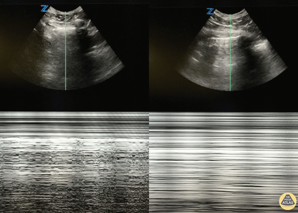
Pneumothorax: M-mode: Seashore vs Barcode
46 y/o M with 20 pack year smoking history with sudden onset right sided chest pain that woke him from sleep. Decreased breath sounds on right side. POCUS with decreased lung slide (right of image) with normal lung slide in left lung (left side of image).
Lung slide can often be appreciated by watching the pleural surfaces move along each other but if you're uncertain, putting the US in m-mode and looking for the classic "seashore sign" (left image) versus the "barcode sign" (right image) can help you figure it out.
Dr. Eric Roseman - Resident Physician, Kings County Emergency/Internal Medicine
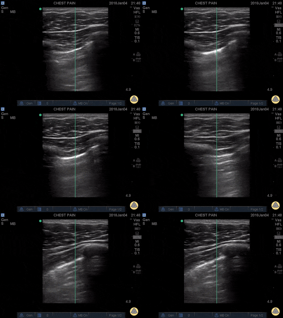
Lung Point: Pneumothorax
Trauma code to the waiting room... 20 y/o male stabbed to the left chest in the midaxillary line. Patient thrown in a wheelchair and pushed to the resuscitation room and POCUS performed immediately revealing this image: the lung sliding disappearing revealing an area without lung slide. Moved up one rib space to the apex, no lung slide at all.
This junction of slide/no slide is the lung point and its pathognomonic for pneumothorax. It represents the exact point where air begins to separate the parietal from visceral pleura, aka the junction of where we normally see the "ants marching" or the "shimmering" aka the lung slide. This is highly specific.
Don't be fooled when you see the sliding with decreased sliding around it. You're looking at a pneumothorax.

Lung Point
A young man presents to the emergency department with multiple thoracic stab woulds. POCUS quickly identifies absent lung sliding as well as a lung point; findings highly sensitive and specific for our diagnosis of pneumothorax.
Renato Tambelli, @R_Tambelli
Emergency Physician Hospital das Clínicas de Marília

Ultrasound Findings in Pulmonary Contusion
In this patient with history of blunt chest trauma, lung ultrasound reveals a focal area of B-lines as well as a hypoechoic wedge shaped subpleural consolidation.
These findings are consistent with pulmonary contusion and can be identified early in the course of a patient’s care. This contrasts with hours-to-short-days that it often takes to fully appreciate the evolution of pulmonary contusion on chest X-ray.
Aaron Inouye, PA-C, North Canyon Medical Center
@PAintheED

Classic Findings in Pneumonia
Pictured here are classic ultrasound findings for pneumonia including a shred sign, lung consolidation with dynamic air bronchograms and a small associated parapneumonic effusion. Note also adjacent B-lines.
A shred sign represents the distinction between the consolidated lung and the aerated lung and is seen in this clip as the irregular “shredded” border just posterior to the consolidation.
Aaron Inouye, PA-C, North Canyon Medical Center
@PAintheED
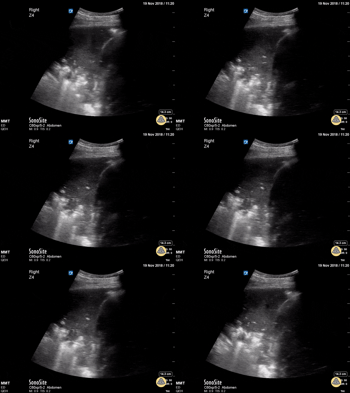
Dynamic Air Bronchograms in Pneumonia
This patient actually had no cough, no crackles and only subtle changes on CXR but in seconds we had diagnosed pneumonia!
Dynamic air bronchograms represent air bubbles moving up and down airways surrounded by alveolar consolidation. Lichtenstein et al compared this finding to static air bronchograms in a 2009 study of 68 ICU patients and found dynamic air bronchograms to be present in 32/52 cases of pneumonia but in only 1/16 cases of atelectasis - a specificity of 94%.
Dr. Trauer
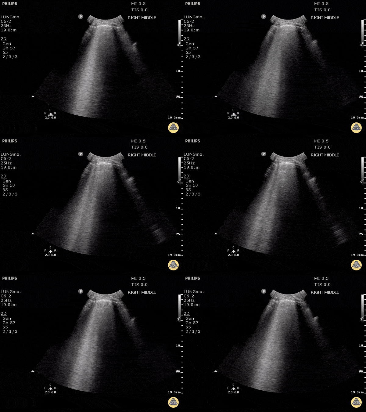
B-Lines
B-lines are vertical artifacts that moves with respiration from the pleural surface. They represent increased water in an area of the lung. In the right clinical context this could represent pulmonary edema. An increase in B-lines correlates with the degree of pulmonary edema.
Keep in mind that in different clinical contexts, they can represent different diagnoses including pulmonary contusions and pneumonia.
Justin Bowra MBBS, FACEM, CCPU Emergency Physician, RNSH et al.
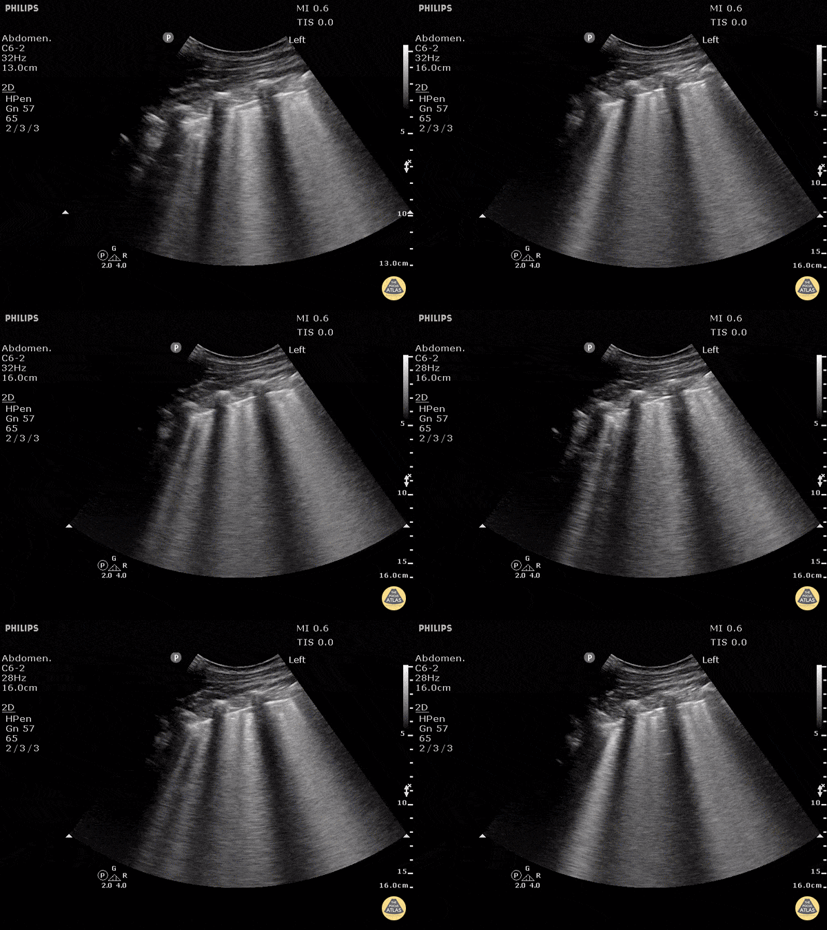
B-Lines - Pulmonary Edema
B-lines obtained with curved probe.
B-lines are vertical artifacts that move with respiration from the pleural surface. They represent increased water in an area of the lung. In the right clinical context this could represent pulmonary edema. An increase in B-lines correlates with the degree of pulmonary edema.
3 B-lines in an intercostal space represent a "positive" region of the lung, and if there are two regions of the lung that are positive, you can diagnose pulmonary edema.
Dr. Justin Bowra et al. (Dr. D Browne and Dr. J Knights)
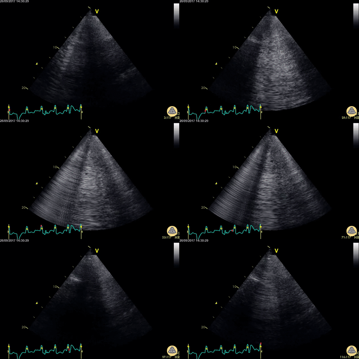
Confluent B Lines
WCUME 2017 Submission for "Best POCUS"
An acutely dyspnoeic patient presents with ventricular tachycardia and has no response to initial chemical cardioversion. Lung POCUS shows widespread bilateral confluent B lines indicating acute pulmonary edema. Unstable tachycardia terminated using synchronized electrical cardioversion.
Dr. Cian McDermott - Dublin, Ireland
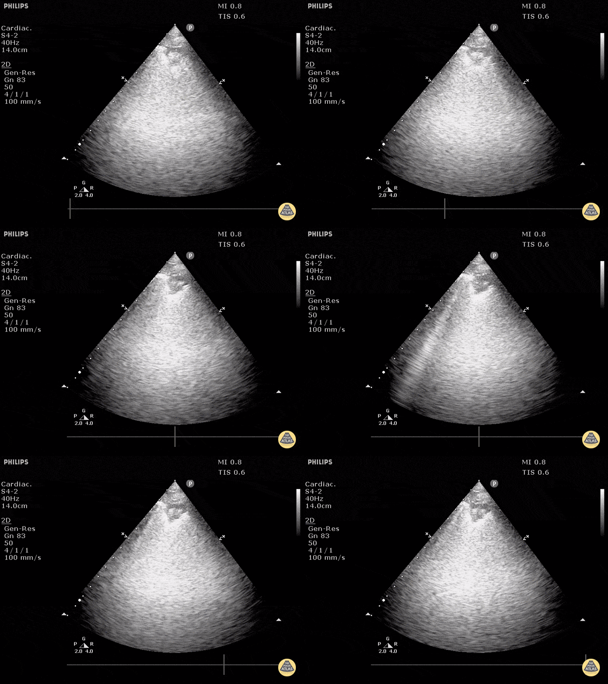
Pulmonary Embolism (Sector Probe)
Pulmonary embolism can be seen by disruption of the pleural line. 0.5cm to 3cm disruptions are typical for PE. Doing DVT studies and echo can help strengthen your diagnosis.
Dr. Justin Bowra et al.
Hydropoint
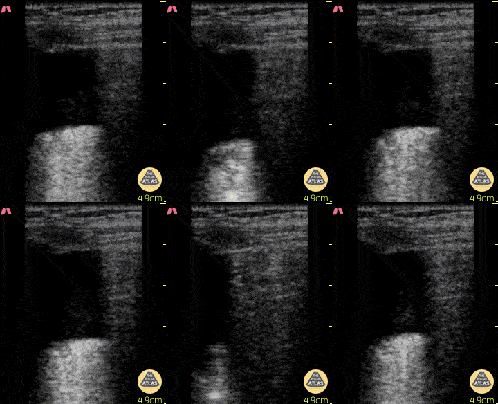
This clip, demonstrating a hydropoint, was taken in a 74 year old M with chest trauma after a fall from 3 meters. A hydropoint shows the air/fluid interface which is suggestive for hemato/hydro/pyo-pneumothorax. It is another sign for diagnosing a pneumothorax described by Volpicelli et al. Critical Ultrasound Journal. 2013
Dr. Van Roosmalen
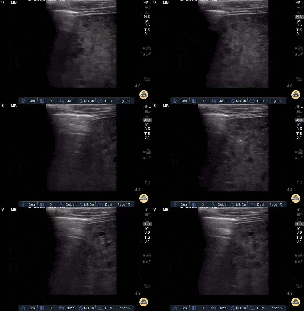
Lung Consolidation
53yoM with 20+ year history of tobacco and alcohol abuse and newly diagnosed SCC presenting with productive cough and cachexia, found to have multifocal pneumonia likely due to aspiration. Mid-axillary ultrasound of the right lung using linear 13-6 MHz probe in the longitudinal plane demonstrating hepatization of the lung (right) and focal consolidation adjacent to normal lung parenchyma (left). US has high SEN (88%) and SPE (86%) for detecting pneumonia when compared to CXR or chest CT.
(Ling 2017)
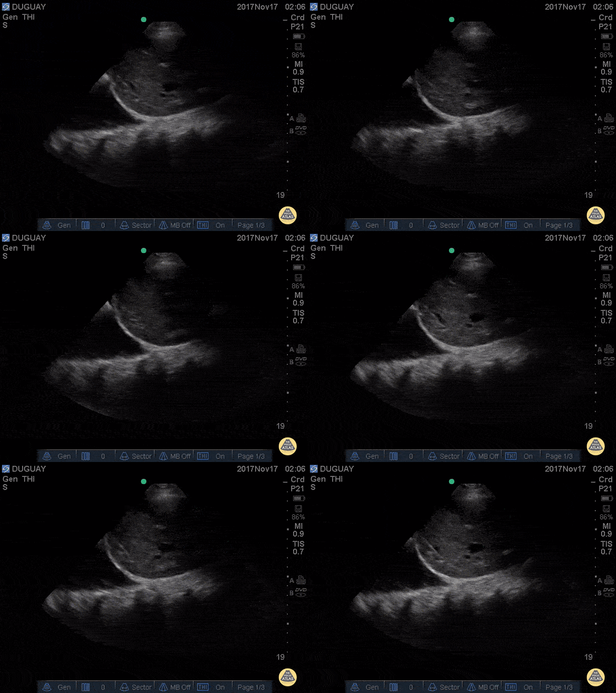
Spine Sign - Pleural Effusion
RUQ scan with large R pleural effusion. Spine sign+ (clear view of several thoracic vertebrae through the effusion)
Gary Duguay

Pleural Effusion
This 80 year old male with a history of Alzheimer's with recent accelerated decline experienced a fall from bed. Though the only visible trauma involved a laceration above the right eye, globally diminished breath sounds were noted, and a prehospital eFAST exam was performed. Pictured is a large, right-sided pleural effusion, with visible spine sign observed during the exam. No other abnormalities were observed.
With the absence of significant chest trauma, the effusion was assumed to be non-traumatic in origin. Due to the patient's recent rapid decline, family elected to manage conservatively, and the patient was admitted to palliative care.
- Tom Hudson, NRP, CCP-C (Richmond Volunteer Rescue Squad)
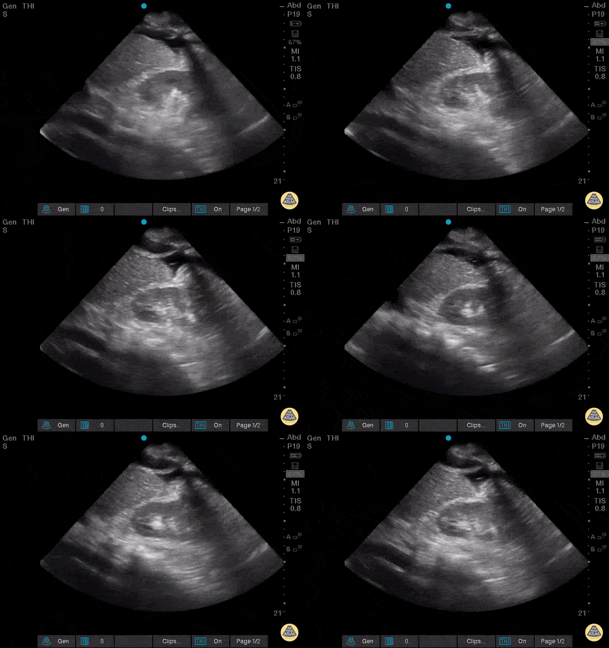
Diaphragmatic Contraction
A 61-year-old female with history of chronic Hepatitis C, end-stage liver disease, and pulmonary hypertension presented to the emergency department complaining of increasing dyspnea and abdominal distension over the last 10 days. Point-of-care ultrasonography of the right upper quadrant showed large anechoic fluid collections in the pleural space and intra-abdominal cavity, with a "spine" sign visible and with distinct “flopping” of the diaphragm over the caudal tip of the cirrhotic liver with each respiratory cycle. A great view of diaphragmatic contraction in a patient with dyspnea!
Scott Brensel, MS-IV, Touro College of Osteopathic Medicine California
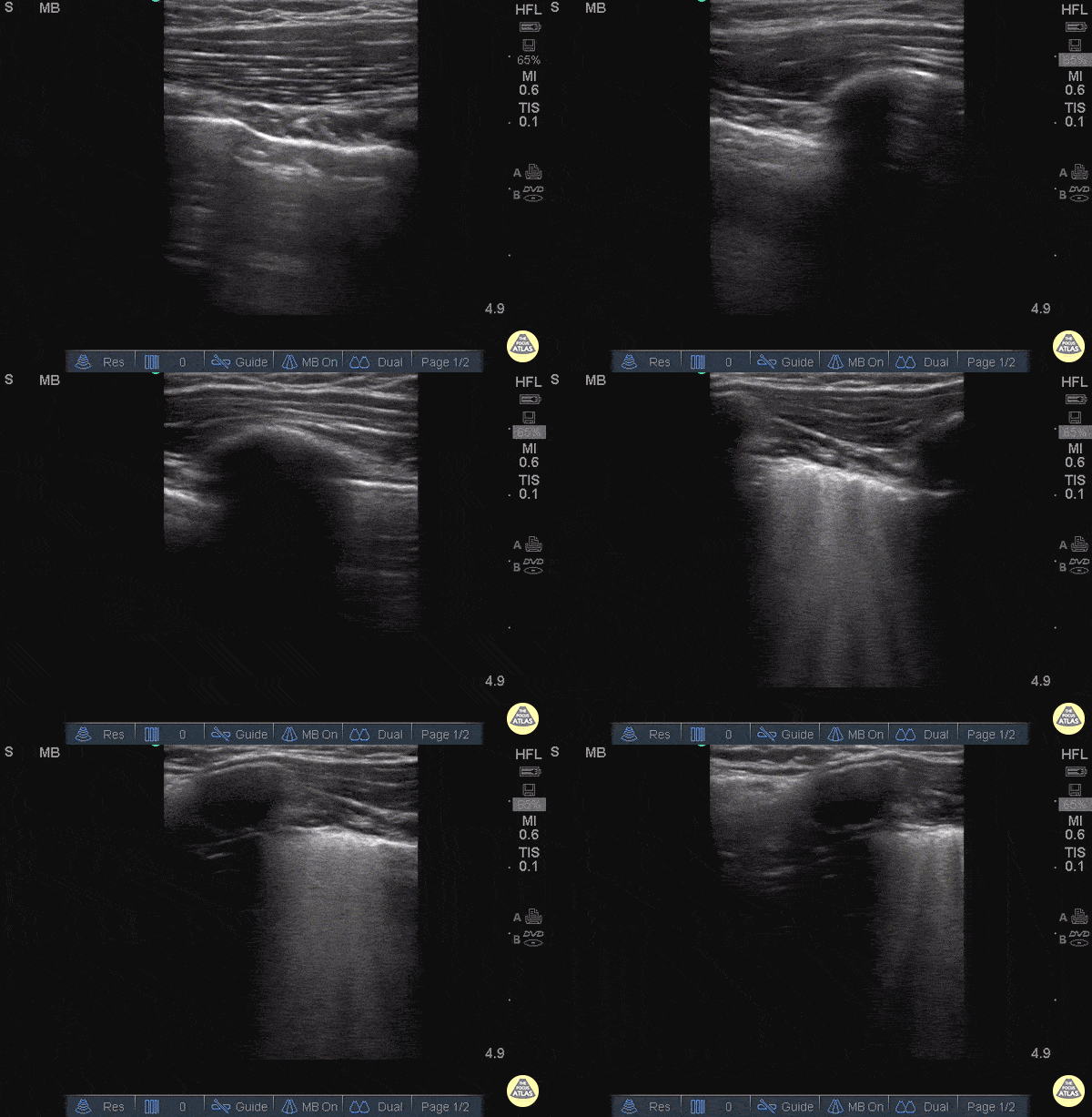
Pulmonary Contusion
25 y/o female in and MVA with hypotension, hypoxia.
Normal lung with A lines can briefly be seen until the sonographer moves the probe superiorly to reveal and area of B lines adjacent to A line.
In the setting of trauma this is consistent with Pulmonary Contusion.
Images: Dr. Catharine Bon - Kings County Hospital Emergency Medicine
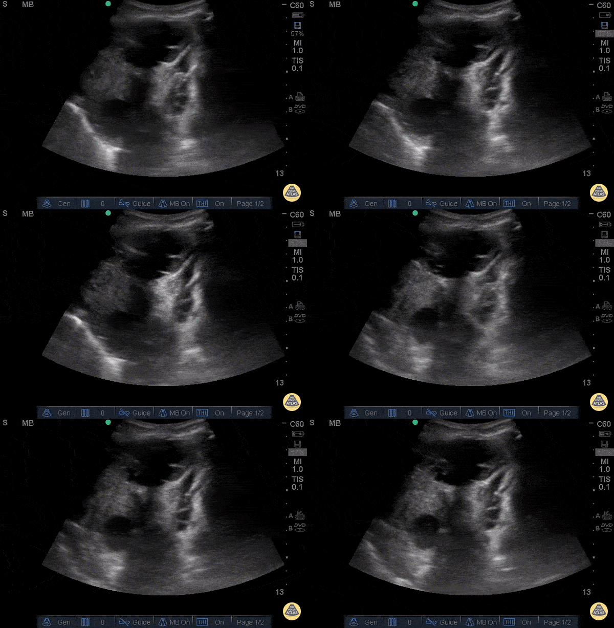
Malignant Pleural Effusion
This is a clip of an elderly gentleman, who was called as a respiratory distress code while walking to clinic without his home oxygen. Initially, patient was tachycardic, tachypnic, and hypoxic to mid 80s on RA, however normotensive in mild respiratory distress, resolved with 3L O2 by nasal canula.
This clip here was obtained by having the patient in the upright position, with the probe in the left lung base. You can see the diaphragm on the left side of the clip, with multiple loculated pleural effusions in left lung base adjacent to compressive atelectasis vs cardiac activity.
The effusion was later drained, found to be a malignant effusion, with a subsequent biopsy showing a Non-Small Cell CA in the left upper lobe and Squamous Cell CA in the right upper lung.
Chris Hanuscin, MD and John F. Kilpatrick, MD
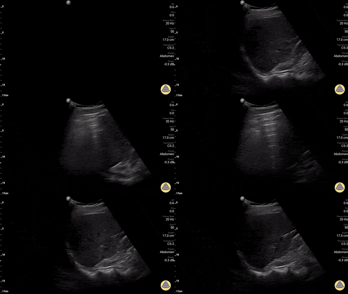
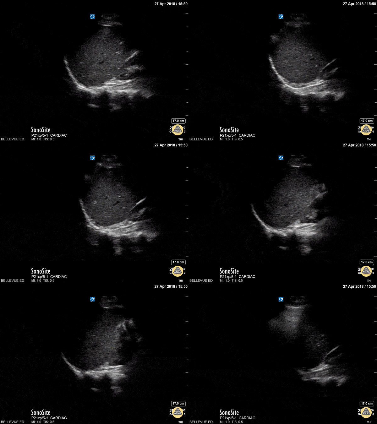
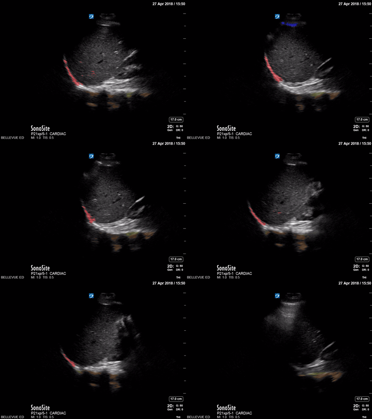
Lung Curtain
Normal lung demonstrating the "lung curtain." As the patient takes a deep breath, the "dirty shadowing" of a normal air-filled lung comes over the liver like a curtain.
Dr. Gordon Johnson
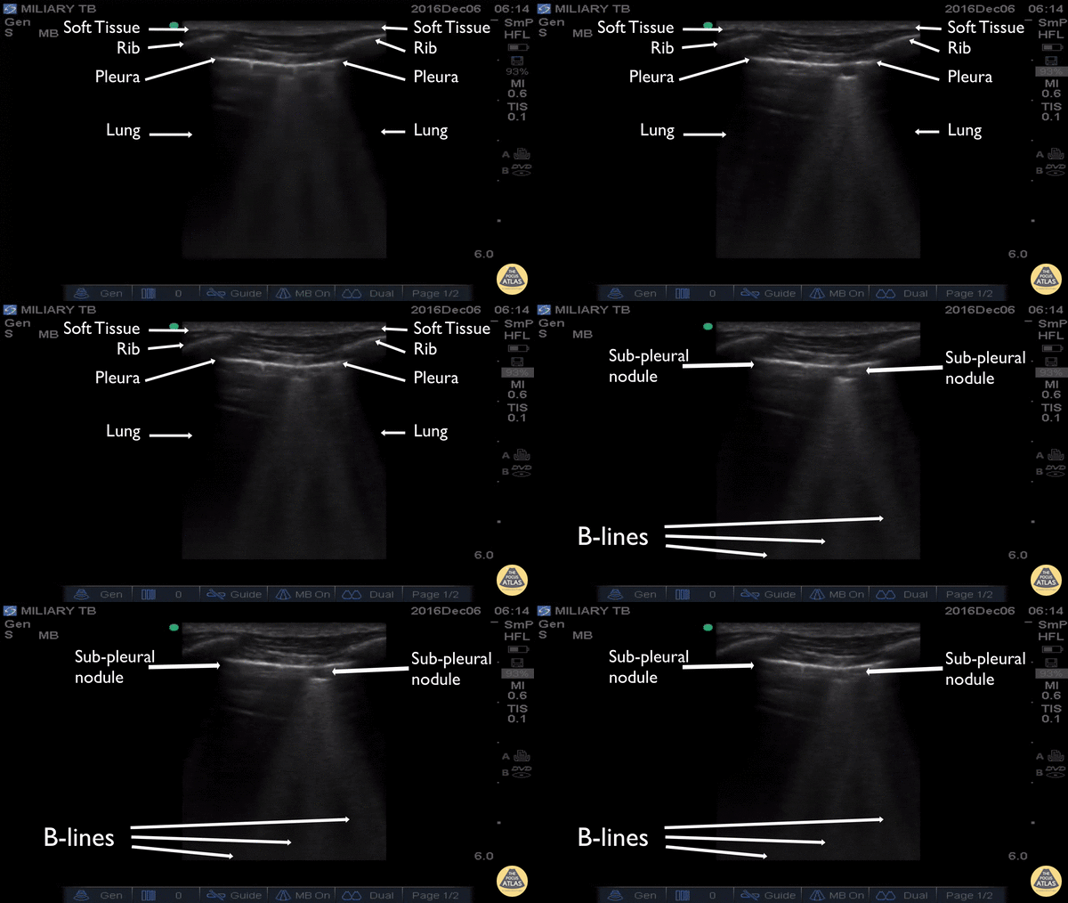
Miliary TB
The clip was captured in rural Uganda on a patient who presented for weight loss, night sweats, and cough. Utilizing the high-frequency, linear transducer the patient’s thoracic pleura and superficial lung were evaluated. The ultrasound demonstrated multiple focal sub-pleural lesions with tripartite B-lines consistent with miliary tuberculosis.
Michael Schick DO, MA
Emergency Medicine Physician
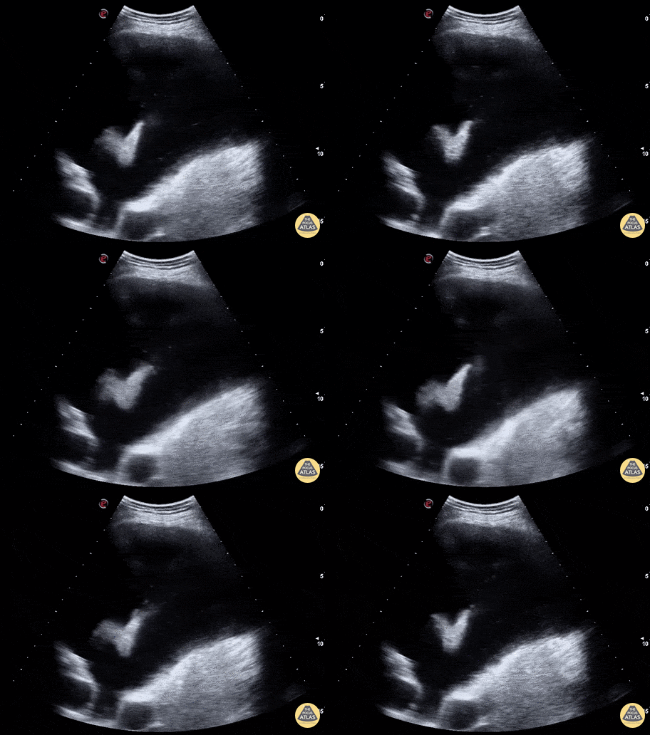
Dove in Pleural Fluid
WCUME 2017 Submission for "Creative Caption"
"In these days of violent extremist and warmongers, can it be a a good omen to find a dove flying in the pleural fluid?"
Marco Garrone, MD - Torino, Italy

B lines - Aspiration Pneumonitis
A 30s M presented to the ED after he was found down in the setting of substance use. He received naloxone from EMS with good response, but on arrival to the ED was hypoxic and in respiratory distress, requiring maximum levels of respiratory support. POCUS was performed, showing extensive confluent B lines in both lungs, though worse on the right, suggestive of the alveolar interstitial syndrome and concerning for aspiration pneumonitis. The patient required intubation for work of breathing, and was admitted for further management.
Dr. James Sutton, PGY2
Denver Health Residency in Emergency Medicine
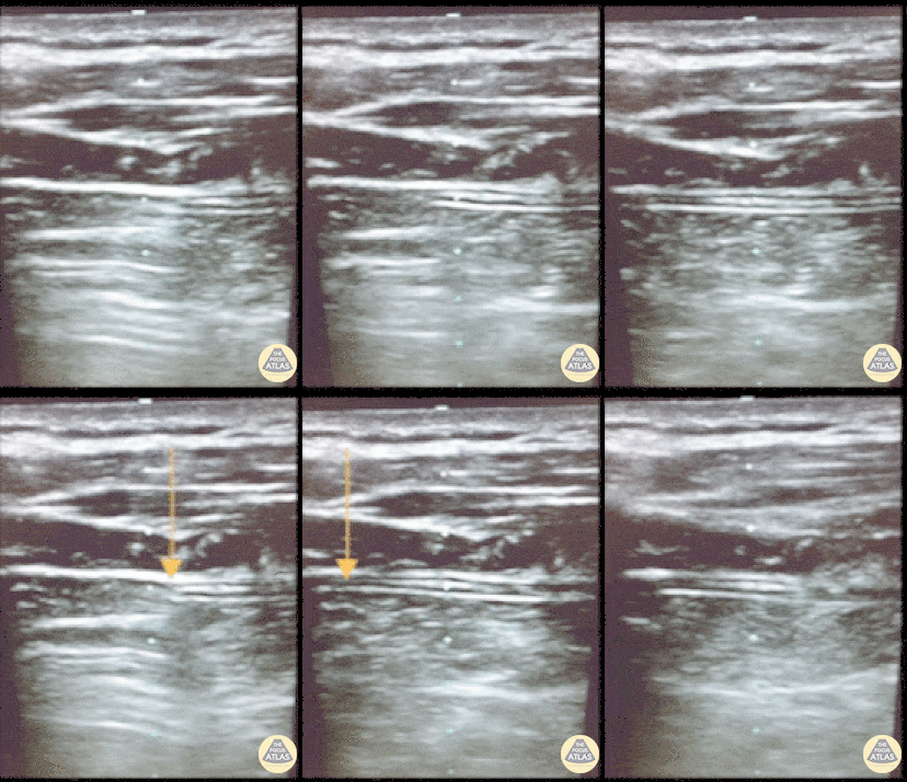
Resolving Pneumothorax
This is an image of a patient's chest wall using a high frequency transducer, with the transducer oriented in a transverse plane between rib spaces. The patient had a pneumothorax and a chest tube was placed. This clip illustrates what happens when the suction is turned 'on'. You will see the pleura slide from right to left as the pneumothorax resolves.
- Jason Tanguay, DO; Ultrasound Leadership Academy Graduate
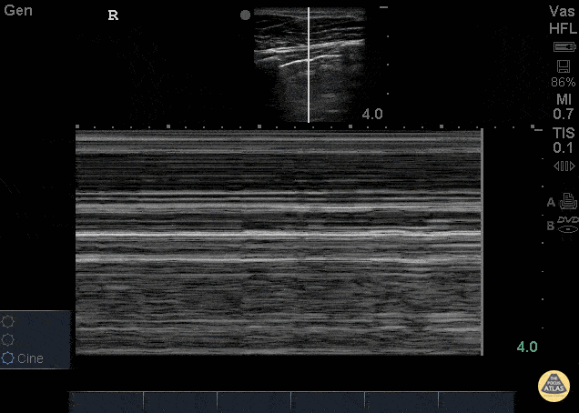
Bar Code Sign - Pneumothorax
26 yo male presents to ED stating he was kicked in the chest. He went home to “try to relax and smoke some weed” now short of breath and with pleuritic chest pain after smoking. POCUS demonstrating decreased lung slide on the left.
This can be seen as decreased lung sliding - In normal lungs, lung sliding refers to the parietal pleura moving against the visceral pleura - described as “ants marching.” M-mode can be used to evaluate lung sliding. Remember, normal lung slide will look like a seashore on M-mode whereas a pneumothorax will appear as horizontal lines termed Bar Code sign (pictured here). Make sure to check in the most anterior fields as well at lateral lung fields.
Dr. Stacey Frisch, Dr. John F Kilpatrick - Kings County Emergency Medicine
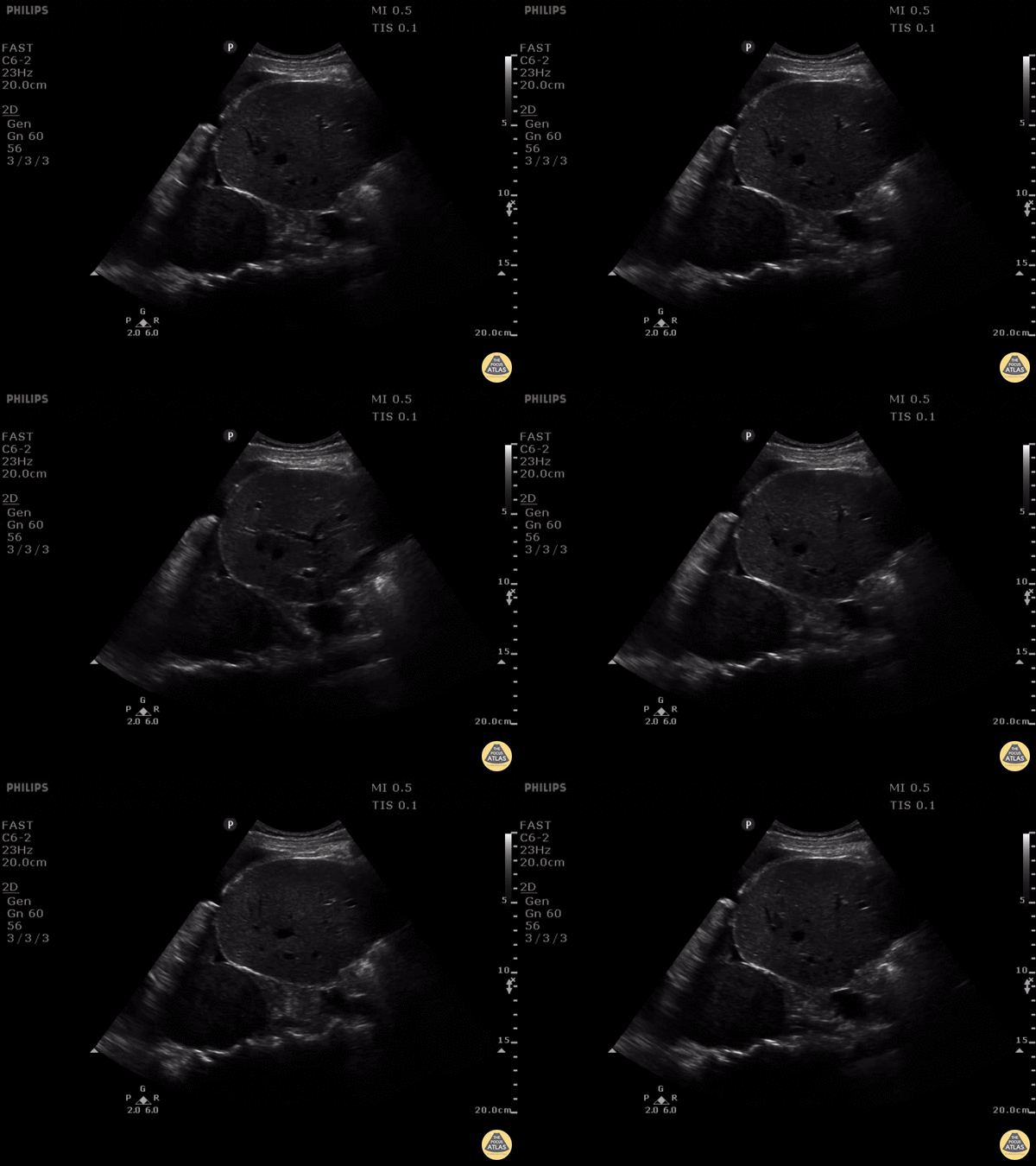
Pleural Effusion, Mass
PLEURAL EFFUSION, THE SPINE SIGN AND SOMETHING ELSE…
This is a clip of a 46 year old woman that presented to ED with gradual onset of severe right-sided chest pain, pleuritic, associated with tachycardia but normal blood pressure. She was mildly tachypneic but not hypoxic, unable to lie down as that exacerbated her pain. The clip shown here is of the patient's right upper quadrant and right lung base. The spine sign is seen along with a right pleural effusion and a circumscribed mass 8 x 6 cm with likely compressive atelectasis and mass effect of the right hemidiaphragm. The effusion was drained obtaining almost 1L of blood, and the CT scan reported very close findings to the ones seen with POCUS a few seconds after patient arrival.
Put the probe on your chest pain patients!
Maria Perez; Emergency Registrar; St Vincent’s Hospital; Melbourne - Australia
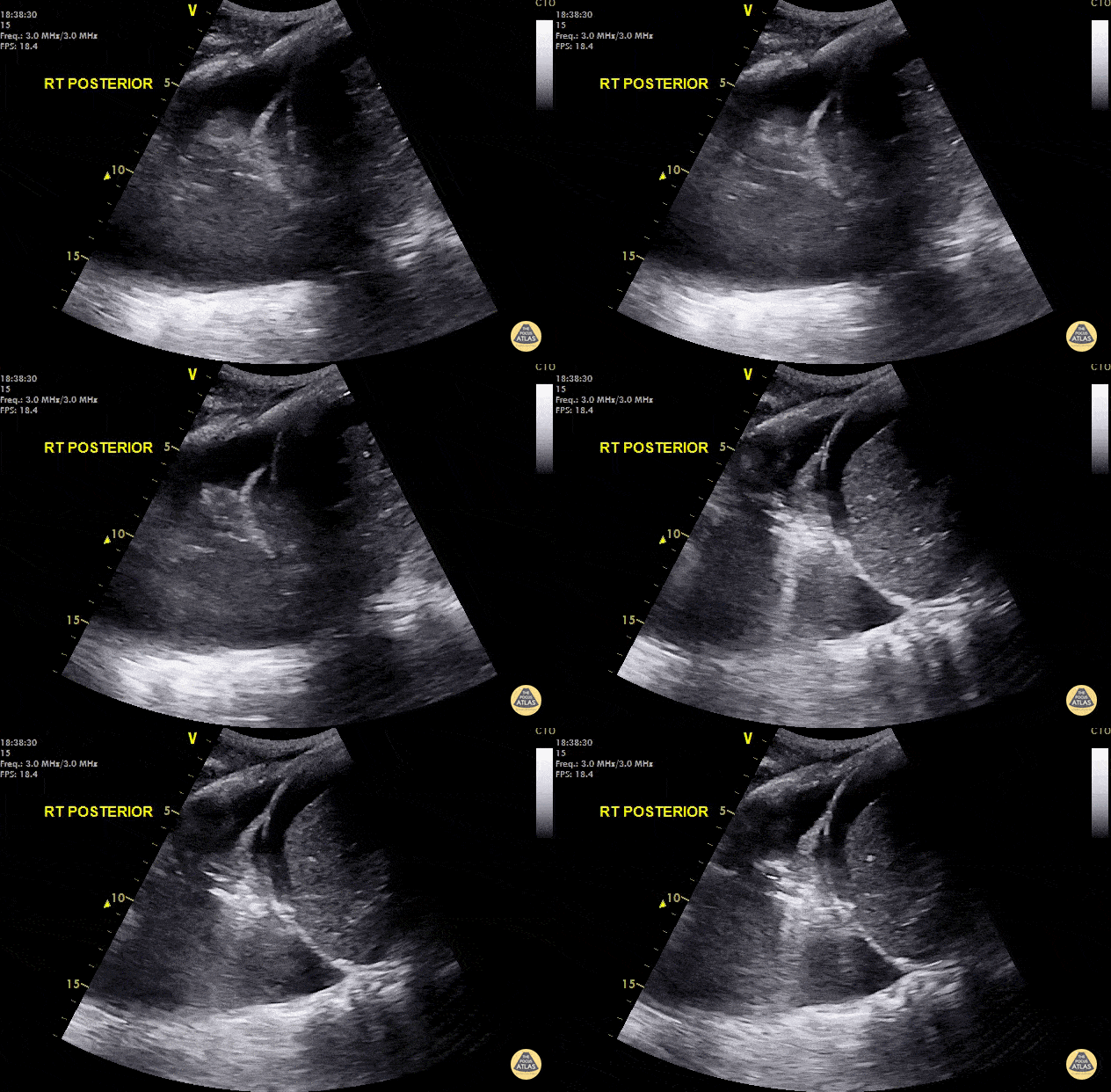
Jellyfish Sign
Hypotensive septic elderly male. Right lung base shows a positive spine sign, jellyfish sign and shred sign indicating right basal pneumonia and a parapneumonic pleural effusion.
Dr. Cian McDermott - Dublin, Ireland
A spine sign occurs when fluid or consolidation in the lung allows for transmission and visualization of the spine superior to the diaphragm.
A jellyfish sign represents compressed and airless (atelectatic) lung tissue floating within an effusion, also known as "lung flapping."
The shred sign is a hyperechoic artifact created at the transition point between solidified (pathologic) and aerated lung.
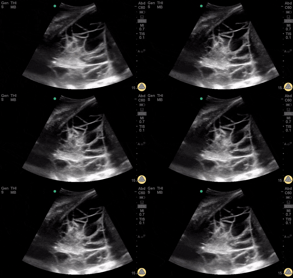
PLAPS
WCUME 2017 Submission for "Best POCUS"
26 y/o M with Hodgkin Lymphoma with a very complicated, septated, fluid-filled PLAPS - posterolateral alveolar or pleural syndrome. Aspiration of the fluid was exudative.
Dr. Yasmin Sadri Savadjani - Firoozgar General Hospital

Large Pleural Effusion
The left lung can be seen freely floating in anechoic fluid on the left side of the screen. Also pictured, the diaphragm, spleen, and edge of the beating heart.
Justin Bowra MBBS, FACEM, CCPU Emergency Physician, RNSH
et al.

Lung Consolidation with Mirror Artifact
A consolidated right upper lung can be seen above the fissure which is to the left (superior) in this image. There is hepatization of the lung with static and dynamic air bronchograms. These represent air with fluid filled alveoli around them. Dynamic air bronchograms are pathognomonic for pneumonia and represent air bubbles moving through the otherwise fluid filled tissue.
Hepatization refers to lung tissue that looks like the liver and represents consolidation.
Mirror artifact can be seen below (to the right) of the fissure as the fissure is a highly reflective surface.
Justin Bowra MBBS, FACEM, CCPU Emergency Physician, RNSH
et al.

Pulmonary Embolism (B mode)
PE with linear probe in B mode.
Pulmonary embolism can be seen by disruption of the pleural line. 0.5cm to 3cm disruptions are typical for PE. Doing DVT studies and echo can help strengthen your diagnosis.
Dr. Justin Bowra et al.
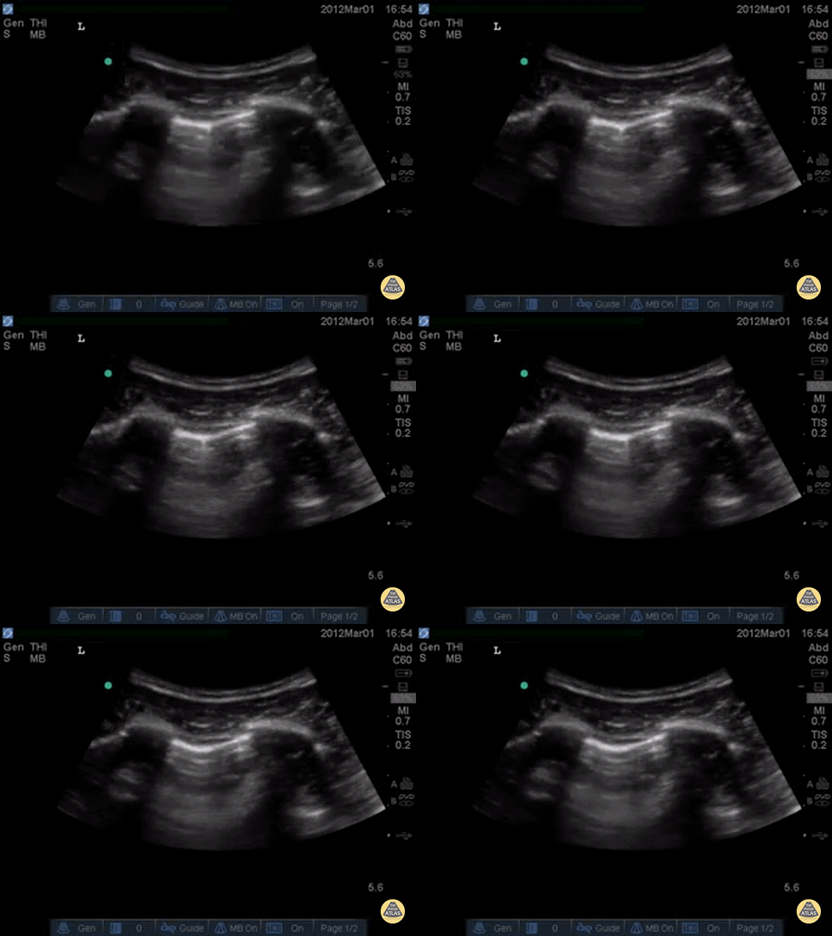
Pneumothorax Lung Point
Decreased lung slide is highly sensitive, it lacks specificity. Lung point however, is a highly specific finding indicating a pneumothorax.
Lung point indicates the transition point between normal pleura with normal lung sliding and where there is air disrupting the pleural space with decreased lung sliding.
In this intercostal space, one can see lung with normal lung slide on the left, and decreased lung slide on the right, and a point where the lung slide changes, which is moving with inspiration. This is the lung point.
Dr. Justin Bowra et al.

Compression Atelectasis
The left lung base can be seen with some b-lines indicating atelectasis, with trace pleural effusion.
Dr. Justin Bowra et al.
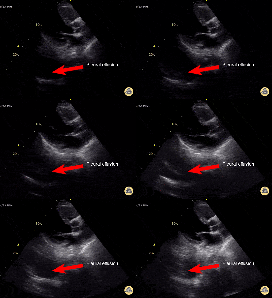
Concealed Pleural Effusion
Don't forget to look behind the heart....
Use a survey sweep to look behind the heart for a multi-organ POCUS exam. In the PLAX view, there is a pleural effusion visible behind the posterior myocardium in the left lobe of the lung. This patient had lung consolidation with a parapneumonic effusion and rapid atrial fibrillation.
Dr. Cian McDermott - Mater University Hospital, Dublin, Ireland
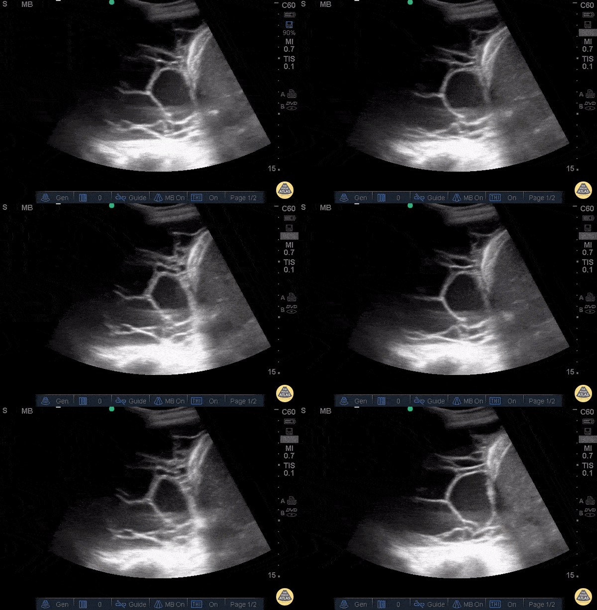
Empyema
Elderly male coming in with generalized weakness, shortness of breath and cough, who had recently been hospitalized for pneumonia. On arrival he was tachypneic and satting in the high 80s on room air.
POCUS in the RUQ and right lung base. Pleura and loculations are visualized along with spine sign which indicates a plueral effusion. Sonographic evidence of septations in the presence of empyema predicts the need for intrapleural fibrinolytic therapy, longer duration of drainage, or possible surgical intervention.
Dr. Gina Pan - Kings County Emergency Medicine

Lung Point
29F in motor vehicle accident w/ right hemo/pneumothoarx. Lung point is demonstrated marking the transition of normal pleural to pneumothorax. This is the most specific sign of pneumothorax.
Greg Powell, MD
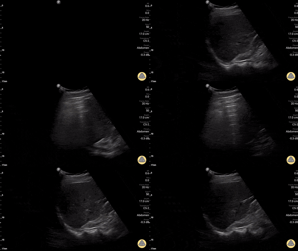
Lung Curtain
Normal lung demonstrating the "lung curtain." As the patient takes a deep breath the "dirty shadowing" of a normal air filled lung, comes over the liver like a curtain.
Dr. Gordon Johnson
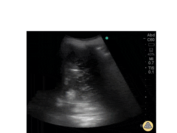
Pleural Effusion
Patient of chronic renal failure with signs of fluid overload and bilateral pleural effusion on chest radiograph.POCUS ---Echogenic material within the fluid. Echogenic material seen within the fluid- Suggestive of Exudative pleural effusion. Transudative pleural effusion will be non echogenic.
Dr. Ramachadran
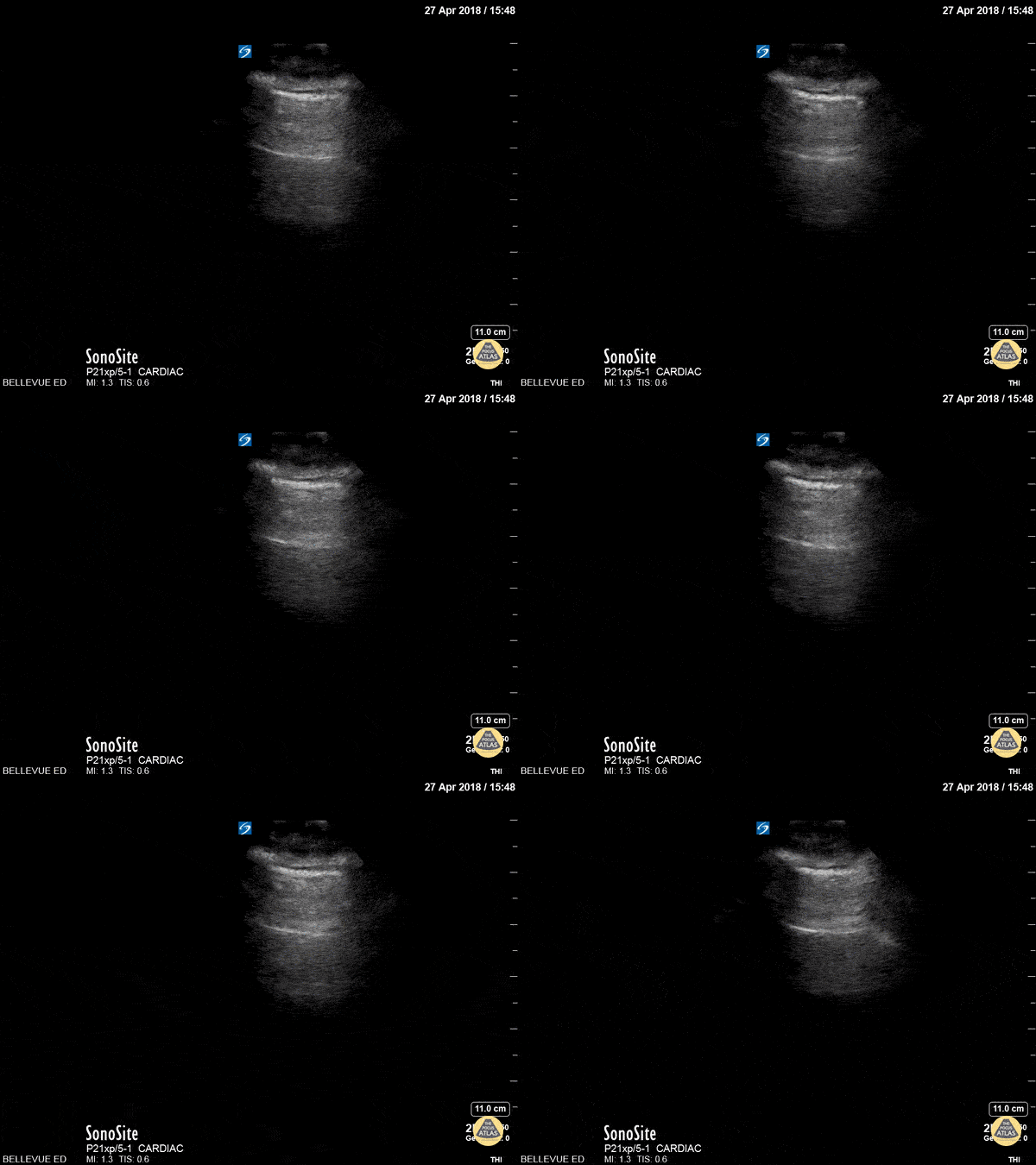
Lung Sliding and A-lines
Lung Sliding and a-lines
This is a clip demonstrating lung sliding. The most superficial hyperechoic layers are the soft tissue and muscular layers of the chest wall. Immediately deep to that is a bright, thin hyperechoic line which appears to be in motion - this is the pleural line. The parietal pleura rubbing against the visceral pleura as the patient breathes creates this shimmery appearance of lung sliding also often described as “ants marching”. The next horizontal hyperechoic line deep to the pleura is an a-line; this is a reverberation artifact created by a reflection of the pleura. The presence of a-lines is a normal finding.
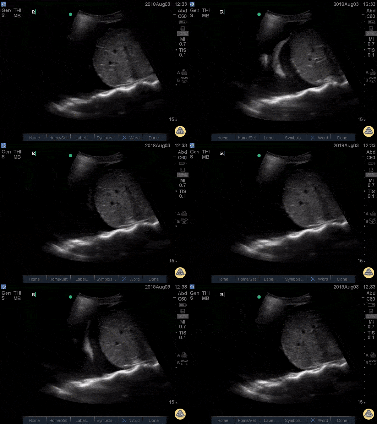
Pleural Space
This is a view of the pleural space in the right upper quadrant. We see the isoechoic liver in the center of the screen and the superior pole of the kidney to the right. The superior edge of the liver is flush against the hyperechoic diaphragm.
Deep to all of this we see the sinuate hyperechoic vertebrae of the spine. Note that the spine appears to stop at the level of the diaphragm – this is due to the fact that US waves do not transmit through the air-filled lungs, and is a normal finding. If the spine did extend beyond the diaphragm (“spine sign”) that would suggest the presence of an effusion.
The area above the diaphragm which comes into view when the patient inhales is the same echotexture as the liver (“mirror image artifact”), further indicating that there is only air above the diaphragm and not effusion or consolidation.
Hannah Kopinksi and Dr. Lindsay Davis - NYU Emergency Medicine
Spine Sign
41 yo M with history of stage 4 lung cancer presents with AMS and dyspnea, normotensive and tachycardia in 140s.
Right upper quadrant view shows large anechoic pleural fluid with spine sign (clear view of several thoracic vertebrae through effusion) and lung flapping i.e. large right pleural effusion.
Nathan Kabariti MS4, Dr. Charles Murchison, Dr. John Riggins, Dr. Donald Doukas - Kings County Emergency Medicine
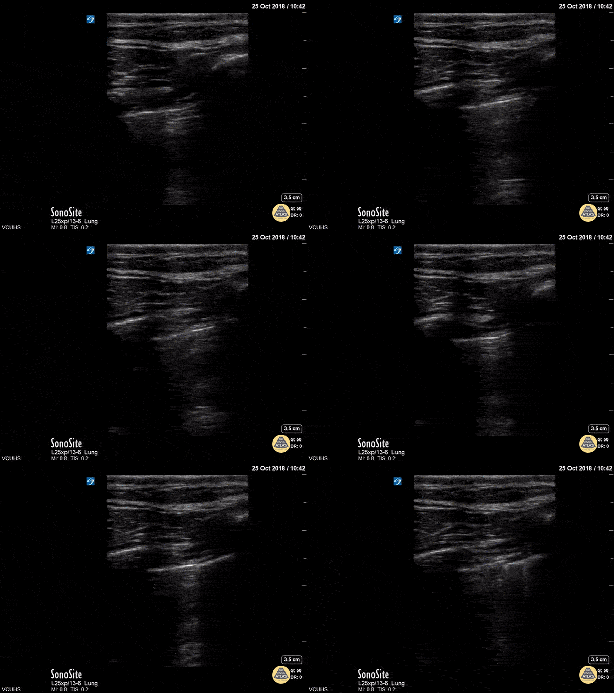
Lung Point
Lung point on a large pneumothorax.
Dr. Stenberg
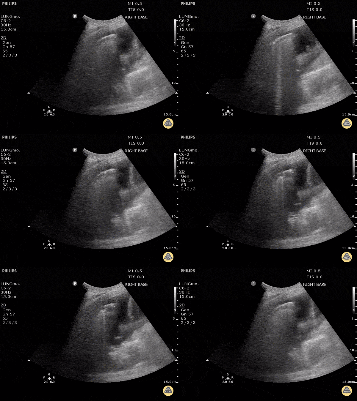
B-lines and Effusion
To the left of the image, the lung can be seen clearly floating in anechoic fluid representing a pleural effusion.
B-Lines can be seen radiating from the surface of the lung to the far left especially as this patient inspired.
B lines (also known as comet tails) are white lines that emanate from the pleural surface of the lung. They have been shown to be highly sensitive for pulmonary edema.
Justin Bowra MBBS, FACEM, CCPU Emergency Physician, RNSH
et al.
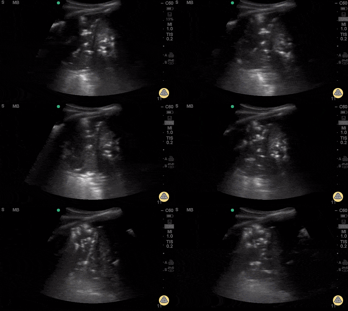
Air Bronchograms
Middle aged female with history of HIV, asthma, and polysubstance use who presents with progressively worsening dyspnea over 3-4 days, found to have diffuse rales, worse in left mid to lower lung fields. AP CXR with bilateral lower lobe patchy infiltrate, left greater than right.
POCUS with curvilinear probe revealed B lines in left mid lung fields and consolidation with air
bronchograms in left lower lung. Air bronchograms is one of the most specific signs for the diagnosis of pneumonia with a specificity (93%) and a positive LR (12.14). Ultimately, CT chest is the gold standard for diagnosis of pneumonia, which was consistent with the CT in this patient.
Priscilla Chao, MD, Matthew Riscinti, MD - Kings County Emergency Medicine
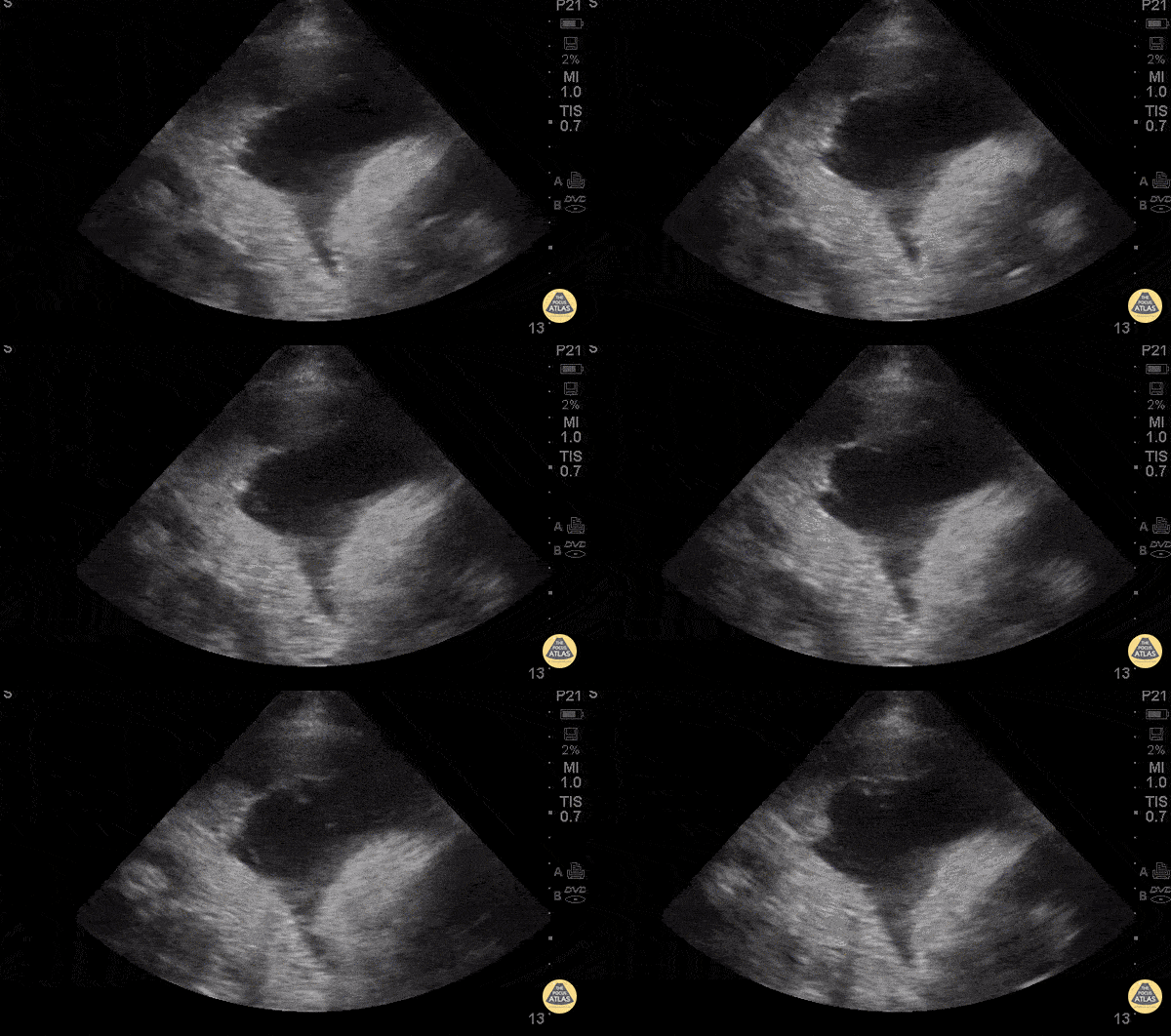
Malignant Effusion
71 y/o F w/ hx of COPD/asthma, lung cancer on chemotherapy with recent admission for malignant pleural effusion (chest tube placed), presenting with for worsening cough/wheezing/shortness of breath for 1 day. No fevers or chest pain.
The clip shows a large fluid collection in the left anterior chest with loculations and mediastinal shift to the right.
Cardiothoracic surgery drained 2.7L of hemorrhagic pleural effusion.
Dr. Vicky Lam - Kings County Emergency Medicine
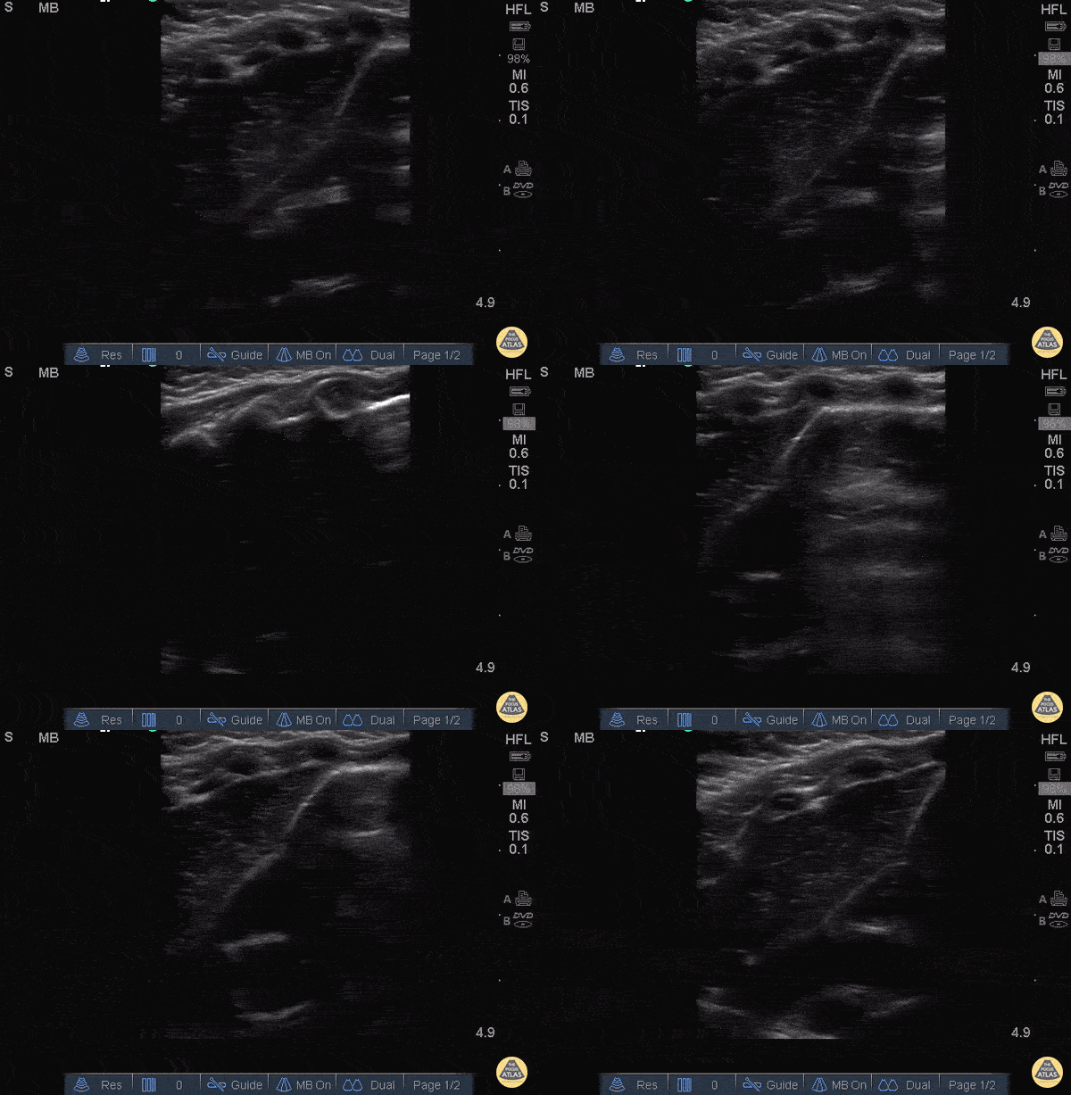
Thymus
Example of a thymus that can often be confused for hepatization! Don’t make this mistake
John F Kilpatrick - Kings County Hospital
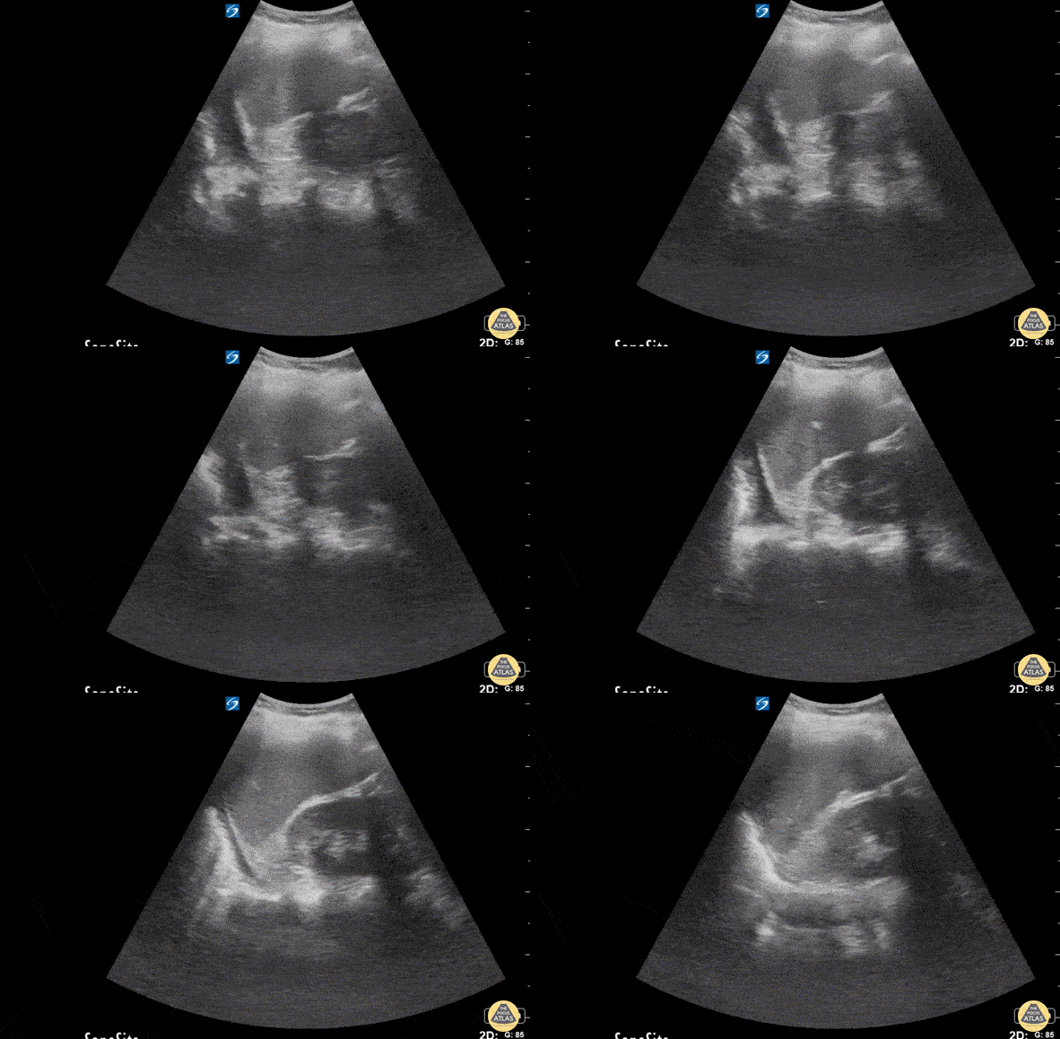
Pleural Effusion After Blunt Trauma
55 y/o female complains of left sided chest pain with cough, SOB, and back pain. History of falls from 8-foot ladder (8 weeks ago) and from standing (2 weeks ago).
With patient in supine, US of LUQ lung in coronal view demonstrated a hypoechoic fluid collection above the left hemi-diaphragm consistent with a L pleural effusion.
It is important to scan above and below the diaphragm to differentiate free fluid in thorax vs fluid in subphrenic space. US is more sensitive than plain radiographs for detecting pleural effusion and can detect smaller amounts of fluid.
Crozer Chester Medical Center.
Raghav Sahni, Dr. Melissa Yu, Dr. Brenton Elliot, Dr. Max Cooper.
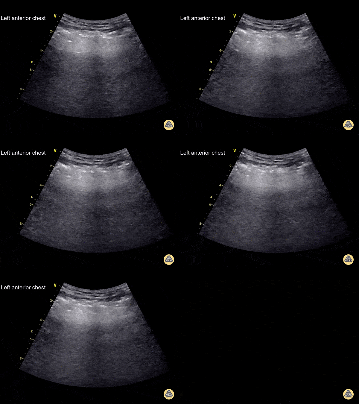
Subcutaneous Emphysema
This patient fell 5m onto a slab of concrete. The curvilinear transducer was used for the EFAST survey of his left anterior chest wall
Hyperechoic irregular lines are seen superficial to the ribs (E lines - subcutaneous emphysema). These arise from air trapped in the soft tissue under the skin and should not be mistaken for the pleural line. Subcutaneous emphysema obscures the rib shadow and pleural line that lie below it
Learning point : the pleural line lies underneath the level of the ribs, subcutaneous emphysema lies above the level of the ribs
This patient had a large left-sided pneumothorax and an intercostal drain was placed followed by a serratus anterior plane block for analgesia
Dr. Cian McDermott - Dublin, Ireland

Hemothorax
A male presented to the ED following a gunshot wound to the left chest. The perisplenic window of the FAST exam revealed a left hemothorax with a large subpleural area of clotted blood.
Image courtesy of Robert Jones DO, FACEP @RJonesSonoEM
Director, Emergency Ultrasound; MetroHealth Medical Center; Professor, Case Western Reserve Medical School, Cleveland, OH
View his original post here

The Light Beam Artifact in COVID-19
This lung ultrasound shows a "light beam" artifact, a single shining band-form from a regular pleura that appears and disappears with spontaneous respirations. Note also the presence of A-lines. This is highly specific of early COVID-19 infection as described by Volpicelli.
Image courtesy of David Hansen, DO & Therese Mead, DO, RDMS, FACEP
Central Michigan University
Twitter handle: @davidbhansen

Lung US Findings in Hypoxic Patient with Suspected COVID-19
This clip demonstrates the presence of focal B lines with pleural irregularity. The patient presented to the ED with cough and O2 saturation varying between 87-89%, with no respiratory distress or significant past medical history. One of the patient’s family members was currently under investigation for COVID-19. The entire lungs were examined and the image above is the PLAPS view. Later the patient had a CT done which showed ground-glass opacity peripherally compatible with areas scanned on the POCUS exam.
Pearl: POCUS may be an excellent tool for the triage of these patients.
Image Courtesy of Dr. Victor Bang (@vmjbang)
Co-Founder of Pocus Jedi

Common Pleural Based Findings in COVID-19
Lung ultrasound performed in a COVID+ patient. Note the clustered B lines, patchy shredding (depression) and thickening of pleural line, and small sub-pleural consolidations.
Image courtesy of Dr. Marco Garrone (@drmarcogarrone)
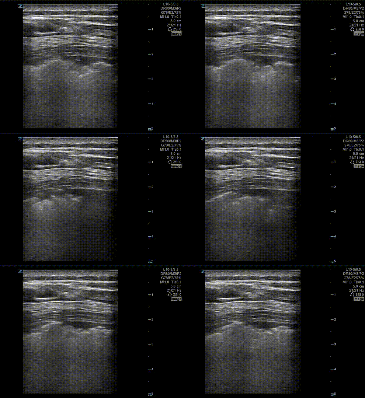
Pleural Findings in COVID-19
This is a lung ultrasound performed on a patient with COVID-19. The patient had no prior pulmonary disease. They presented with mild tachypnea and hypoxia. A chest x-ray revealed diffuse interstitial and patchy airspace densities. This ultrasound clip shown demonstrates an irregular pleural line with subpleural nodular consolidation and waterfall B-lines.
Image courtesy of Dr. Eric Abrams (@eabramsMD)
![A COVID-19 Patient with 3 days of Symptoms [1/3]](https://images.squarespace-cdn.com/content/v1/58118909e3df282037abfad7/1584923302263-T9LBTKNFW294TEL8OU0L/ezgif.com-optimize+%2843%29.gif)
A COVID-19 Patient with 3 days of Symptoms [1/3]
This is an ultrasound clip of the right upper lobe of a patient with confirmed COVID-19 pneumonia. The patient presented to the emergency department on day 3 of symptoms with fever. They were found to be tachypneic and with mild hypoxia. [Clip 1/3]
Lung ultrasound demonstrates confluent B lines associated with thickening and irregularity of the pleural line. A thin parapneumonic effusion can also be appreciated here.
Image courtesy of Fritz Fuller (@POCUS_Society)
![A COVID-19 Patient with 3 days of Symptoms [2/3]](https://images.squarespace-cdn.com/content/v1/58118909e3df282037abfad7/1584923302390-3HOKDDGEEXWXTE1ZKHPK/ezgif.com-optimize+%2844%29.gif)
A COVID-19 Patient with 3 days of Symptoms [2/3]
This is an ultrasound clip in a patient with confirmed COVID-19 pneumonia. The patient presented to the emergency department on day 3 of symptoms with fever. They were found to be tachypneic and with mild hypoxia. [Clip 2/3]
Lung ultrasound demonstrates patchy B lines associated with thickening and irregularity of the pleural line.
Image courtesy of Fritz Fuller (@POCUS_Society)
![A COVID-19 Patient with 3 days of Symptoms [3/3]](https://images.squarespace-cdn.com/content/v1/58118909e3df282037abfad7/1584923302992-4BOINIWQM3AMUCPWU745/ezgif.com-optimize+%2845%29.gif)
A COVID-19 Patient with 3 days of Symptoms [3/3]
This is an ultrasound clip of the right posterior lung field of a patient with confirmed COVID-19 pneumonia. The patient presented to the emergency department on day 3 of symptoms with fever. They were found to be tachypneic and with mild hypoxia. [Clip 3/3]
Lung ultrasound demonstrates confluent B lines associated with a small subpleural consolidation.
Image courtesy of Fritz Fuller (@POCUS_Society)
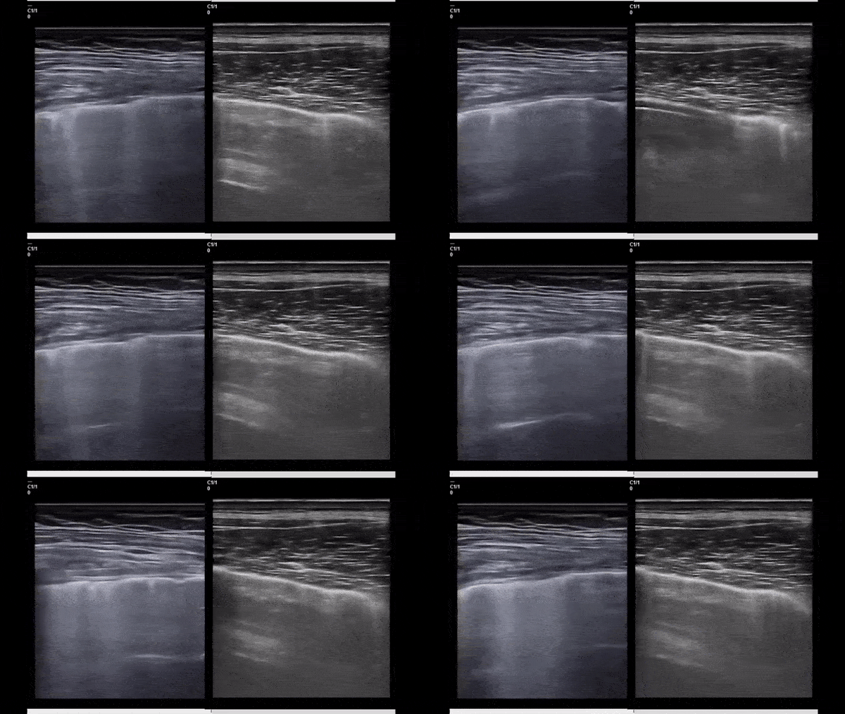
B-lines in COVID-19 Versus CHF
This side by side ultrasound clip compares B-lines in a patient with COVID-19 (left) and a patient with CHF (right). While subtle, the difference lies where the B-lines connect to the pleura! In COVID-19 many of the B-lines initiate from depressed/irregular areas of the pleura (imagine little holes being punched in the pleural line) while in CHF, the B-lines initiate from a smooth pleural line. These pleural defects are not specific to COVID -19 and can be seen in other viral pneumonia as well.
Take Home: Not all B-lines are created equal!
Image courtesy of Dr. Marco Garrone (@drmarcogarrone)
![Hospitalized COVID-19+ Patient on Day 9 of Symptoms [1/2]](https://images.squarespace-cdn.com/content/v1/58118909e3df282037abfad7/1585424289744-RG5K8531BSVL0L2IF4NT/ezgif.com-optimize+%2848%29.gif)
Hospitalized COVID-19+ Patient on Day 9 of Symptoms [1/2]
These clips are taken from a patient admitted with COVID-19 pneumonia. The patient was on day 9 of symptoms. His cough had improved and fever resolved however he had hypoxia requiring supplemental oxygen.
Left clip: Lung ultrasound of right lower lobe demonstrating thickened irregular pleura, diffuse b lines (confluent) with scattered puntate subpleural consolidation and small effusion overlying pleural effusion.
Right clip: Lung ultrasound of right upper lobe demonstrating a moderate subpleural consolidation with air bronchograms present.
Image courtesy of Fritz Fuller (@POCUS_Society)
![Hospitalized COVID-19+ Patient on Day 9 of Symptoms [2/2]](https://images.squarespace-cdn.com/content/v1/58118909e3df282037abfad7/1585424468095-8K5EKTWKAPOC1EYA9OAN/ezgif.com-optimize+%2849%29.gif)
Hospitalized COVID-19+ Patient on Day 9 of Symptoms [2/2]
These clips are taken from a patient admitted with COVID-19 pneumonia. The patient was on day 9 of symptoms. His cough had improved and fever resolved however he had hypoxia requiring supplemental oxygen.
Lung ultrasound of left lower and left upper lobes demonstrating thickened irregular pleura, diffuse b lines (confluent) with subpleural consolidations.
Image courtesy of Fritz Fuller (@POCUS_Society)
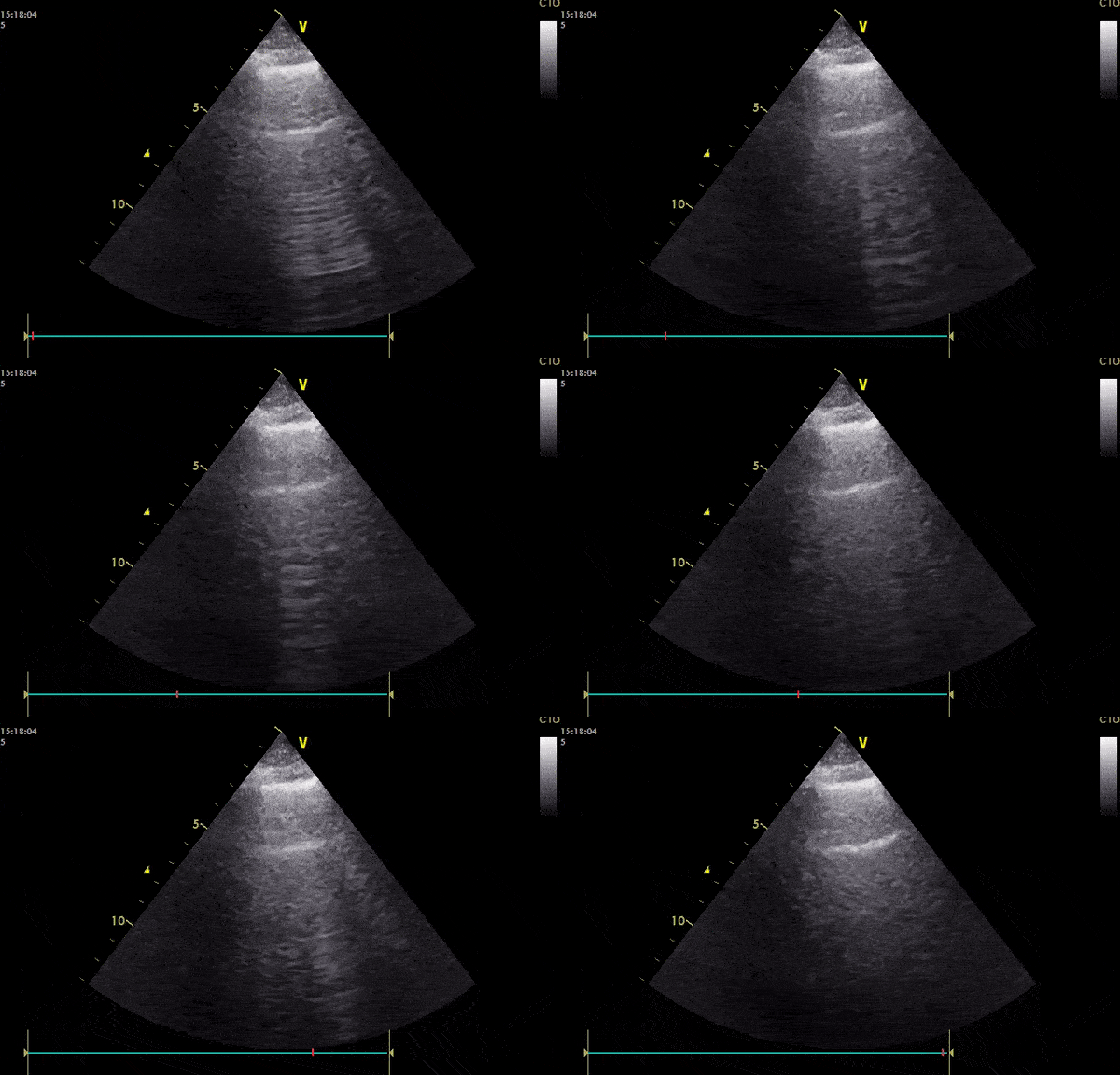
A Hypoxic COVID-19 Patient
This is an ultrasound clip performed in a 57 year old male patient with known COVID-19 pneumonia. The patient presented with dyspnea and fever, he had an oxygen saturation of 94% on room air. The clip demonstrates an irregular pleural line with numerous B lines present. Interestingly, A-lines are also present that appears to be intermittently erased where B lines cross.
Image courtesy of Pierre Bernatas (@pb2316)
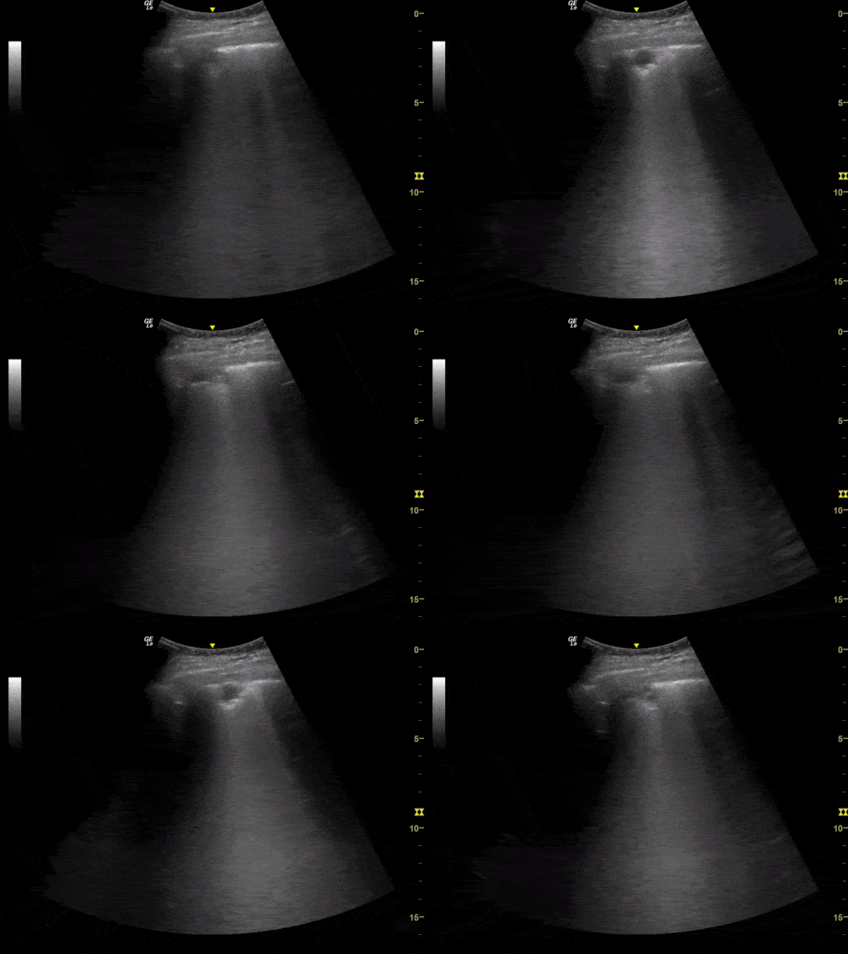
Subpleural Consolidation in Suspected COVID-19 Pneumonia
This lung ultrasound clip demonstrates a subpleural consolidation in a patient with suspected pulmonary involvement by COVID-19. The clip is taken from an exam performed on an elderly male with flu-like symptoms for 14 days with progressive respiratory failure.
Image courtesy of Renato Melo, Emergency Physician at Hospital das Clinicas de Marília-SP, Brazil. PocusJedi (@JediPocus) associated.
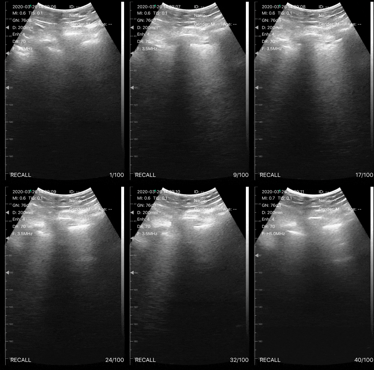
A Young Man with Hypoxia and Cough
This lung ultrasound illustrates a small subpleural consolidation with associated waterfall b-lines in a patient with suspected COVID-19. The patient was a male in his mid 30s who presented to the ED with cough, but no fever. On admission the patient had an O2 saturation of 85%, with no respiratory distress.
Image courtesy of Dr. Victor Bang (@vmjbang)
Co-Founder of Pocus Jedi
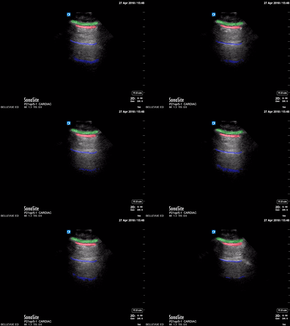
Lung Sliding - Colorized
Lung sliding
Green: Subcutaneous tissue, Red: Pleural space, Blue: A lines
Images: Dr. Lindsay Davis, Dr. Hannah Kopinski. Image Editing: Michael Amador and Dr. Matthew Riscinti
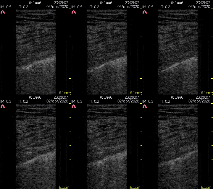
Pleural Findings in COVID 19
This lung ultrasound clip demonstrates multiple scattered b lines emanating from an irregular pleural line in a patient with known COVID-19.
Image courtesy of Dr. Jaime Alejandro Sánchez Gutiérrez (@pleuralpocus)
Pleural Space - Colorized
Pleural space
Red: Diaphragm, Blue: Pleural space, Green: A lines, Orange: Spine
Images: Dr. Lindsay Davis, Dr. Hannah Kopinski. Image Editing: Michael Amador and Dr. Matthew Riscinti


![A COVID-19 Patient with 3 days of Symptoms [1/3]](https://images.squarespace-cdn.com/content/v1/58118909e3df282037abfad7/1584923302263-T9LBTKNFW294TEL8OU0L/ezgif.com-optimize+%2843%29.gif)
![A COVID-19 Patient with 3 days of Symptoms [2/3]](https://images.squarespace-cdn.com/content/v1/58118909e3df282037abfad7/1584923302390-3HOKDDGEEXWXTE1ZKHPK/ezgif.com-optimize+%2844%29.gif)
![A COVID-19 Patient with 3 days of Symptoms [3/3]](https://images.squarespace-cdn.com/content/v1/58118909e3df282037abfad7/1584923302992-4BOINIWQM3AMUCPWU745/ezgif.com-optimize+%2845%29.gif)

![Hospitalized COVID-19+ Patient on Day 9 of Symptoms [1/2]](https://images.squarespace-cdn.com/content/v1/58118909e3df282037abfad7/1585424289744-RG5K8531BSVL0L2IF4NT/ezgif.com-optimize+%2848%29.gif)
![Hospitalized COVID-19+ Patient on Day 9 of Symptoms [2/2]](https://images.squarespace-cdn.com/content/v1/58118909e3df282037abfad7/1585424468095-8K5EKTWKAPOC1EYA9OAN/ezgif.com-optimize+%2849%29.gif)


