
Normal Cardiac Anatomy

Normal Parasternal Long Axis (PLAX) View
Parasternal long axis view with normal ejection fraction
Nigist Taddese MBChB. Division of Hospital Medicine, John H Stroger Hospital of Cook County

Normal Parasternal Short Axis View
Normal PSAX view at a level of the papillary muscles. In the center of the screen is the muscular walled LV, which forms a perfect circle. The smaller, thin-walled RV is seen superficially and wrapped around the LV.
Dr. Felipe Urriola, Puerto Aysen Hospital, Emergency Department, Chilean Patagonia.

Normal PLAX
This is a normal parasternal long axis (PLAX) view. The right ventricle (RV) is at the top of the screen. Further down and from left to right: left ventricle (LV), outflow tract, aortic valve, ascending aorta. The actively moving mitral valve separates the LV from the left atrium (LA). At the bottom of the screen, the circular, anechoic image is the descending aorta.
Dr. Felipe Urriola. Puerto Aysen Hospital Emergency Department, Chilean Patagonia.

Normal Parasternal Long
Seen here is a parasternal long axis view of the heart highlighting normal anatomy. You can see the left atrium, aorta, and right ventricular outflow tract, descending thoracic aorta, mitral valve and interventricular septum. Also notice the presence of a small amount of physiologic pericardial fluid. Pericardial fluid in this image is the hypoechoic shadow anterior to the descending thoracic aorta. There is no evidence of pleural effusion, however when present it would appear as a hypoechoic shadow inferior to the descending thoracic aorta.
Shahad Al Chalaby, MD. PGY3, Highland Hospital, Alameda Health System Internal Medicine Residency Program
@shahad_Chalaby

Normal subcostal view
Seen here is a normal subcostal view of the heart. Note the hyperechoic strings (chordae tendenae) attaching to the cusps of mitral and tricuspid valves as well as to the ventricular walls via the papillary muscles. There is also notable absence of any hypoechoic pericardial fluid or effusion.
Shahad Al Chalaby, MD. PGY2, Internal Medicine
Highland Hospital, Alameda Health System Internal Medicine Residency Program. CA, USA
@shahad_Chalaby
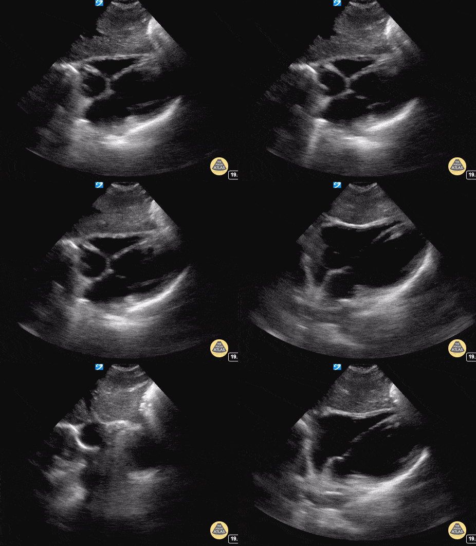
Subxiphoid / Subcostal - Normal
The most superficial structure we see is the liver. Immediately deep to that we see the heart separated from the liver by the diaphragm. Closest to the liver is the right atrium, tricuspid valve, right ventricle. Deeper to that, we see the left atrium, mitral valve, and left ventricle.
Hannah Kopinski - MS4, Dr. Lindsay Davis - NYU/Bellevue Department of Emergency Ultrasound, Dr. Matthew Riscinti - Kings County Emergency Medicine
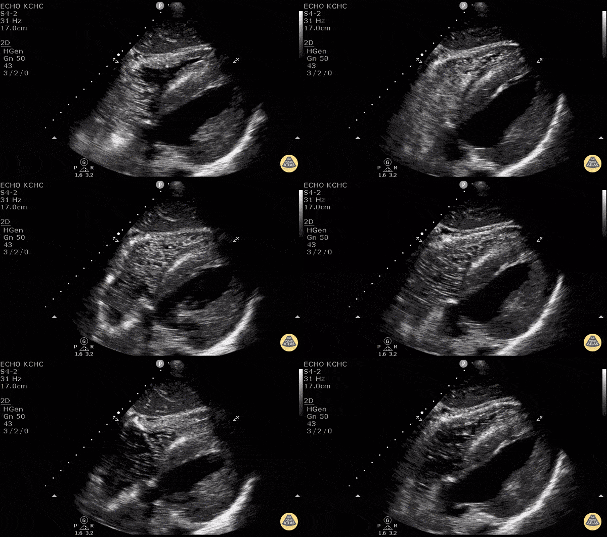
Central Line Confirmation
After placing a central line, POCUS can be used to rapidly and accurately assess correct venous placement as well as exclusion of pneumothorax prior to confirmation via a CXR. Simply attach an agitated flush to the central line, get a good view of the right side of the heart (here we used the subxiphoid view) then flush rapidly. If you see bubbles pass from the right atrium to the right ventricle of the heart, you know you’re good to go. POCUS has a sensitivity of 86.8% and specificity of 100% for identifying correct central venous catheter placement. See our evidence atlas for more information. PMID: 28123616
If you placed this under direct visualization you know you aren’t likely to have caused a pneumothorax… but POCUS can rule that out too! (Look for lung sliding, lung point, B lines, and lung pulse.)
Devki Joshi MS4 - SUNY Downstate Medical School, Dr. Matthew Riscinti and Dr. Isaac Gordon - Kings County Emergency Medicine
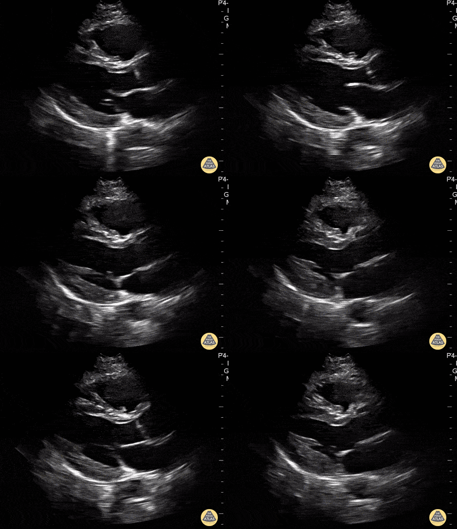
Parasternal Long Axis - Normal
In this view we see the muscular left ventricle and the smaller left atrium separated by the mitral valve near the bottom of the screen. The aortic outflow tract (valve and root) comes off the left ventricle superficial to the left atrium. The most superficial chamber seen in this view is the right ventricle, separated from the left ventricle by the interventricular septum. The heart is surrounded by the bright hyperechoic pericardium. At the very bottom of the screen, the round structure outside the pericardium is the descending thoracic aorta in transverse view.
Hannah Kopinski - MS4, Dr. Lindsay Davis - NYU/Bellevue Department of Emergency Ultrasound, Dr. Matthew Riscinti - Kings County Emergency Medicine
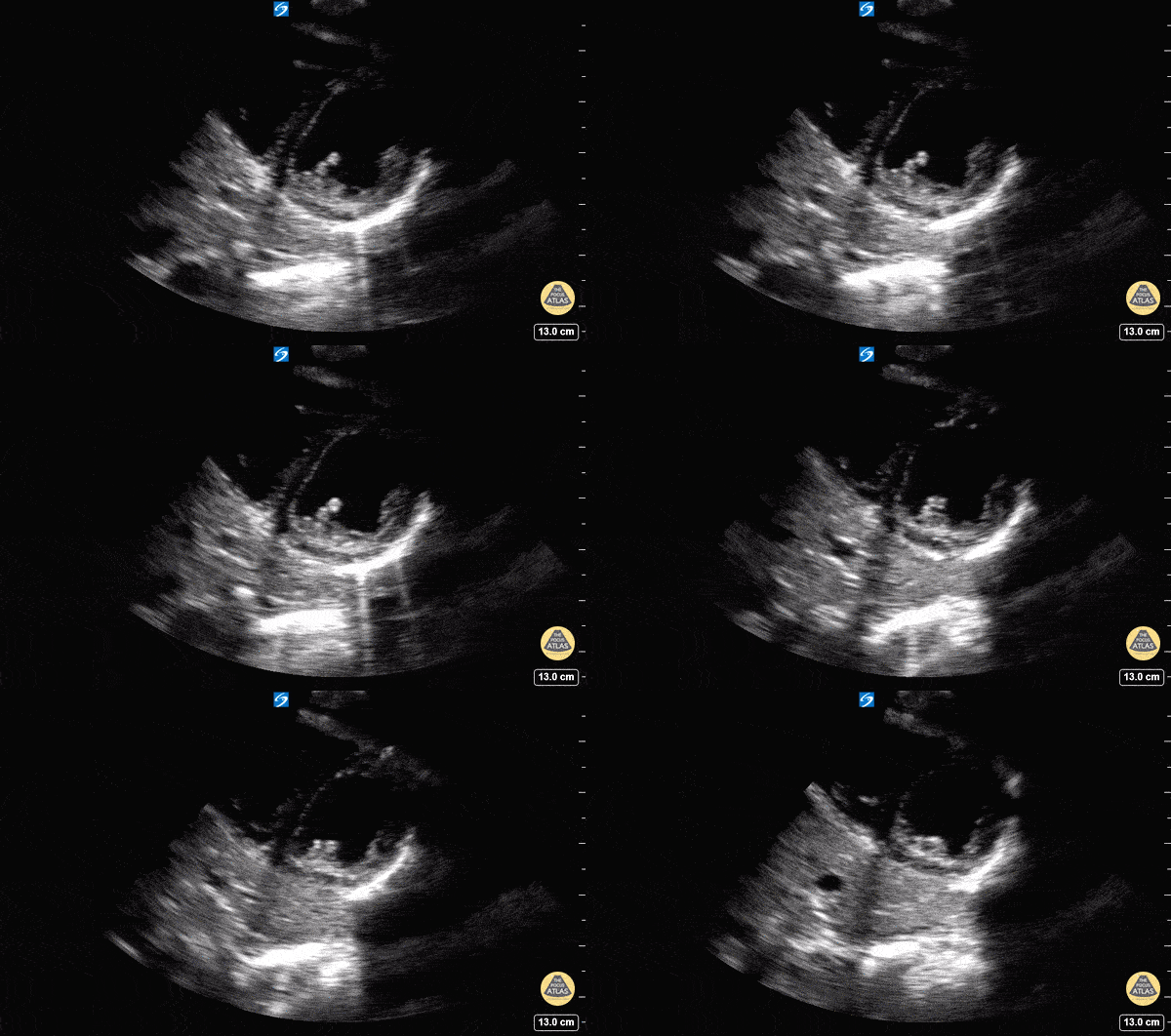
Parasternal Short Axis - Normal
In this view we are evaluating the heart in cross-section. In the center of the screen is the muscular walled left ventricle, which should form a perfect circle. This is at a level below the mitral valve and we can see the papillary muscles come into view. The smaller, thin walled, crescent shaped right ventricle is seen superficially and to the left of the screen.
Hannah Kopinski - MS4, Dr. Lindsay Davis - NYU/Bellevue Department of Emergency Ultrasound, Dr. Matthew Riscinti - Kings County Emergency Medicine
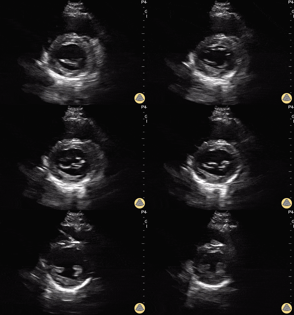
Parasternal Short Axis (Mitral Valve) - Normal
In this view we are evaluating the heart in cross-section. In the center of the screen is the muscular walled left ventricle, which should form a perfect circle. At the beginning of the clip, we see the “fish mouth” appearance of the mitral valve as it opens and closes. Then the probe is tilted inferiorly towards the apex and the papillary muscles come into view. The smaller, thin walled, crescent shaped right ventricle is seen superficially and to the left of the screen.
Hannah Kopinski - MS4, Dr. Lindsay Davis - NYU/Bellevue Department of Emergency Ultrasound, Dr. Matthew Riscinti - Kings County Emergency Medicine
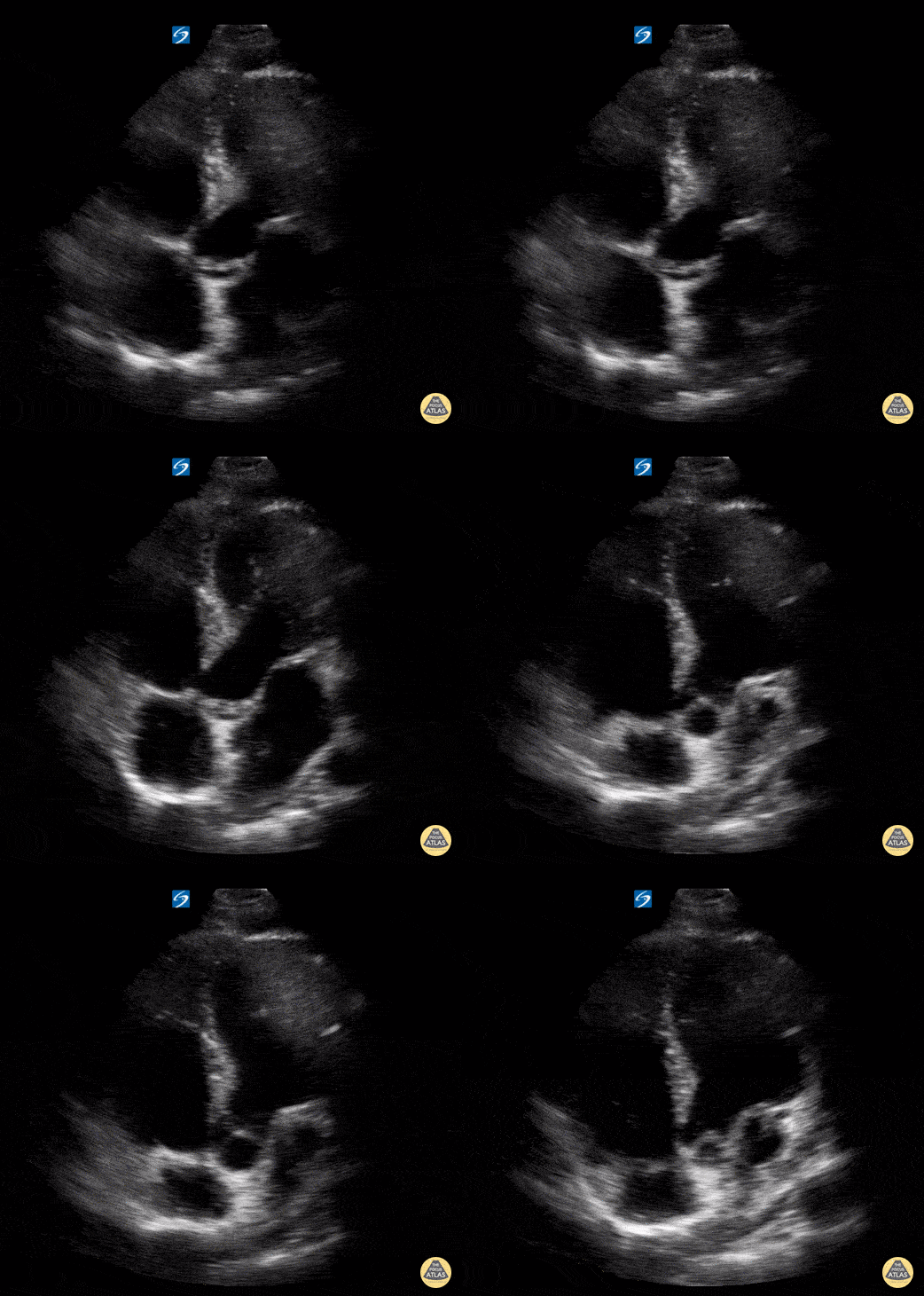
Apical 5 Chamber - Normal
In this view we are able to visualize all four chambers of the heart. Clockwise from the top left of the screen we see the right ventricle, left ventricle, left atrium, and right atrium. We can also see the tricuspid valve between the RA and RV and the mitral valve between the LA and LV. The “fifth chamber” is the aortic valve/root which can be appreciated in the center of the image.
Hannah Kopinski - MS4, Dr. Lindsay Davis - NYU/Bellevue Department of Emergency Ultrasound, Dr. Matthew Riscinti - Kings County Emergency Medicine
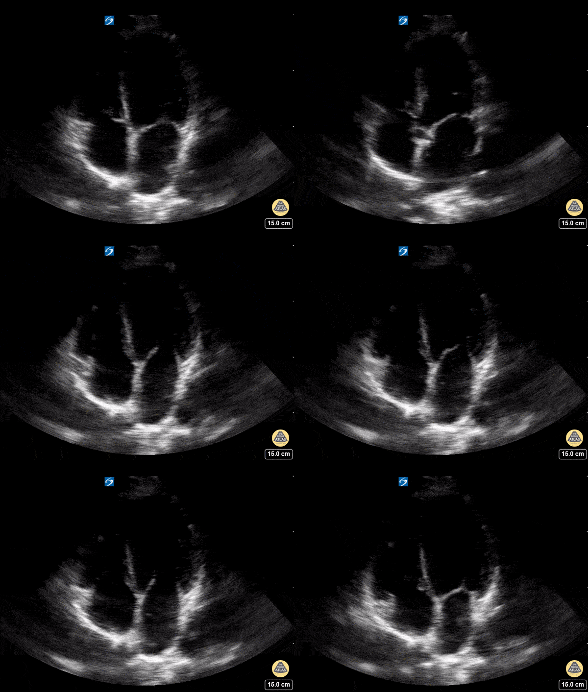
Apical Four Chamber - Normal
In this view we are able to visualize all four chambers of the heart. Clockwise from the top left of the screen we see the right ventricle, left ventricle, left atrium, and right atrium. We can also see the tricuspid valve between the RA and RV and the mitral valve between the LA and LV.
Hannah Kopinski - MS4, Dr. Lindsay Davis - NYU/Bellevue Department of Emergency Ultrasound
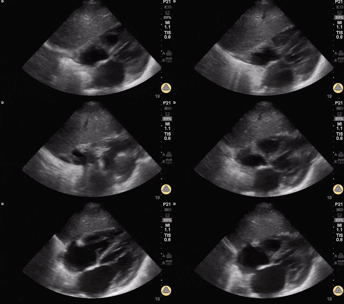
Subxiphoid / Subcostal - Normal Anatomy
The most superficial structure we see is the liver. Immediately deep to that we see the heart separated from the liver by the diaphragm. Closest to the liver is the right atrium, tricuspid valve, right ventricle. Deeper to that, we see the left atrium, mitral valve, and left ventricle.
Hannah Kopinski - MS4, Dr. Lindsay Davis - NYU/Bellevue Department of Emergency Ultrasound, Dr. Matthew Riscinti - Kings County Emergency Medicine
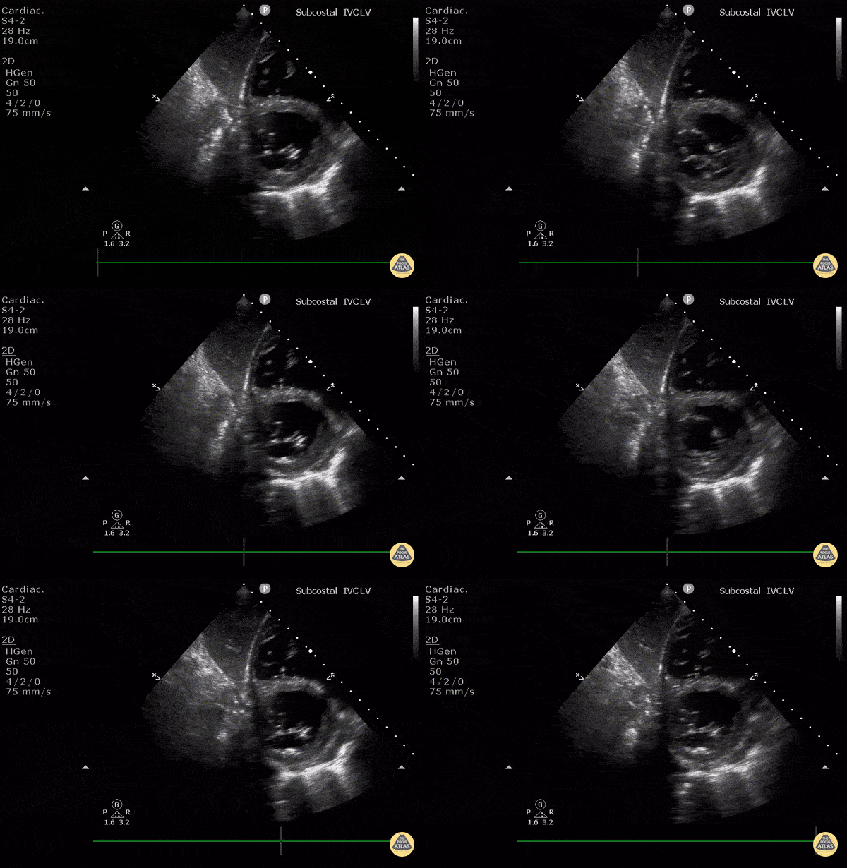
Subcostal Short Axis
WCUME17 Submission for "Novel Indication"
Subcostal short axis view of the left ventricle can be useful if PSAX window is poor. A good alternative to estimate LV function.
This image shows: LV in short axis at level of MV and the RV in short axis, and the interventricular septum. The liver is overlying.
Technique: Start with IVC in long axis at entrance to RA and tilt transducer cephalad. Slide transducer to patient's right, fan transducer out to patient’s left.
Dr. Cian McDermott - Dublin, Ireland
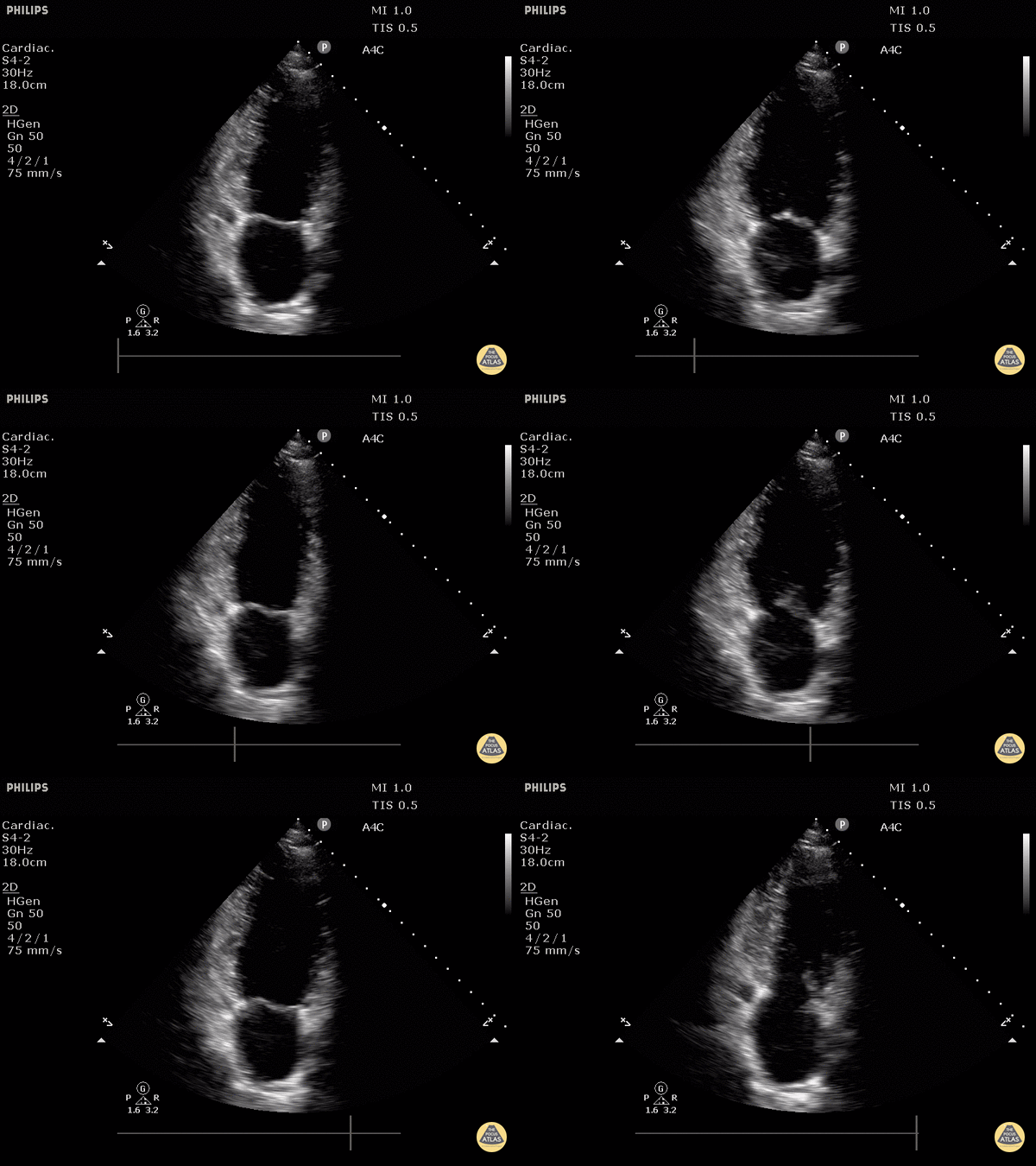
Normal Apical 2 Chamber View
Justin Bowra MBBS, FACEM, CCPU Emergency Physician, RNSH et al.
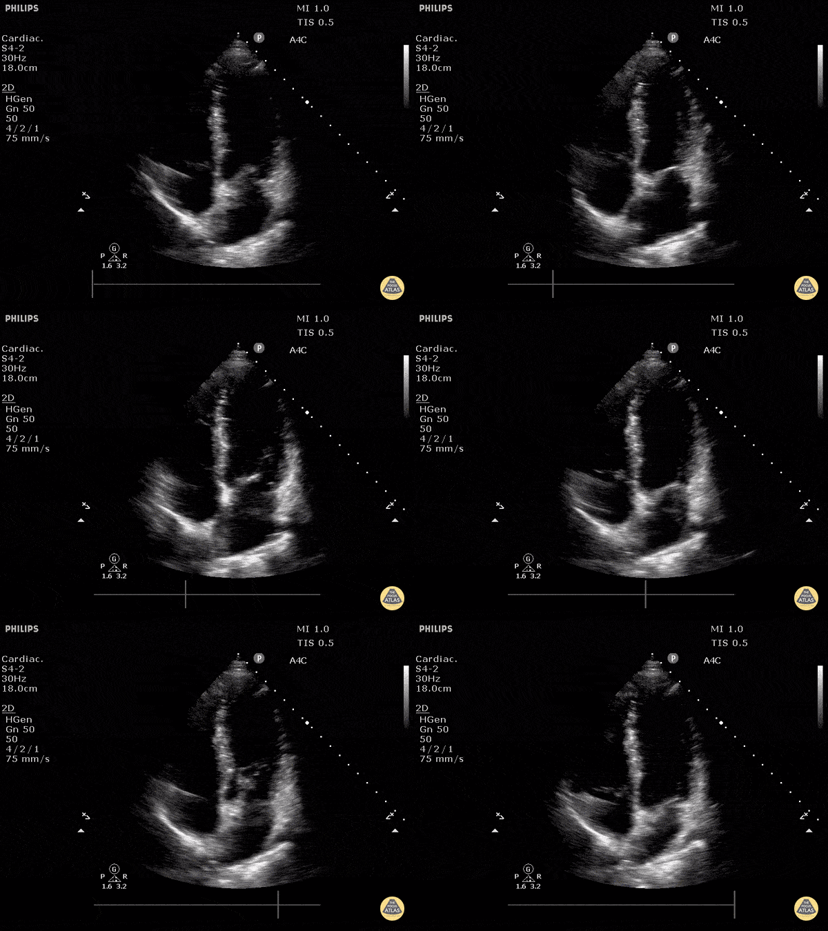
Normal Apical 4 Chamber
Justin Bowra MBBS, FACEM, CCPU Emergency Physician, RNSH et al.
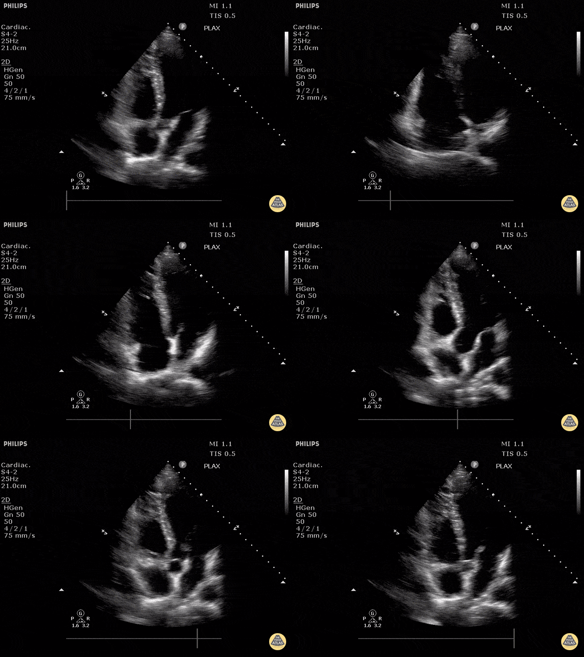
Apical 5 Chamber View
Justin Bowra MBBS, FACEM, CCPU Emergency Physician, RNSH et al.

Normal Subcostal View
Justin Bowra MBBS, FACEM, CCPU Emergency Physician, RNSH et al.
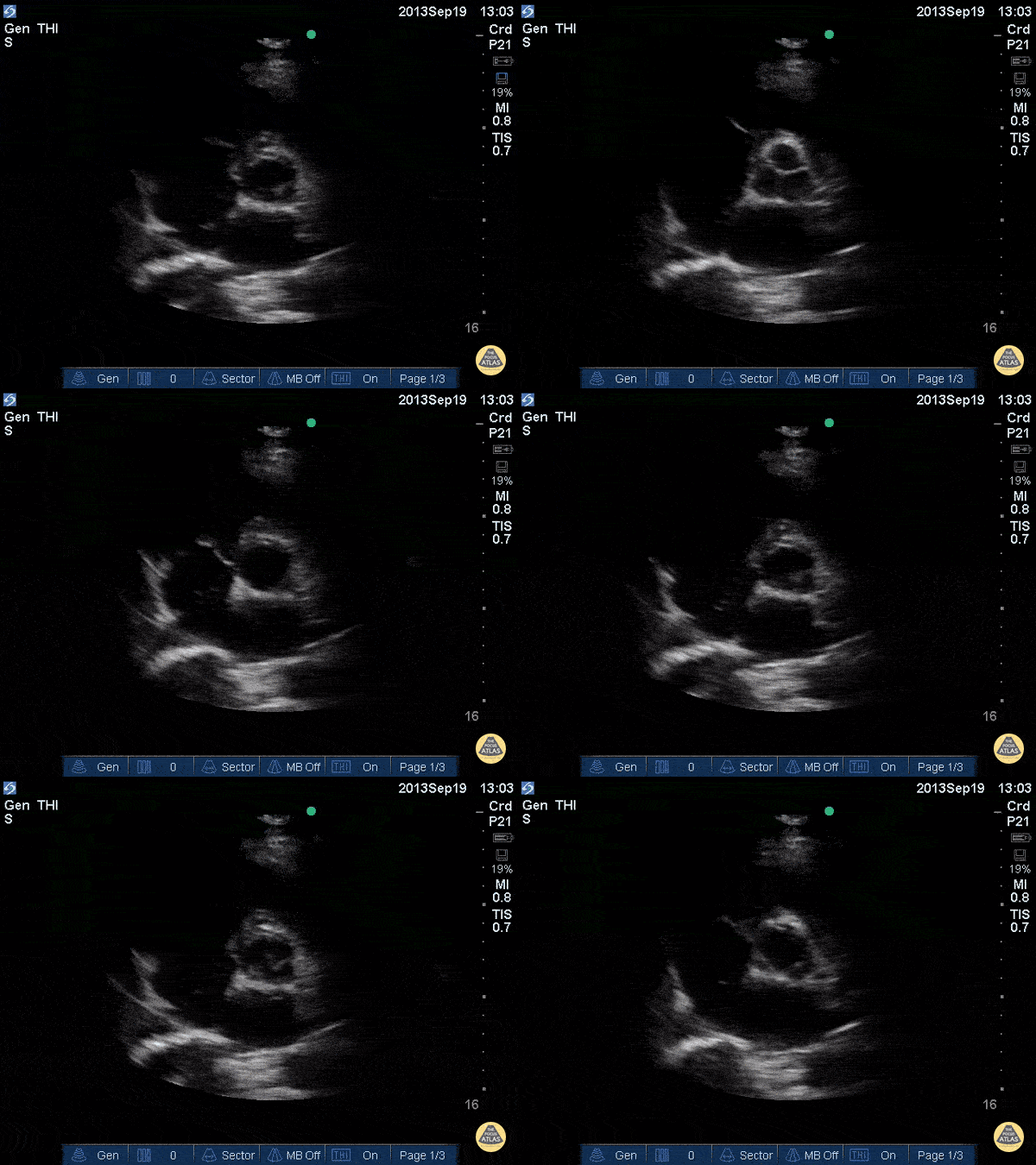
Aortic Valve (Short)
Justin Bowra MBBS, FACEM, CCPU Emergency Physician, RNSH et al.
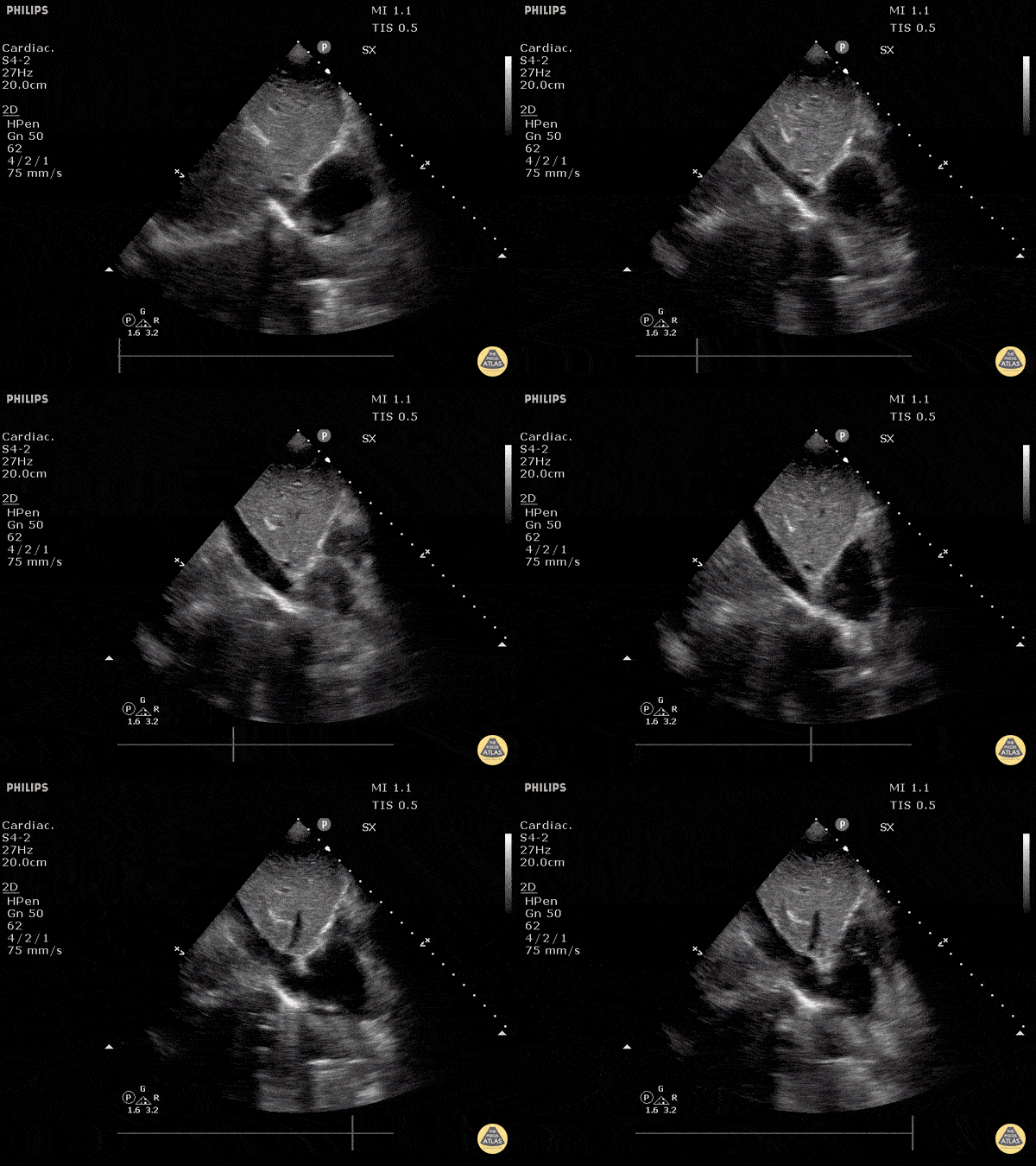
Inferior Vena Cava Emptyting into Right Atrium
Normal IVC seen in longitudinal view, emptying into the right atrium.
Justin Bowra MBBS, FACEM, CCPU Emergency Physician, RNSH et al.
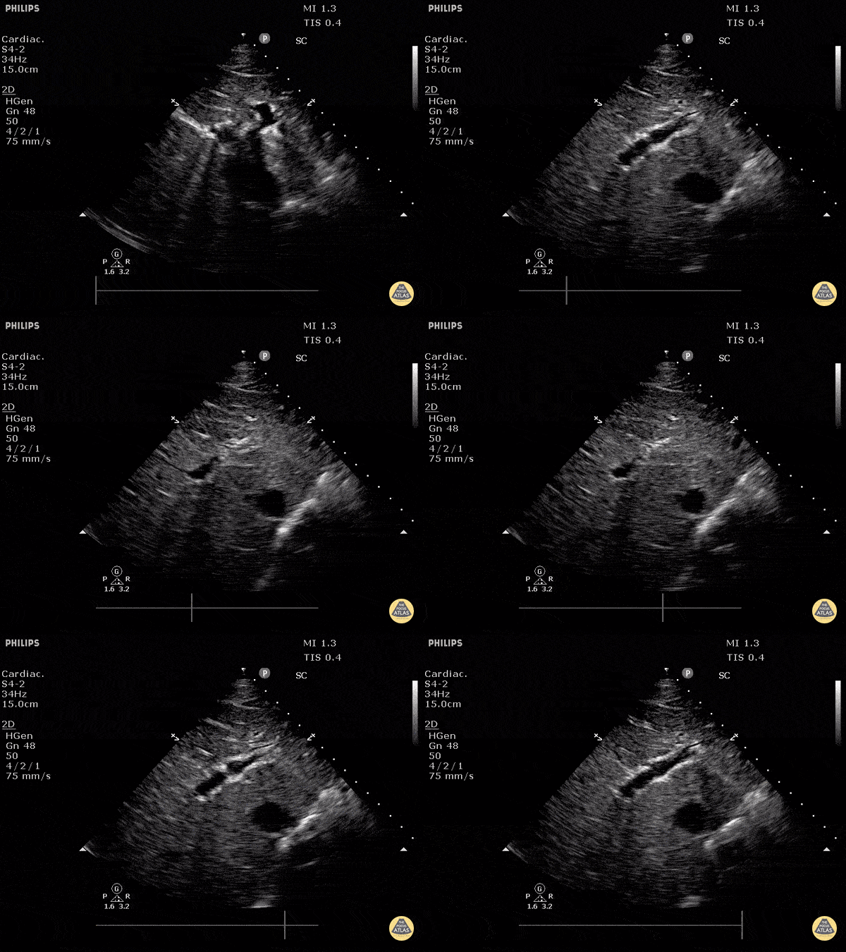
Normal Inferior Vena Cava
Normal IVC seen in transverse plane using a subxiphoid approach.
Justin Bowra MBBS, FACEM, CCPU Emergency Physician, RNSH et al. (Dr. S Fares)
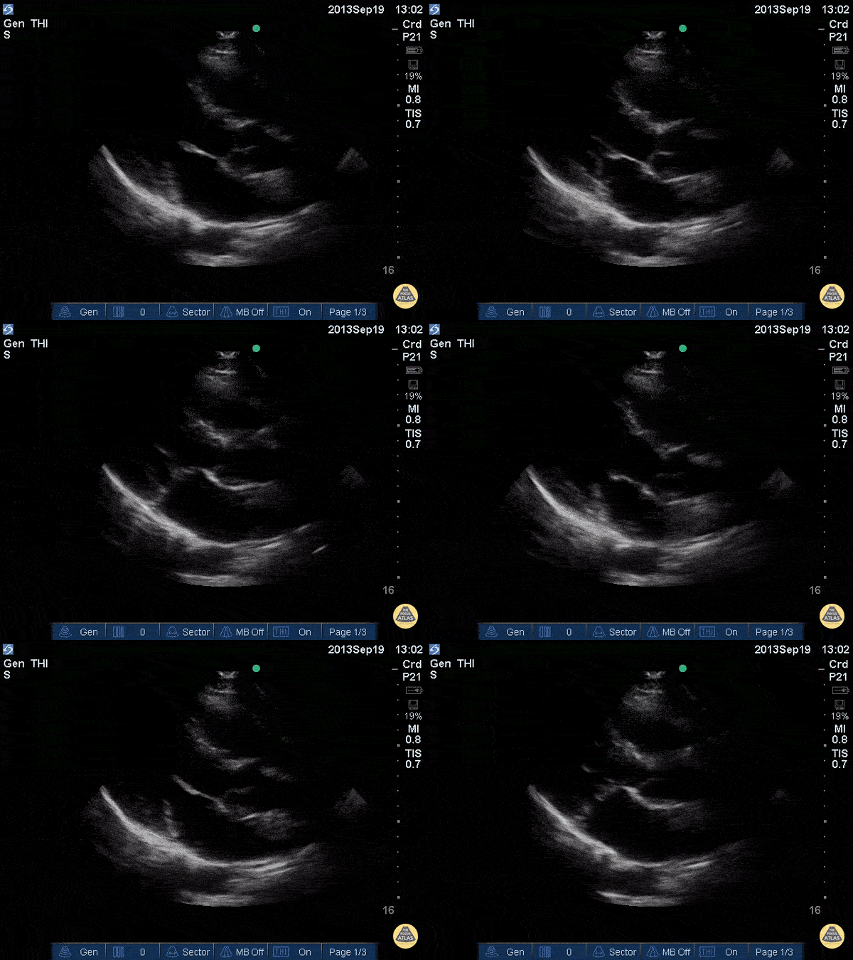
Mitral Valve Parasternal Long-Axis View
Justin Bowra MBBS, FACEM, CCPU Emergency Physician, RNSH et al.
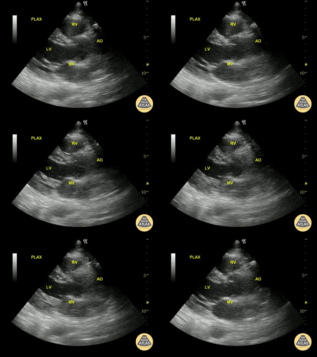
Normal Parasternal Long-Axis View
Justin Bowra MBBS, FACEM, CCPU Emergency Physician, RNSH et al.
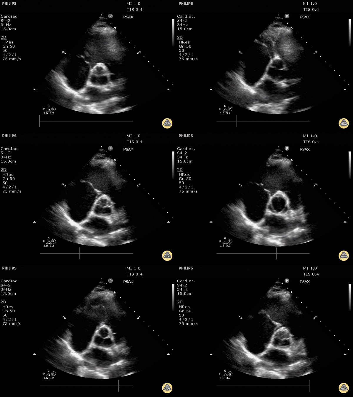
Normal Aortic Valve (Parasternal Short-Axis View)
Justin Bowra MBBS, FACEM, CCPU Emergency Physician, RNSH et al.
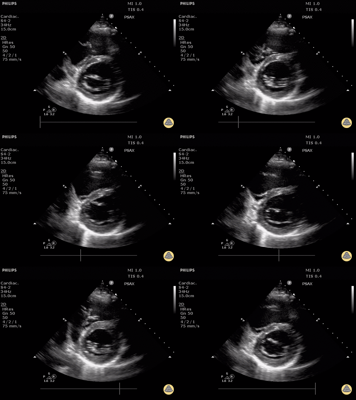
Normal Mitral Valve (Parasternal Short-Axis View)
Justin Bowra MBBS, FACEM, CCPU Emergency Physician, RNSH et al.


