
OB/Gyn

Dichorionic Diamniotic Twin Intrauterine Pregnancy
A 27-year-old female presented for evaluation of cough, congestion, decreased appetite, muscle aches, and fatigue. Patient was unsure of the date of her last menstrual period.
This image is a transabdominal ultrasound of the uterus demonstrating a dichorionic diamniotic twin intrauterine pregnancy. The pregnancy was dated at approximately 8 weeks and 2 days, with fetal heart rates of 167 bpm and 174 bpm.
Arthur Sieron BMBS, PGY-1 Emergency Medicine, Central Michigan University; Eric Spencer DO, PGY-3 Emergency Medicine, Central Michigan University; Halimah Hamidu, MS4 Central Michigan University College of Medicine.

Biparietal diameter (BPD) measurement in a 26-week pregnancy.
BPD should be measured in the axial plane, perpendicular to the falx cerebri, and from the outer edge of the near calvarial wall to the inner edge of the far calvarial wall.
Contributors:
Stephen Holihan (MD); Dillon Nerland (MD); Madison Waddell (MS4)

Caesarean Section Scar Ectopic Pregnancy
32 year old female at 8-9 weeks gestation by LMP referred to the ED for pregnancy of unknown location. She reported 1-2 weeks of mild pelvic cramping and vaginal spotting.
Bedside trans-abdominal imaging demonstrated a round, heterogenous structure without a clear yolk sack or fetal pole, but with several hallmarks of caesarean scar ectopic pregnancy: position in the anterior uterine wall; in the lower (closer to the cervix) half of the uterus; bulging into the neighboring bladder; with thin interposed myometrium. After confirmatory formal ultrasound, she was admitted for methotrexate termination, which proved successful.
Contributors: Nicholas Maurer, MD, MPH (PGY-1); Megan Chenworth (PGY-3)
Emergency Medicine, Northwestern McGaw Medical Center

US Confirmation of Appropriate IUD Location
Transvaginal ultrasound showing the IUD correctly placed in both sagittal and transverse plane.
Contributor: Dr. Nicolay B. Werner
Akershus University Hospital, Norway

Fetal Pole with Cardiac Activity
This is a transverse uterus view in early pregnancy demonstrating a a fetal pole with visualized cardiac activity.
Mike Macias, MD, Emergency Physician, @emedcurious

Uterus with Leiomyomas (fibroids)
Heterogeneous uterus with multiple well defined hypoechoic masses consistent with leiomyomas (fibroids).
Dimitri Livshits DO, Ultrasound Fellow, Kings County/SUNY Downstate; Jane Belyavskaya MD, Ultrasound Fellow, Kings County/SUNY Downstate; Chris Hanuscin MD, Ultrasound Division Director, Kings County/SUNY Downstate;

Ectopic Pregnancy
30s female currently 7 week pregnant with lower abdominal pain and tenderness to palpation to lower abdomen. POCUS showed a gestation sac containing a yolk sac and fetal pole outside of uterus. Also noted is a thickened endometrial stripe (to the right of the gestational sac). No free fluid was noted in the RUQ. The patient was taken to the OR for definitive management of ectopic pregnancy.
Dimitri Livshits DO, Ultrasound Fellow, Kings County/SUNY Downstate; Jane Belyavskaya MD, Ultrasound Fellow, Kings County/SUNY Downstate; Farnam Kazi MD, Ultrasound Faculty, Kings County/SUNY Downstate;

Molar Pregnancy
This is a saggital view of the uterus belonging to a patient who returned to the emergency department after persistent vomiting. An initial urine pregnancy test performed yielded a negative result however this patient’s ultrasound scan ultimately revealed a molar pregnancy. As Dr. Jones explains, this patient false-negative urine pregnancy test is explained by a phenomenon known as the High Dose Hook Effect.
Image courtesy of Robert Jones DO, FACEP @RJonesSonoEM
Director, Emergency Ultrasound; MetroHealth Medical Center; Professor, Case Western Reserve Medical School, Cleveland, OH
View his original post here

Tubal Ectopic Pregnancy
Notice to the left of the screen the presence of an adnexal mass separate to the ovaries which in this case indicates a right tubal ectopic pregnancy. Also noted here within the endometrium is an outer echogenic layer, middle hypoechoic layer and an inner hyperechoic stripe, which makes up the classic trilaminar pattern that is usually observable during the proliferative phase of menstruation.
Image courtesy of Robert Jones DO, FACEP @RJonesSonoEM
Director, Emergency Ultrasound; MetroHealth Medical Center; Professor, Case Western Reserve Medical School, Cleveland, OH
View his original post here

IUD in Transverse Plane
Patient came to the ED due to flank pain. Renal ultrasound was performed by me with the US team. Upon looking for the bladder, I saw my first IUD via US which appeared hyperechoic. ParaGard has been shown to be more than 99% effective.
Mehtab Galeh, MD, @GalehMehtab

IUD in Sagittal Plane
Patient came to the ED due to flank pain. Renal ultrasound was performed by me with the US team. Upon looking for the bladder, I saw my first IUD via US which appeared hyperechoic. ParaGard has been shown to be more than 99% effective.
Mehtab Galeh, MD, @GalehMehtab

Ovarian Hyperstimulation Syndrome (OHSS)
20 y/o female s/p egg retrieval for egg donation 3 days prior presents for diffuse abdominal pain and bloating. In the clip (bladder sagittal view), you can see massive enlargement of the ovary and its multiple follicles. FAST revealed free fluid throughout the abdomen likely secondary to leakage of fluid from these follicles, a process referred to as ovarian hyperstimulation syndrome (OHSS). The patient was admitted to GYN for possible drainage.
Jennifer Kaminsky, MD PGY-2; @jen_kaminskyMD
Pamela Santivanez, MD PGY-1
Sean Beckman, Rocky Vista University COM OMS-4
Joshua Greenstein, MD, Director of ED Ultrasound
Northwell Health - Staten Island University Hospital

Fetal Pole with Cardiac Motion - Sagittal
Sagittal view of uterus demonstrating fetal pole. A subtle flicker within the fetal pole can be seen consistent with cardiac motion.
Michael Macias

Yolk Sac
A transabdominal scan with a curvilinear probe failed to show an intrauterine yolk sac however, when using a linear probe a yolk sac became visible.
Image courtesy of Robert Jones DO, FACEP @RJonesSonoEM
Director, Emergency Ultrasound; MetroHealth Medical Center; Professor, Case Western Reserve Medical School, Cleveland, OH
View his original post here

Pelvic Cystic Mass
A middle-aged female with a history of colon cancer presented to the ED with constipation and abdominal pain. A recent colonoscopy had revealed rectosigmoid adenocarcinoma. Her physical exam was notable for a distended abdomen that was tender to palpation. POCUS revealed the presence of a large, complex, cystic structure within the pelvis. Subsequent CT confirmed a loculated fluid collection within the pelvis compressing the rectum and sigmoid colon; this mechanical obstruction was likely contributing to patient’s constipation. Differential diagnosis included loculated ascites versus cystic tumor.
Kyla Walworth, MS-4 & Matthew McDowell, PGY-1
Central Michigan University College of Medicine

IUD Foreign Body
Transvaginal US showing a gestational sac of about 6 weeks by dates with a fragment of an old IUD embedded in the endometrium shown as the hyperechoic line with posterior acoustic shadow. Patient had an IUD removal 10 months prior.
Image courtesy of Robert Jones DO, FACEP @RJonesSonoEM
Director, Emergency Ultrasound; MetroHealth Medical Center; Professor, Case Western Reserve Medical School, Cleveland, OH
View his original post here

Ovarian Torsion
A 38-year-old female with PMH ulcerative colitis presented with several hour history of RLQ abdominal pain. Pain was described as sharp and constant; it woke her from her sleep. Vitals were WNL. Physical exam was notable for right adnexal tenderness and RLQ abdominal tenderness to palpation.
Pelvic ultrasound revealed an enlarged right ovary with peripheralized follicles, heterogenous ovarian stroma, and peri-ovarian free fluid; findings concerning for ovarian torsion. It is important to remember that while ovarian torsion remains a clinical diagnosis, US grey scale findings are some of the most reliable radiographic adjunct predictors of the diagnosis as Doppler imaging often remains normal.
Reference: Grunau GL, Harris A, Buckley J, Todd NJ. Diagnosis of Ovarian Torsion: Is It Time to Forget About Doppler? Journal of Obstetrics and Gynaecology Canada. 2018;40(7):871-875.
Devin Peuser, @DevinPeuser
Brooklyn, NY

Uterine Perforation
A patient presented to the ED 4 days s/p pregnancy termination via D&C with a fever and profound hypotension. POCUS revealed pelvic free fluid with rising gas bubbles indicative of a uterine perforation.
Image courtesy of Robert Jones DO, FACEP @RJonesSonoEM
Director, Emergency Ultrasound; MetroHealth Medical Center; Professor, Case Western Reserve Medical School, Cleveland, OH
View his original post here

Fetal Pole with Cardiac Motion - Transverse
Transverse view of uterus demonstrating fetal pole. A subtle flicker within the fetal pole can be seen consistent with cardiac motion.
Michael Macias

Left Ovarian Cyst
A young female presented to the ED with sudden pelvic pain. She has no PMH and a negative hCG. Bedside sagittal transabdominal ultrasound revealed a large right ovarian cyst mimicking the urinary bladder. Notice the bladder decompressed with a foley balloon.
Image courtesy of Robert Jones DO, FACEP @RJonesSonoEM
Director, Emergency Ultrasound; MetroHealth Medical Center; Professor, Case Western Reserve Medical School, Cleveland, OH
View his original post here

Yolk Sac
This trans abdominal ultrasound reveals a yolk sac.
John Joseph, MD. University of Michigan

Intrauterine Pregnancy
21 year old female presented to the ED reporting lower abdominal discomfort. HPI notable for absent trauma, dysuria, hematuria, constipation, and fever. Last menstrual period was 3 months prior to presentation. POCUS was faster than urine pregnancy test to clinch the diagnosis!
Dr. Victor Bang. Emergency Physician at Hospital das Clínicas de Marília. Co-founder of Pocus Jedi.
@vmjbang
Ruptured Ectopic With Positive Fast
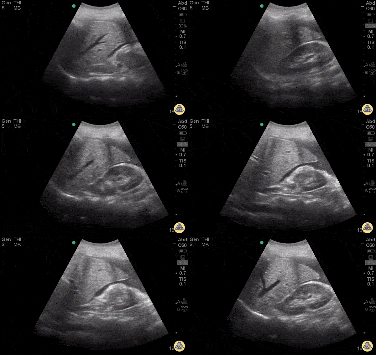
30 y/o F presented to ED for abdominal pain stating she had a recent miscarriage. Now she is having vaginal bleeding for the last 2 weeks. Borderline hypotensive and in severe distress due to pain. Abdomen diffusely tender with guarding. POCUS demonstrated +FAST with blood in the hepatorenal space. Remember to fan all the way to liver tip in FAST scan to fully evaluate for free fluid, it can be subtle and not simply in morrison’s pouch. The patient was rushed to OR by OBGYN for ex-lap based on this scan and was found to have ruptured tubal ectopic pregnancy.
Dr. Stacey Frisch - Kings County Emergency Medicine
Transabdominal Right Adnexa with Ovarian Cyst
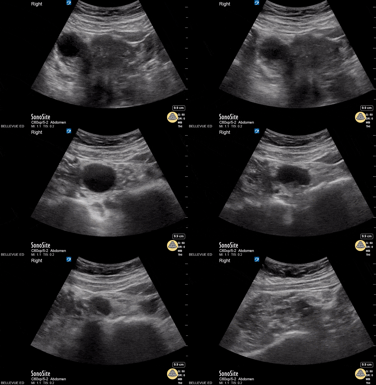
In this transabdominal view of the uterus and adnexa we see a thin walled, ovoid anechoic structure on the left side of the screen. This is a simple cyst on the right ovary. As the probe fans, the uterine fundus comes in and out of view medial to the ovary .
Transabdominal Uterus Transverse
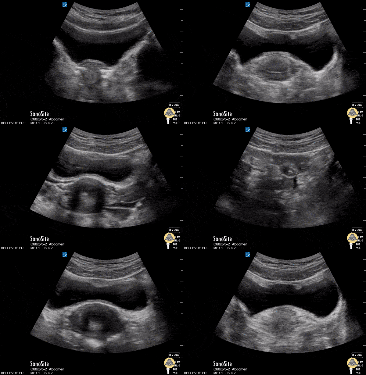
This is a clip of the uterus in transverse view using the transabdominal approach. The large anechoic structure in the center is the bladder in transverse view, and immediately deep to the bladder is the round uterine fundus. As the probe fans through the uterus we see some dark shadowing within it, likely due to artifact created by an IUD. The fallopian tubes are also faintly visible bilaterally branching off the uterus.
Molar Pregnancy
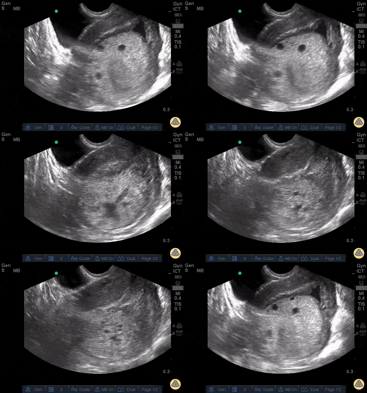
23 y/o F, gestational age 10 weeks by LMP referred to ER for suspicion of molar pregnancy. TV US shows hydropic vesicles within uterus, represented by anechoic regions scattered throughout a hyperechoic mass in uterine cavity. This is classically described as a “bunch of grapes” or “snowstorm pattern”.
Patients diagnosed with molar pregnancy can present with vaginal bleeding or symptoms similar to hyperemesis gravidarum, making ER POCUS a useful evaluation tool that can expedite disposition.
Dr. Bryan Flores, Dr. Teresa Smith - Kings County Emergency Medicine
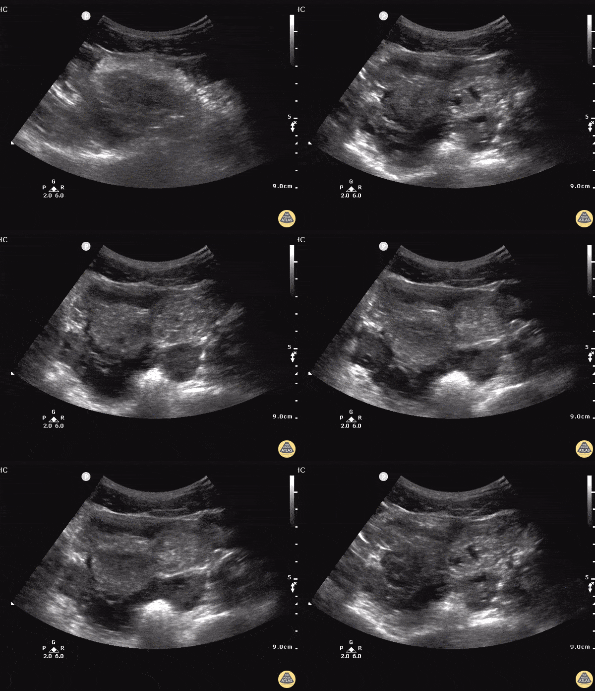
Hematocolpos
13yoF with worsening abdominal pain and back pain x3 months, found to have large bilobed pelvic mass consistent with hematocolpos/hematometra due to imperforate hymen. Transabdominal ultrasound using a 5-1MHz phased array probe in the transverse plane 2cm above pubic symphysis. A large hypoechoic structure within the uterus is seen displacing the bladder superoanteriorly with posterior acoustic enhancement. US can demonstrate retained old blood as a hypoechoic cystic structure and monitor resolution after hymenectomy.
(Hassani 1978)
Ectopic - Ruptured Cornual Ectopic Pregnancy
29 y/o female with intermittent lower abdominal and vaginal pain and nausea x3 days. LMP 3.5 weeks prior. Transabdominal ultrasound demonstrates pregnancy in cornu of the uterus. Symptoms progressed over 24 hours and patient became hypotensive and was taken to OR for exploratory laparotomy and found to have ruptured cornual ectopic with hemoperitoneum.
Stacey Frisch, MD Juliana Jaramillo, MD Stephan Rinnert, MD - Kings County/SUNY Downstate Emergency Medicine
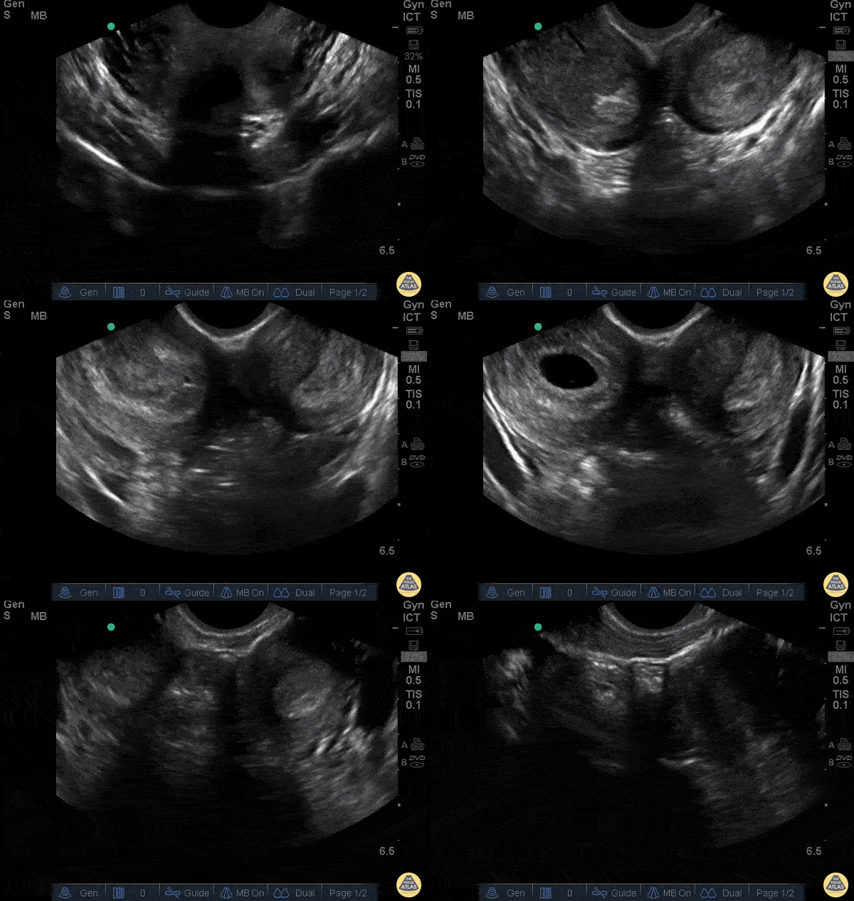
Bicornuate Uterus with IUP
WCUME 2017 Submission for "Best POCUS"
23 y/o female, pelvic pain, no vb or discharge, g1p0 found to be pregnant at this ER visit. Approximately 4 weeks by dates.
Found to be pregnant and found to have a bicornuate uterus with IUP on the right side.
Carl Alsup, MD - Sierra Nevada Memorial Hospital
Ectopic - Ruptured Cornual Ectopic Pregnancy - Longitudinal
24 y/o F presents after a brief syncopal episode. Endorses heavy vaginal bleeding for 2 weeks. On exam, tachycardic and tender to palpation in lower abdomen. Urine HCG was positive.
Transabdominal longitudinal sonogram view of the pelvis showed a large hypoechoic collection suggestive of free pelvic fluid in the proximity of a solid hyperechoic mass in the left adnexal. Just inferior to the mass is the uterus a small amount of hypoechoic fluid in the endometrium but no clear intrauterine pregnancy.
GYN was consulted immediately and the ultimate operative note for this patient described a ruptured cornual ectopic pregnancy. A cornual pregnancy or interstitial pregnancy is a type of ectopic pregnancy located outside of the uterine cavity in the distal fallopian tube as it penetrates into the muscular wall of the uterus. This type of ectopic pregnancy has the potential to grow to larger sizes than standard tubal ectopic pregnancies and carries a higher mortality risk.
Dr. Tareq Azad and Dr. Scott Kendall - Kings County Emergency Medicine

Ectopic - Ruptured Cornual Ectopic Pregnancy - Transverse
24 y/o F presents after a brief syncopal episode. Endorses heavy vaginal bleeding for 2 weeks. On exam, tachycardic and tender to palpation in lower abdomen. Urine HCG was positive.
Transabdominal tranverse sonogram view of the pelvis showed again a hyperechoic mass in the left adnexal abbuting an empty uterus, ovaries in view, and a significant amount of free fluid beneath these structures.
GYN was consulted immediately and the ultimate operative note for this patient described a ruptured cornual ectopic pregnancy. A cornual pregnancy or interstitial pregnancy is a type of ectopic pregnancy located outside of the uterine cavity in the distal fallopian tube as it penetrates into the muscular wall of the uterus. This type of ectopic pregnancy has the potential to grow to larger sizes than standard tubal ectopic pregnancies and carries a higher mortality risk.
Dr. Tareq Azad and Dr. Scott Kendall - Kings County Emergency Medicine
Anembryonic Pregnancy
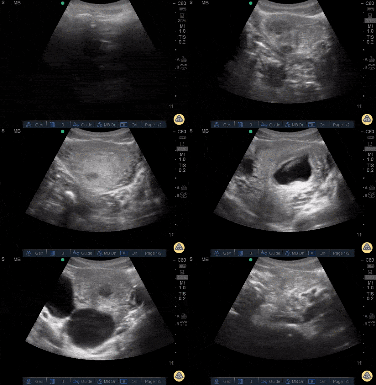
Anembryonic pregnancy
27 y/o G8P2 (5 prior D&Cs) presenting at 14 weeks pregnant by LMP for left lower quadrant pain radiating to her back "feels like mini-contractions) for 2 hours and vaginal bleeding.
POCUS demonstrates intrauterine gestational sac with a mean sac diameter 3.1cm (correlating to 8 weeks) without yolk sac or fetal pole, consistent with an anembryonic pregnancy. The patient left AMA and returned 2 days later with sharp, 10/10, intermittent, contraction-like pain in the lower abd radiating to the back and heavy vaginal bleeding. Physical exam at that time demonstrated cervical dilation to 2cm with tissue in the os and pooling of blood within the vaginal vault. The patient underwent a dilation and curettage in the operating room.
Stacey Frisch, MD, Sage Wiener, MD - Kings County/SUNY Downstate Emergency Medicine
Ectopic - Tubal Ectopic Pregnancy
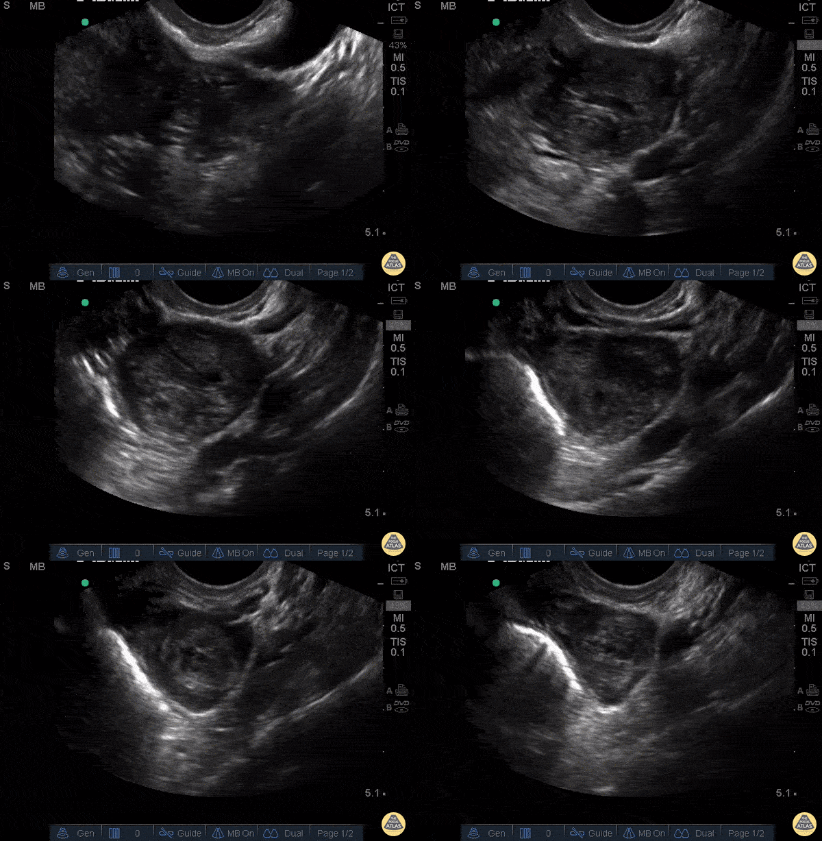
31 y/o G1P0 presenting at unknown gestational age for pelvic pain and vaginal bleeding. Described increasing vaginal bleeding for 7 days and intermittent sharp lower abdominal and pelvic pain, mostly left sided, 6/10 in severity. Pelvic exam was significant for cervical motion tenderness and left adnexal tenderness.
Pelvic ultrasound shows left adnexal complex heterogeneous structure and associated free fluid in the pelvis. Patient underwent diagnostic laparoscopy with left salpingectomy for left tubal ectopic pregnancy.
Stacey Frisch, MD Aleksandr Gleyzer, MD - Kings County/SUNY Downstate Emergency Medicine
Hemorrhagic Ovarian Cyst
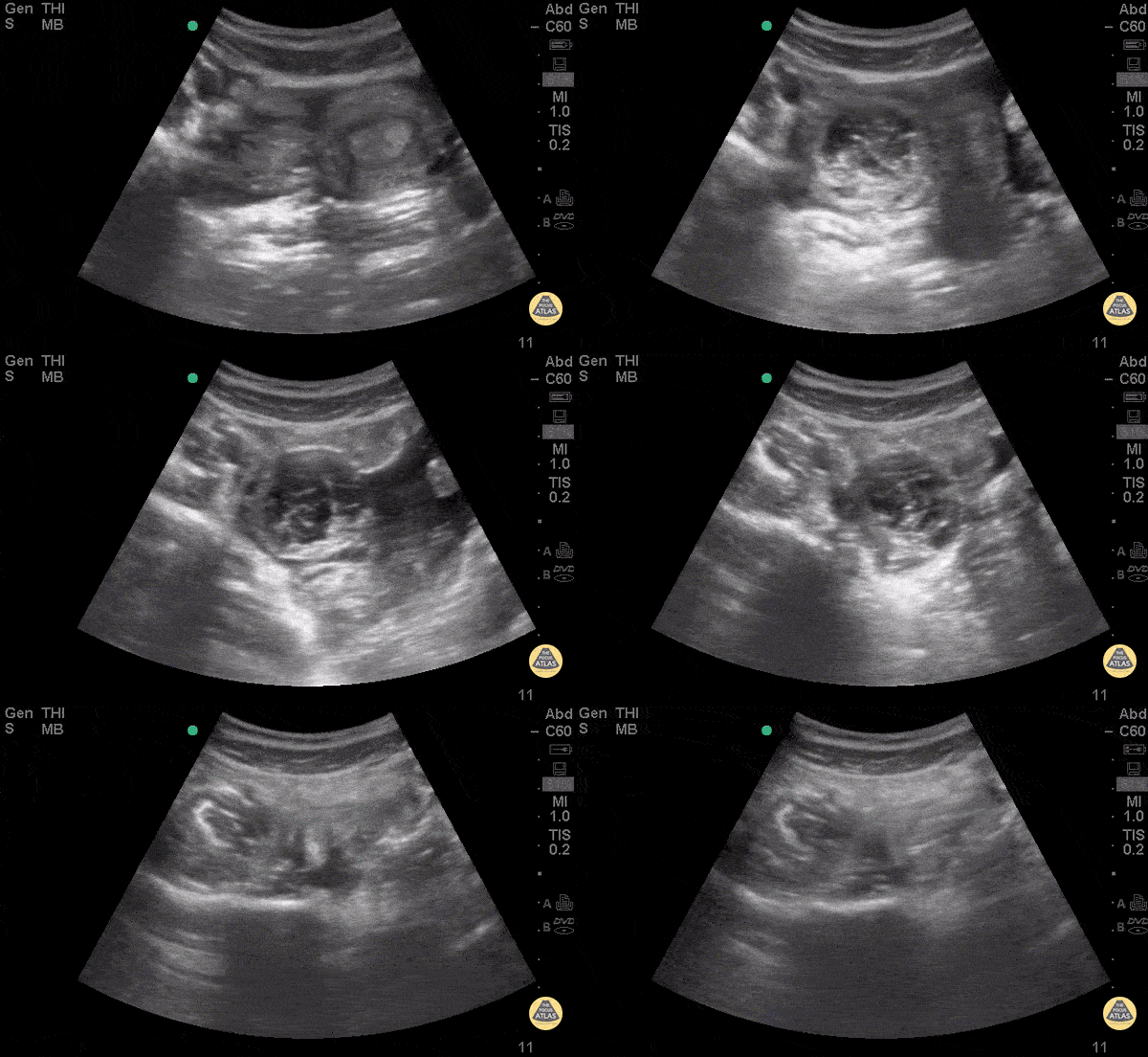
18yo F with 1 month history of abdominal pain presents with acute worsening of abdominal pain for 2 days. Exam revealed diffuse disproportionate pain to palpation of abdomen and right adnexal tenderness.
POCUS demonstrates a hemorrhagic ovarian cyst defined by a cystic structure adjacent to the uterus with a well-defined wall and lacy/fishnet pattern within the structure. A hyper-acoustic shadowing can be seen distally along with free fluid surrounding the HOC.
HOC can also be mistaken for a neoplastic ovarian mass. A distinguishing feature would be changes in fluid collection and changes in size of diameter of the cyst.
Dr. Praneetha Chaganti, Dr. Andrew Aherne Kings County/SUNY Downstate Medical Center
Ruptured Ovarian Cyst - Hematoma
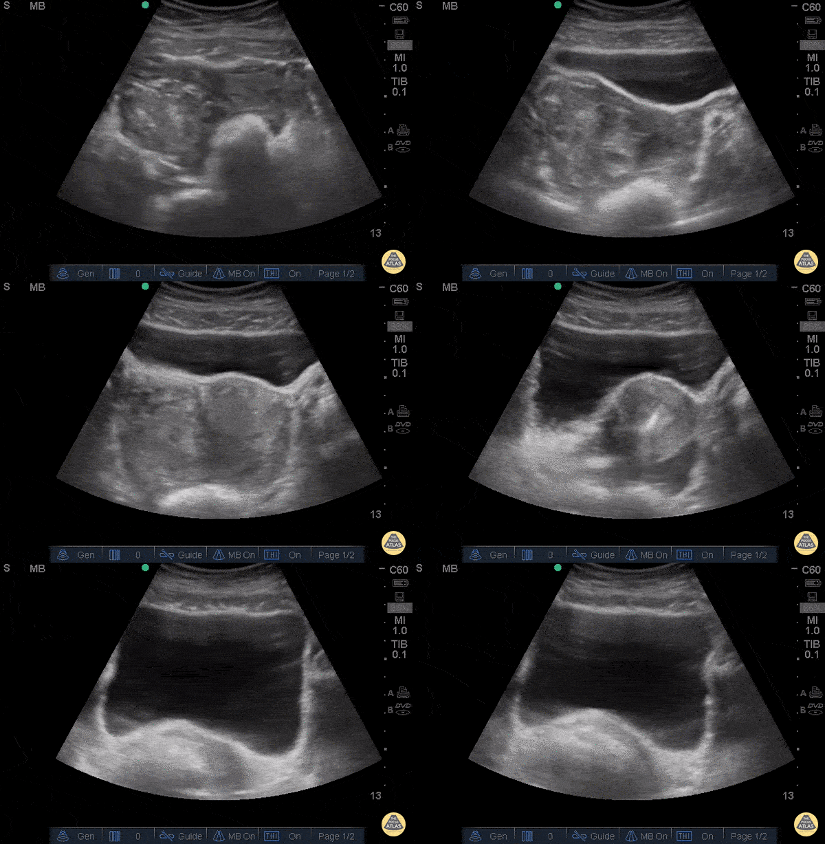
28 year-old female was BIBEMS after a witnessed syncopal episode at home. The patient endorsed abdominal pain that started during intercourse that morning and had been getting worse.
On arrival, the patient appeared pale and diaphoretic. The patient’s FAST exam was performed immediately and showed free fluid in the RUQ and LUQ. The suprapubic view showed a large pelvic hematoma. The patient was evaluated by the GYN service and was taken emergently to the OR where she was found to have a ruptured cyst.
Don't forget, the FAST can be used for more than trauma.
Dr. Guru Shan and Dr. Catherine Bon - Kings County Emergency Medicine
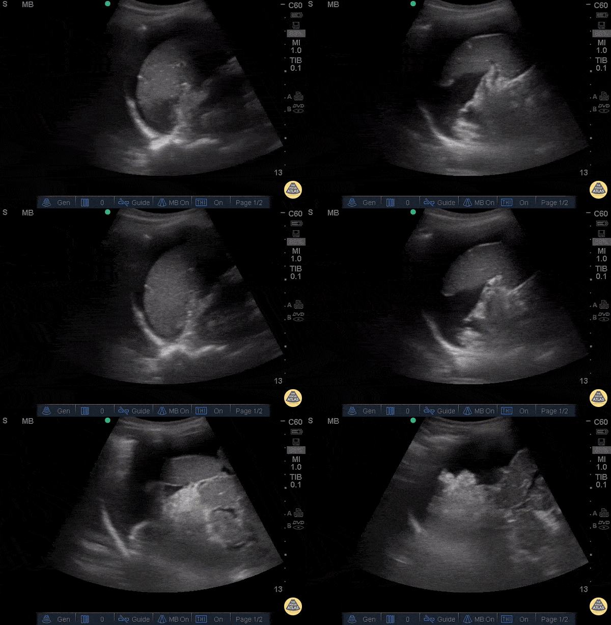
Positive FAST in LUQ - Ruptured Ovarian Cyst
28 year-old female was BIBEMS after a witnessed syncopal episode at home. The patient endorsed abdominal pain that started during intercourse that morning and had been getting worse.
On arrival, the patient appeared pale and diaphoretic. The patient’s FAST exam was performed immediately and showed free fluid in the RUQ and LUQ. The suprapubic view showed a large pelvic hematoma. The patient was evaluated by the GYN service and was taken emergently to the OR where she was found to have a ruptured cyst.
Don't forget, the FAST can be used for more than trauma.
Dr. Guru Shan and Dr. Catherine Bon - Kings County Emergency Medicine
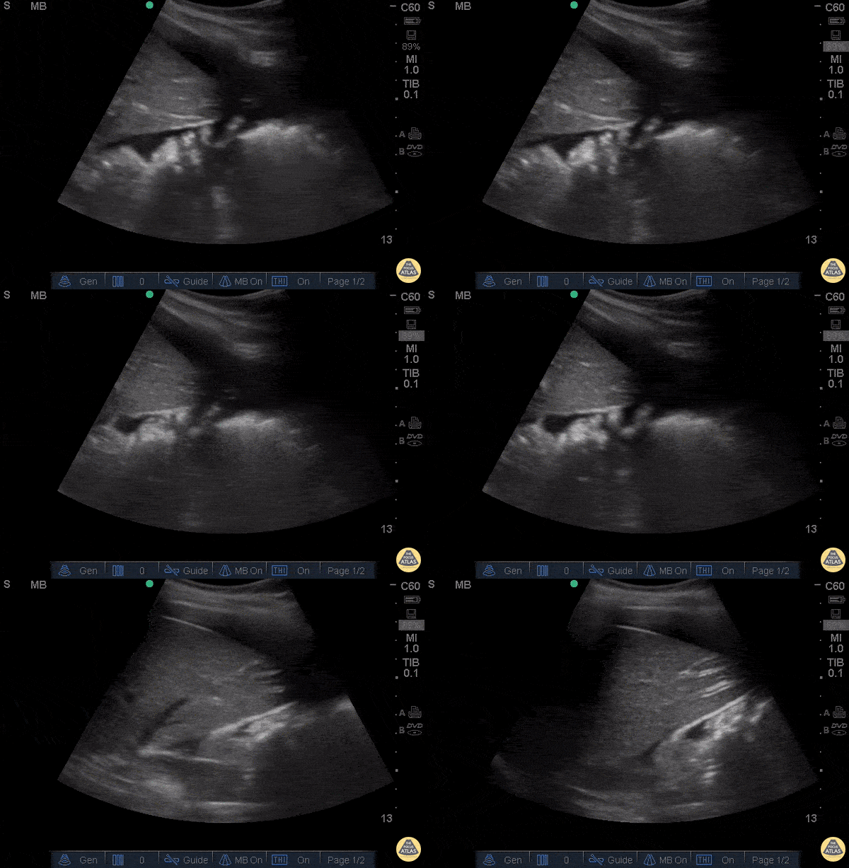
Positive FAST in RUQ - Ruptured Ovarian Cyst
28 year-old female was BIBEMS after a witnessed syncopal episode at home. The patient endorsed abdominal pain that started during intercourse that morning and had been getting worse.
On arrival, the patient appeared pale and diaphoretic. The patient’s FAST exam was performed immediately and showed free fluid in the RUQ and LUQ. The suprapubic view showed a large pelvic hematoma. The patient was evaluated by the GYN service and was taken emergently to the OR where she was found to have a ruptured cyst.
Don't forget, the FAST can be used for more than trauma.
Dr. Guru Shan and Dr. Catherine Bon - Kings County Emergency Medicine
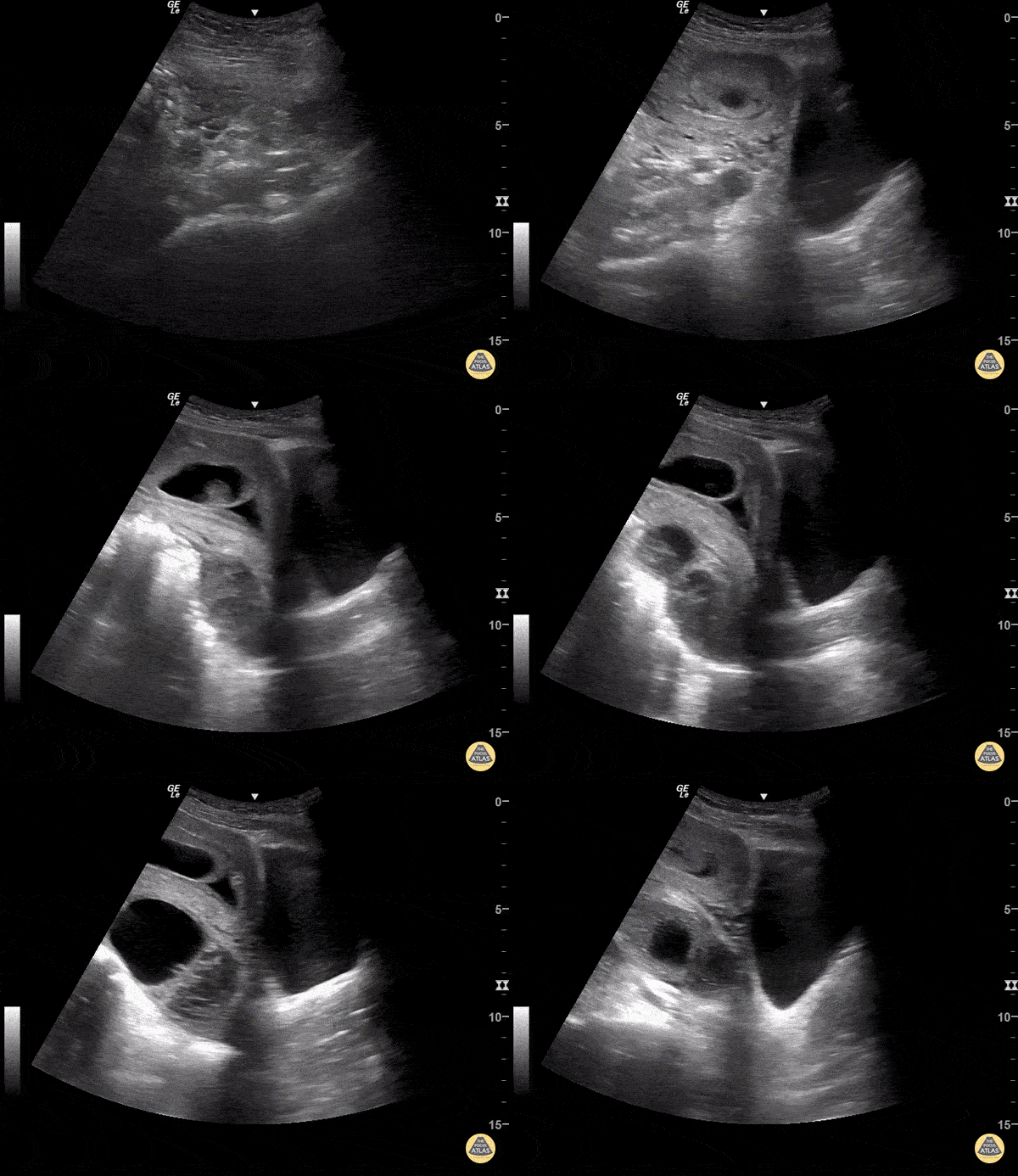
Hemorrhagic Cyst and Subchorionic Hemorrhage
IUP with hemorrhagic cyst and subchorionic hemorrhage.
Sukh Singh, MD
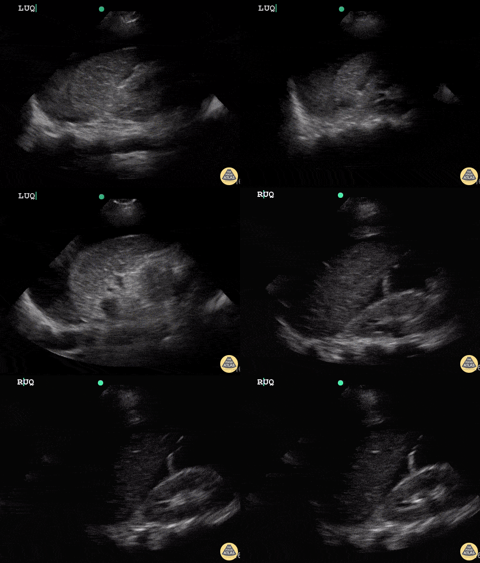
Uterine Rupture (Positive FAST)
21 year old female that was having prolonged labour and pain, presented in shock and delivered a non-viable fetus with minimal amount of blood loss from vagina. Continued to be hypotensive and became altered requiring intubation and crash central line. RUSH (including FAST) exam performed to determined etiology of undifferentiated shock.
FAST revealed free fluid in abdomen and pt was taken to the OR with GYN and Trauma Surgery. Found to have uterine rupture in OR.
Dr. Sathya Subramaniam, Pediatric EM Fellow - Kings County/SUNY Downstate
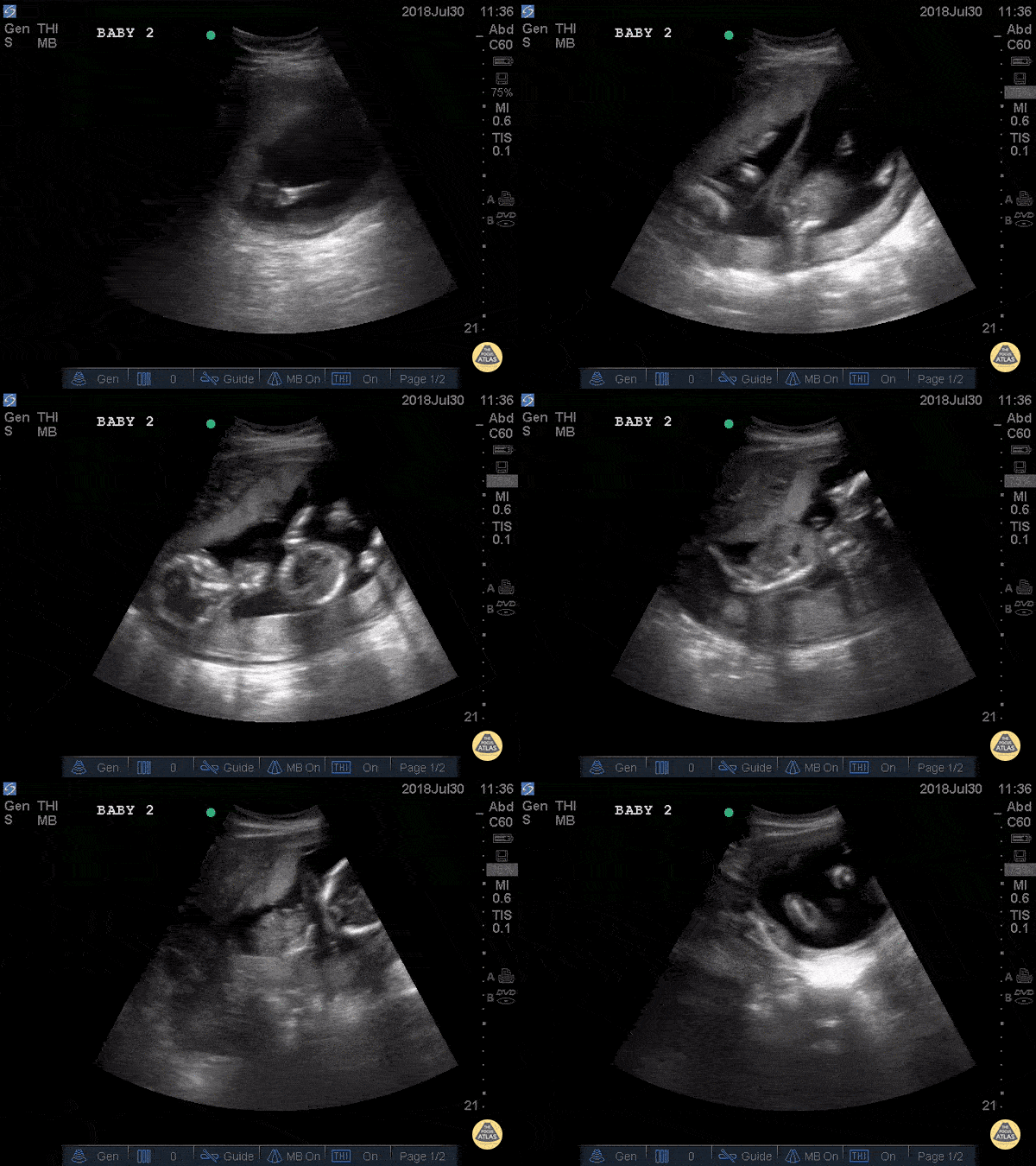
Twin IUP
Young female patient at 19 weeks gestation presented s/p syncopal event. POCUS performed and patient had a negative FAST. This image is a transabdominal ultrasound of an intrauterine twin gestation. A placenta can be visualized as the echogenic material superior to the fetuses and the hyperechoic umbilical cord can be visualized in the center of the gestational sac.
A normal fetal heart rate (FHR) ranges from 120-170 and is expected as early as 6 week gestational age. This scan shows FHRs of 150 and 158. Fetal movement can also be appreciated on this scan, which is expected at 9-10 weeks gestational age.
Dr. Eli Madden, Dr. Julianna Jaramillo - Kings County Emergency Medicine
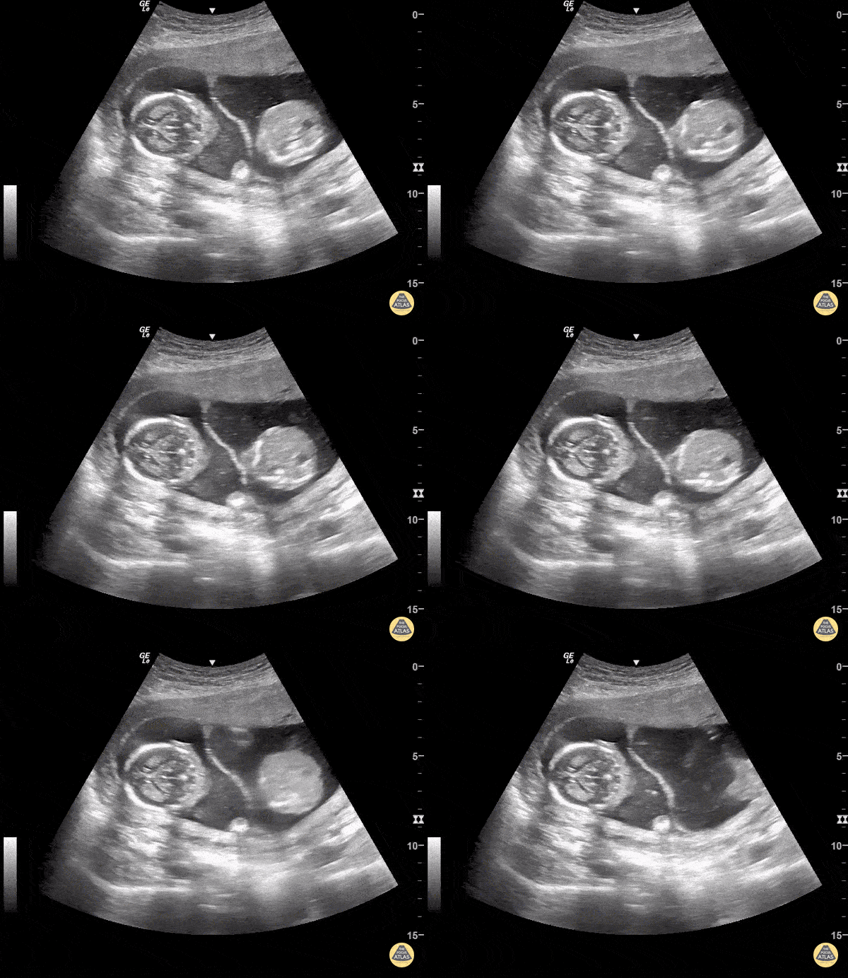
Twins
This is a normal transabdominal POCUS of twins separated by a membrane.
Sukh Singh, MD

Ovarian Cyst
Sukh Singh, MD
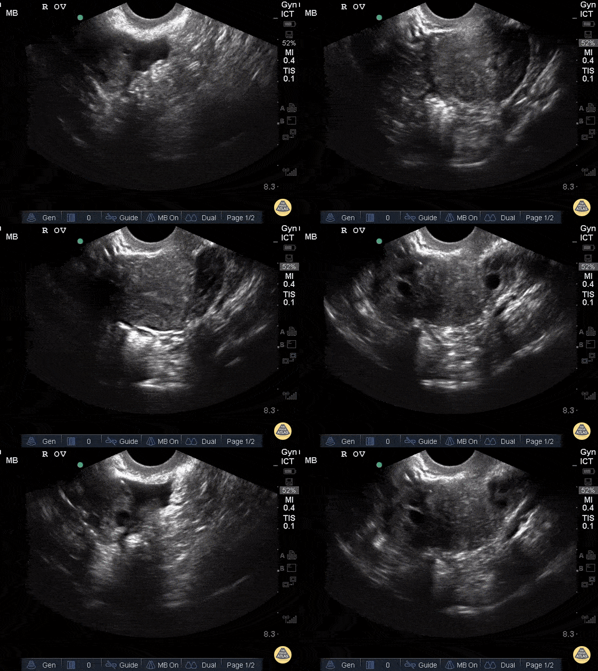
Normal Ovaries
Nulliparous patient.
Sukh Singh, MD
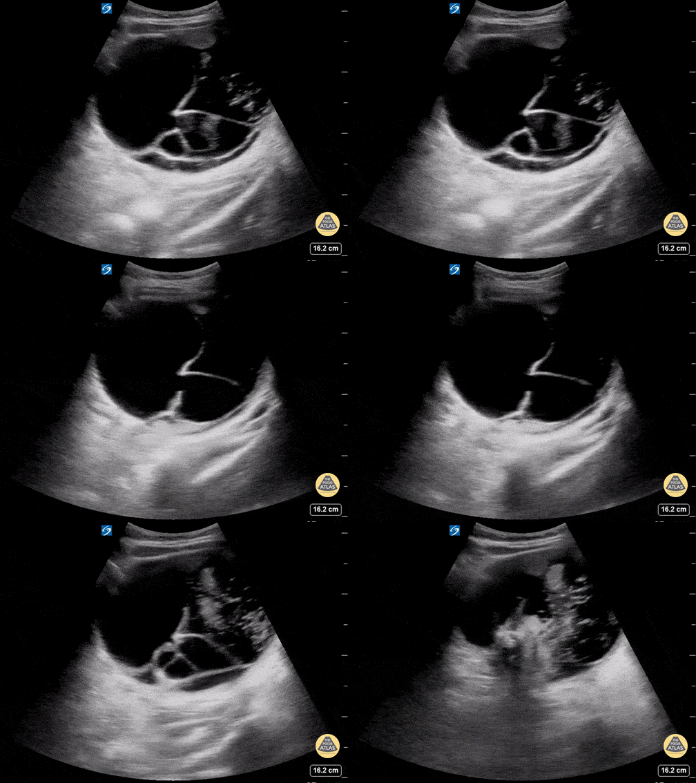
Ovarian Teratoma
This is a transverse view of the RLQ in a young female who presented with dysuria and a history of constipation. On physical exam, a visible mass was noted to the right of her umbilicus. Urinalysis and urine pregnancy test were negative. Bedside transabdominal ultrasound revealed a septated mass containing heterogeneous material with scattered hyperechoic foci most consistent with an ovarian teratoma.
Allison Perkins MD, PGY-1, Jared Toupin MD, PGY-2
Carnegie Mellon University Emergency Medicine Residency

Crown Rump Length Measurement
This image demonstrates caliper measurement of the crown rump length in an early pregnancy. Note that when using the proper setting and measurement function, most cart based ultrasound machines will automatically calculate the gestational age based off measurement obtained.
Michael Macias
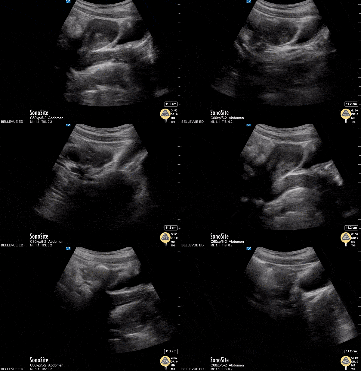
Transabdominal Left Adnexa
In this transabdominal view of the uterus in transverse, we see the left fallopian tube branching off the uterus leading to the left ovary in which multiple small hypoechoic follicles are visible, giving the ovary its typical “chocolate chip cookie” appearance. At the end of the clip, one of the left iliac vessels is seen in long axis just deep and lateral to the ovary. The bright hyperechoic lines with dark shadowing in the surrounding fields are bowel gas.
Hannah Kopinksi and Dr. Lindsay Davis - NYU Emergency Medicine
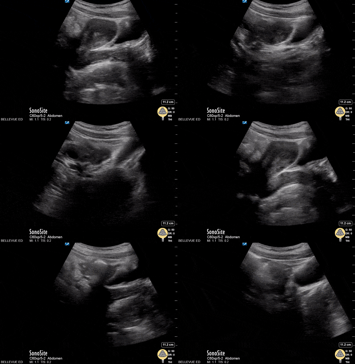
Transabdominal Uterus Long Axis
This is a transabdominal image of the uterus in long (sagittal) axis. On the far right of the screen we see the anechoic urine filled bladder. Immediately to the left of the bladder is the anteverted uterus which can be followed down and to the right as it curves and transitions into cervix and vagina. The thin, hyperechoic line in the center of the uterus is the endometrial stripe.
Hannah Kopinksi and Dr. Lindsay Davis - NYU Emergency Medicine
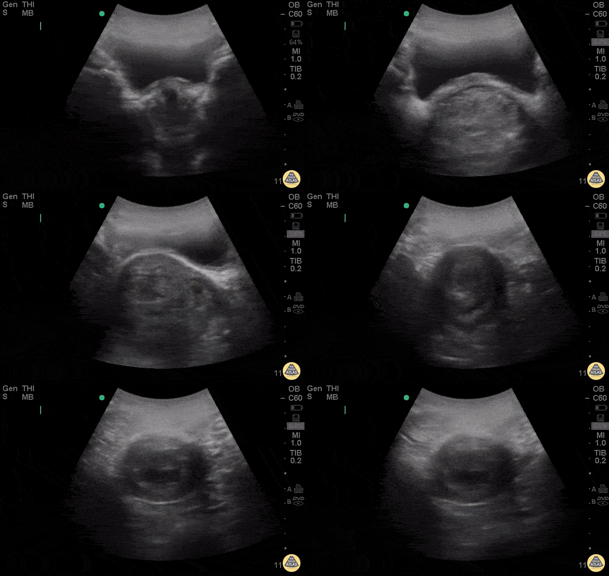
Spontaneous Abortion
28yo F G5P4 presenting at 8 weeks pregnant by LMP for pelvic pain and vaginal bleeding for 2 days.
POCUS demonstrates an anechoic gestational sac without a visible yolk sac of fetal pole, progressing past the cervix. The patient had a spontaneous abortion in the ER, passing products of conception shortly after the POCUS.
Esther Kwak, MD, Ian Desouza, MD- Kings County/SUNY Downstate Emergency Medicine
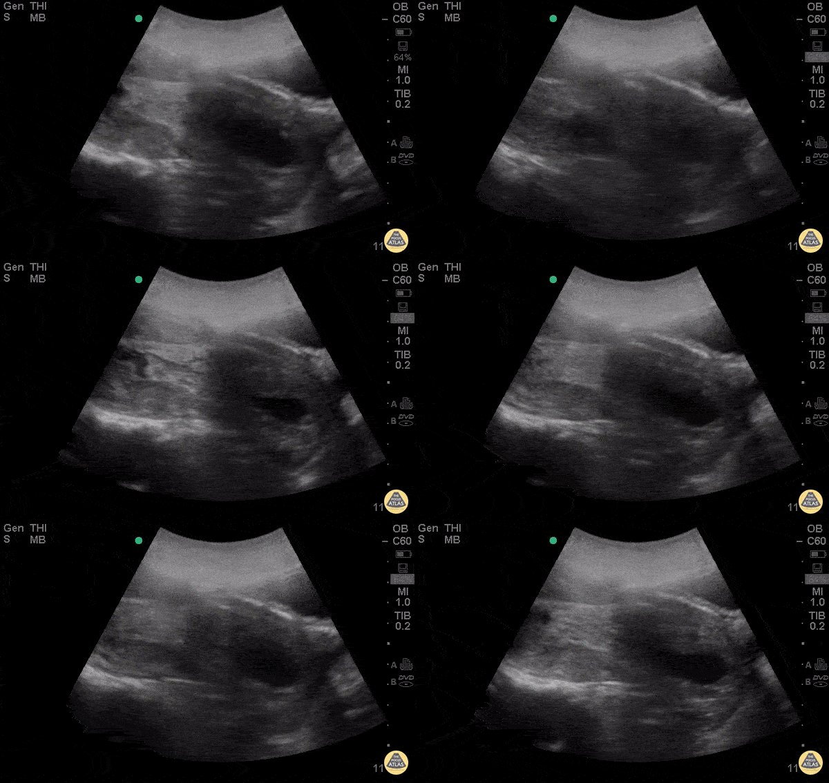
Spontaneous Abortion Sagittal
28yo F G5P4 presenting at 8 weeks pregnant by LMP for pelvic pain and vaginal bleeding for 2 days.
POCUS demonstrates an anechoic gestational sac without a visible yolk sac of fetal pole, progressing past the cervix. The patient had a spontaneous abortion in the ER, passing products of conception shortly after the POCUS.
Esther Kwak, MD, Ian Desouza, MD- Kings County/SUNY Downstate Emergency Medicine

Early IUP with Fetal Motion and Cardiac Activity
This is a sagittal view demonstrating a late first trimester pregnancy with fetal movement and cardiac motion.
Michael Macias
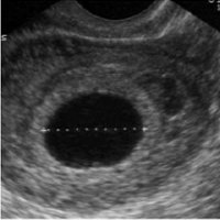
Early Pregnancy: Gestational Sac
Seen at 4-6 weeks:
- Not diagnostic for IUP
- Ectopic may have pseudogestational sac, though this is rare
- It might be early pregnancy but they will need close follow up

Early Pregnancy: Yolk Sac
Seen at 5-7 weeks:
- First evidence of IUP
- Appears as ring like structure within the gestational sac

Measuring Fetal Heart Rate with M-mode
In this image M-mode (motion mode) is used to calculate the fetal heart rate. Notice in this image, the calipers span two cardiac cycles. Depending on machine settings, the number of cardiac cycles to measure to obtain fetal heart rate varies.
Michael Macias

Early Pregnancy: Fetal Pole
Seen at 6-8 weeks:
- Tissue with similar echogenicity as uterus seen within the gestational sac
- If you see a fetal pole, you should see fetal cardiac activity
- Early on, the yolk sac and fetal pole may be present simultaneously
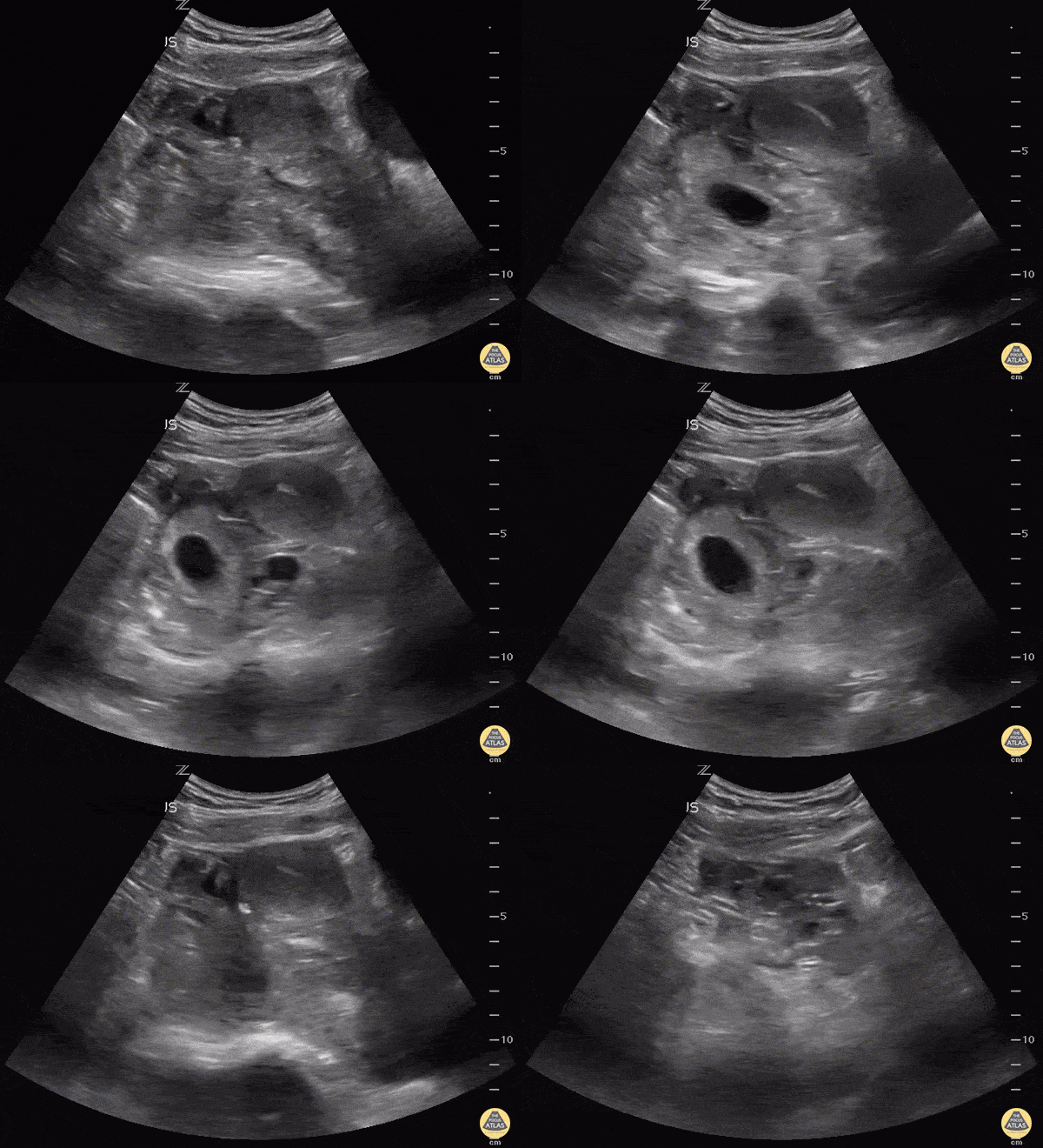
Ectopic Pregnancy
Always make sure the "IUP" is actually in the uterus by observing the uterus superior to the bladder in the sagittal view. This young lady could have been diagnosed easily with IUP and her abdomen pain dismissed as pain with pregnancy instead of a 10 week ectopic.
Matt Rutz, MD
@IUEM_Ultrasound
Indiana University School of Medicine
Department of Emergency Medicine
Division of Ultrasound
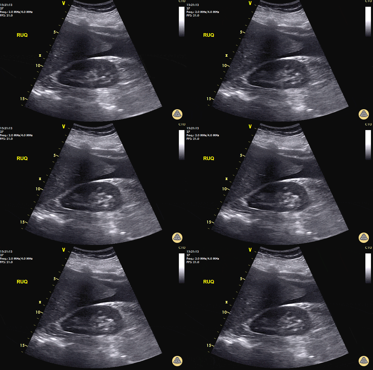
Positive FAST - Ruptured Ectopic
40 y/o F with abdominal pain, syncopal, +HCG. POCUS reveals free fluid in RUQ at inferior pole of the kidney and caudal tip of liver. Patient taken immediately to the OR and 800mL of blood evacuated. A left fallopian ectopic pregnancy was found.
Always look for free fluid at the inferior pole of the kidney. POCUS saves lives.
Dr. Cian McDermott - Dublin, Ireland

Ruptured Ectopic Pregnancy
A 20s F presented with syncope in the setting of multiple days of abdominal pain which acutely worsened. She arrived hypotensive, tachycardic, and on FAST exam, had free fluid in the right and left upper quadrants as well as suprapubic window. Serum beta-hCG testing was positive. A detailed examination of the adnexa is shown here, demonstrating a ruptured ectopic pregnancy. This patient received blood via massive transfusion protocol and was taken emergently to the OR, where an exploratory laparotomy demonstrated an ectopic pregnancy and more than 2L of hemoperitoneum.
Dr. Will Dewispelaere, PGY2, and Dr. Greg Wiener, PGY4
Denver Health Residency in Emergency Medicine

Bicornuate Uterus - Pregnant
“Bicornuate uterus” is not actually a dichotomous diagnosis - patients exist along a morphologic spectrum, and can actually get pregnant without much difficulty in some cases.
Dr. Elias Jaffa, MD, MS, FACEP

+FAST Ectopic Pregnancy with IUD
30s F with PMH prior ectopic pregnancy with IUD in place presented with positive home pregnancy test and lower abdominal pain. POCUS demonstrated a visible IUD still in place with free fluid surrounding the uterus. As the patient was hemodynamically stable, she had a consultative TVUS which confirmed these findings. Gynecology took the patient to the OR which confirmed a tubal ectopic pregnancy with a small amount of hemoperitoneum.
Dr. Michael MacGillivray, PGY4
Denver Health Residency in Emergency Medicine
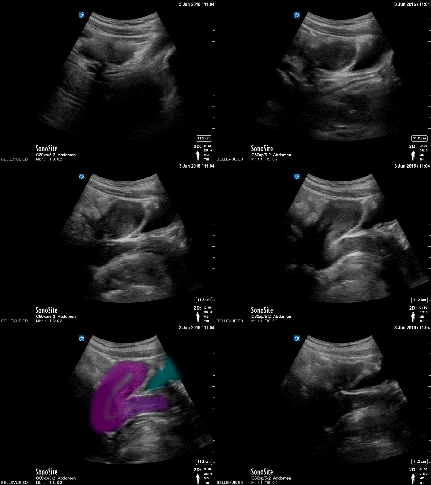
Transabdominal Uterus Sagittal View - Colorized
Transabdominal Uterus (sagittal view)
Pink: Uterus, Purple: Cervix, Teal: Bladder
Images: Dr. Lindsay Davis, Dr. Hannah Kopinski. Image Editing: Michael Amador and Dr. Matthew Riscinti
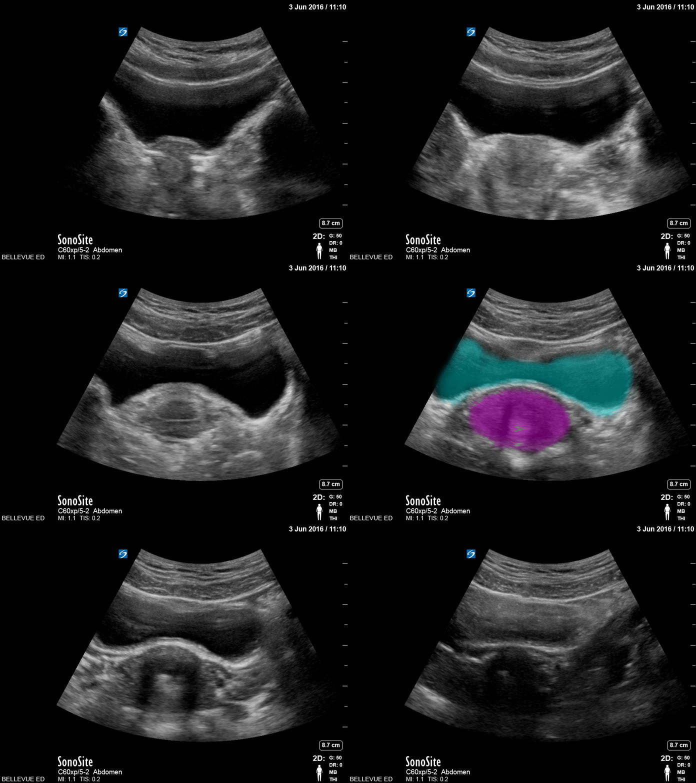
Transabdominal Uterus Transverse View - Colorized
Transabdominal Uterus (transverse)
Teal: Bladder, Pink: Uterus
Images: Dr. Lindsay Davis, Dr. Hannah Kopinski. Image Editing: Michael Amador and Dr. Matthew Riscinti

Dilated Cervix with intact membranes protruding out
17 YOF, G1P0 and 18 weeks pregnant, who presents to the ER with the complaint of "feeling like something was coming out" of her vagina since this morning. Mild lower back pain. Denies pelvic pain, vaginal bleeding, or leaking fluid. US shows dilated cervix with intact membranes protruding out.
Vicky Lam
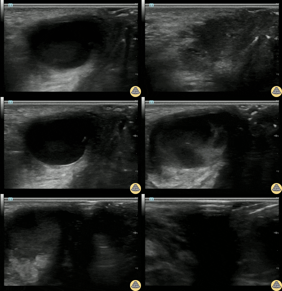
Bartholin's Abscess
20s F with past medical history of multiple bartholin gland abscesses requiring drainage presented with genital pain and swelling. I&D of the abscess was attempted which was initially unsuccessful, so POCUS was performed to confirm the location of the abscess. Gynecology was then consulted for drainage and was able to successfully drain the abscess.
Alexandrea Netto PA, Denver Health and Hospital Authority
Katie McCabe MD, Attending Physician, Denver Health Residency in Emergency Medicine

Twin Gestation
30s F G1P0 at ~11-12 weeks by LMP presented to the ED with vaginal spotting and abdominal cramping. POCUS was performed and demonstrated a twin gestation with two viable fetuses, each measuring about 11 weeks by CRL.
Tyler LaCoste, PA
Dr. Anna Engeln
Denver Health Medical Center


