
Peds-Lung

Lack of Lung Sliding Seen in M-Mode
Pediatric patient presenting with shortness of breath. Found to have a large left sided pneumothorax. M-mode tracing shows lack of lung sliding
Contributor: Kathryn Pade, MD, Rady Children's Hospital San Diego

Normal Lung Sliding During FAST Exam
11 year old female presented to the emergency department with a puncture wound to her left chest wall. FAST exam did not reveal any intraabdominal free fluid, and there was lung sliding bilaterally, suggesting no pneumothorax. The patient had her wound explored, repaired, and was discharged home.
Contributed by: Zach Boivin, MD, @ZachBoivinMD

Esophageal Intubation
Intubation attempt into the esophagus of an 8 year old male with respiratory distress, notice the soft tissue posterior to the trachea moving with the provider attempting the pass the tube.
Image contributed by: Zach Boivin, MD, @ZachBoivinMD

Normal Lung Slide
Normal lung sliding using linear probe
Contributor: Peter Gutierrez, MD, FAAP, Emory University School of Medicine/Children's Healthcare of Atlanta, @pocuspete

Confluence of B-Lines
Toddler with pneumonia; multiple B-lines noted with adjacent area of consolidation.
Contributor: Peter Gutierrez, MD, FAAP, Emory University School of Medicine/Children's Healthcare of Atlanta, @pocuspete
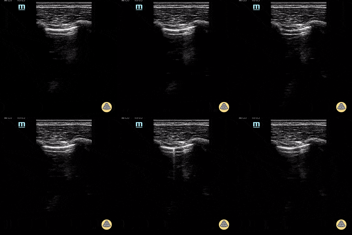
Lung Point
Lung point indicative of pneumothorax.
Contributor: Peter Gutierrez, MD, FAAP, Emory University School of Medicine/Children's Healthcare of Atlanta, @pocuspete
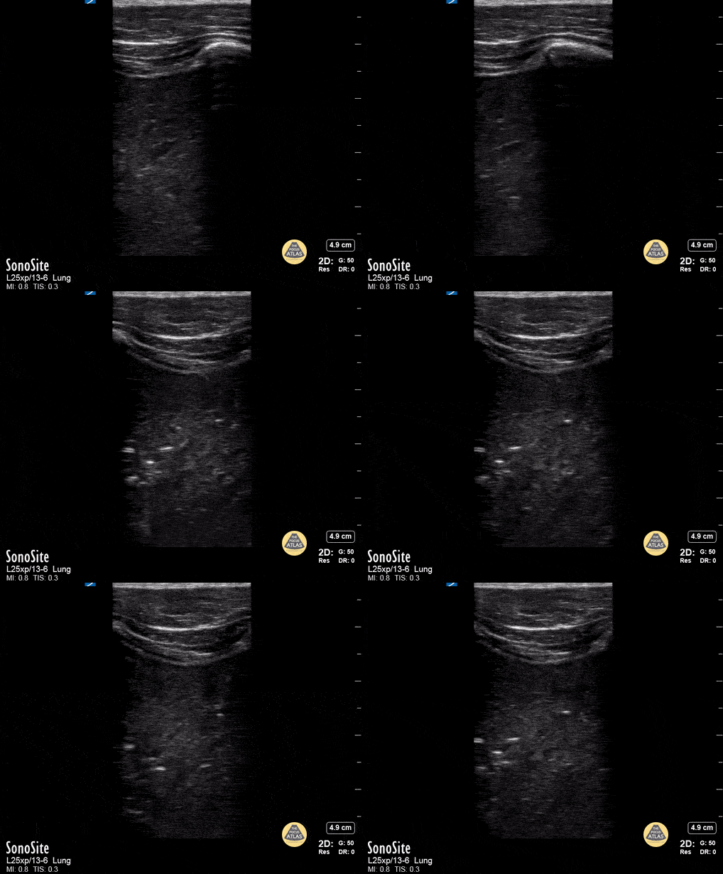
Lung Consolidation
Lung consolidation in a child with fever and cough; faint dynamic air bronchogram seen
Contributor: Peter Gutierrez, MD, FAAP, Emory University School of Medicine/Children's Healthcare of Atlanta, @pocuspete

Large Pleural Effusion
Large pleural effusion in a teenager
Contributor: Peter Gutierrez, MD, FAAP, Emory University School of Medicine/Children's Healthcare of Atlanta, @pocuspete
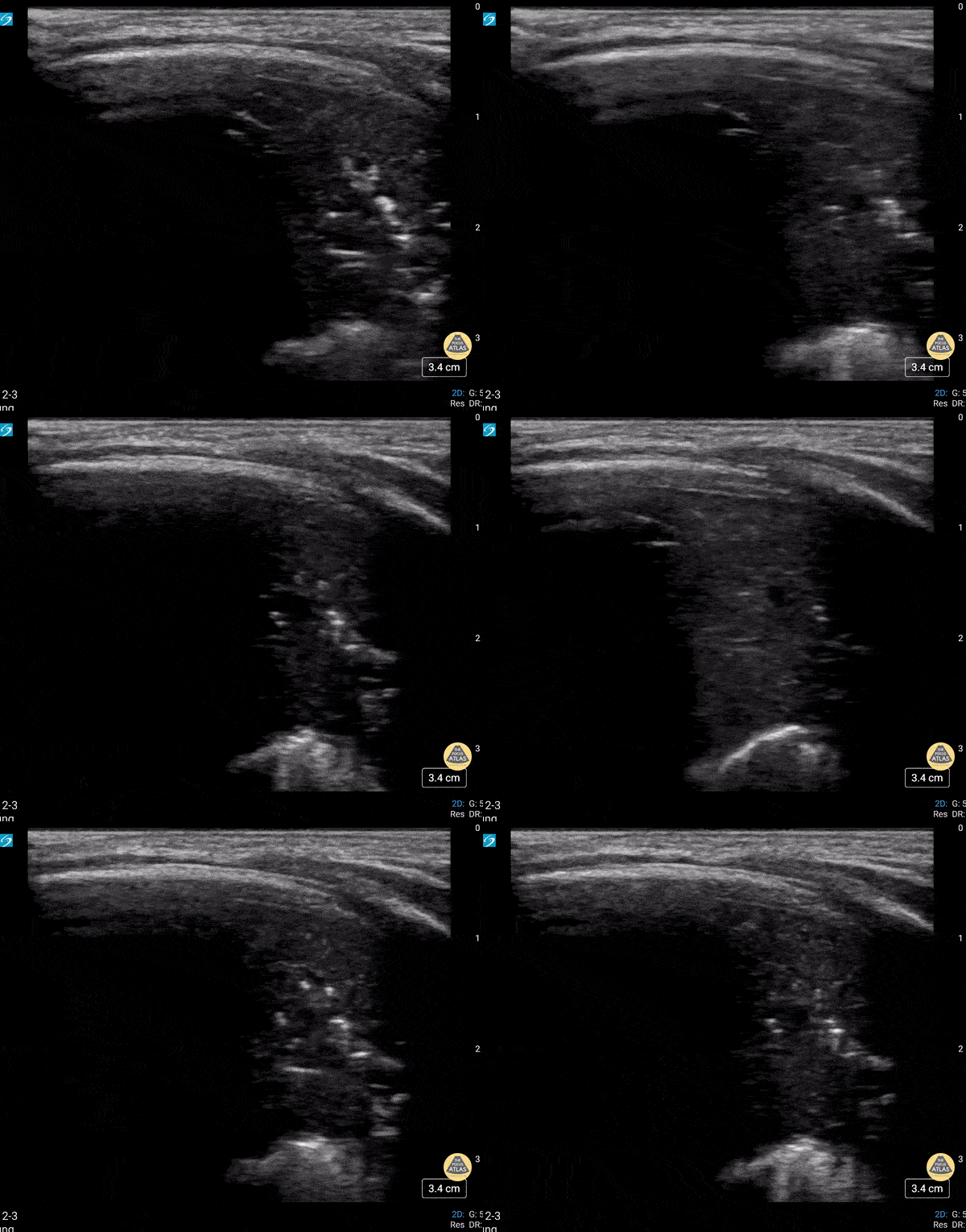
Dynamic Air Bronchograms
Neonate in respiratory failure status post intubation. The presence of dynamic air bronchograms helps point away from atelectasis.
Contributor: Peter Gutierrez, MD, FAAP | Emory University School of Medicine/Children's Healthcare of Atlanta | @pocuspete

Lack of Lung Sliding
Lack of lung sliding.
Contributor: Peter Gutierrez, MD, FAAP | Emory University School of Medicine/Children's Healthcare of Atlanta | @pocuspete
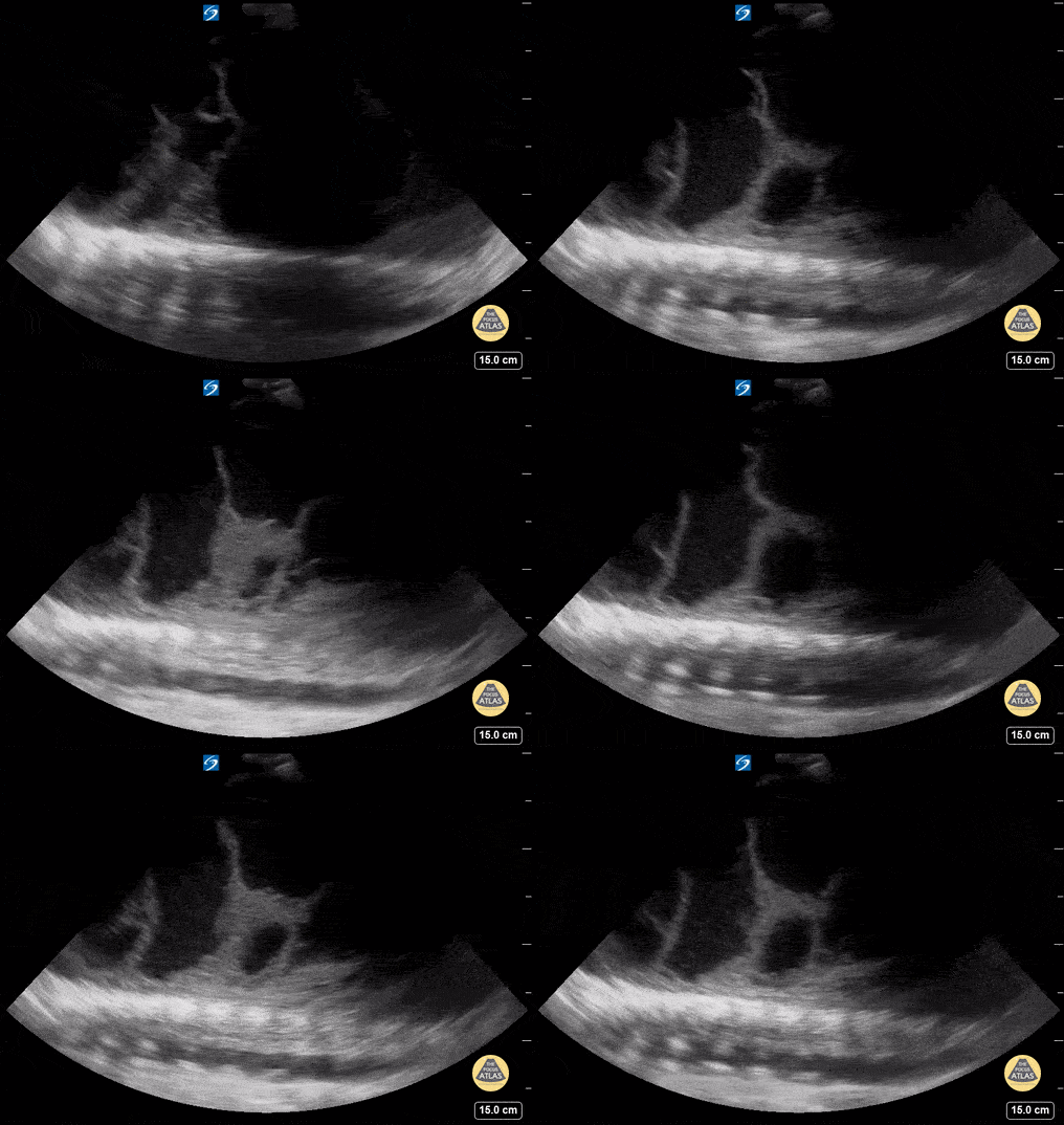
Loculated Pleural Effusion
16 yo with initial diagnosis of pneumonia. Worsening shortness of breath. Found to have a loculated pleural effusion on POCUS.
Contributor: Kathryn Pade, MD, Rady Children's Hospital San Diego
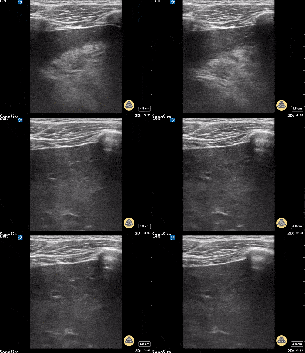
5 Year Old with Pneumonia
5 y/o with pneumonia- the image demonstrates hepatization of the lung consistent with pneumonia/consolidation.
Contributor: Kathryn Pade, MD, Rady Children's Hospital San Diego
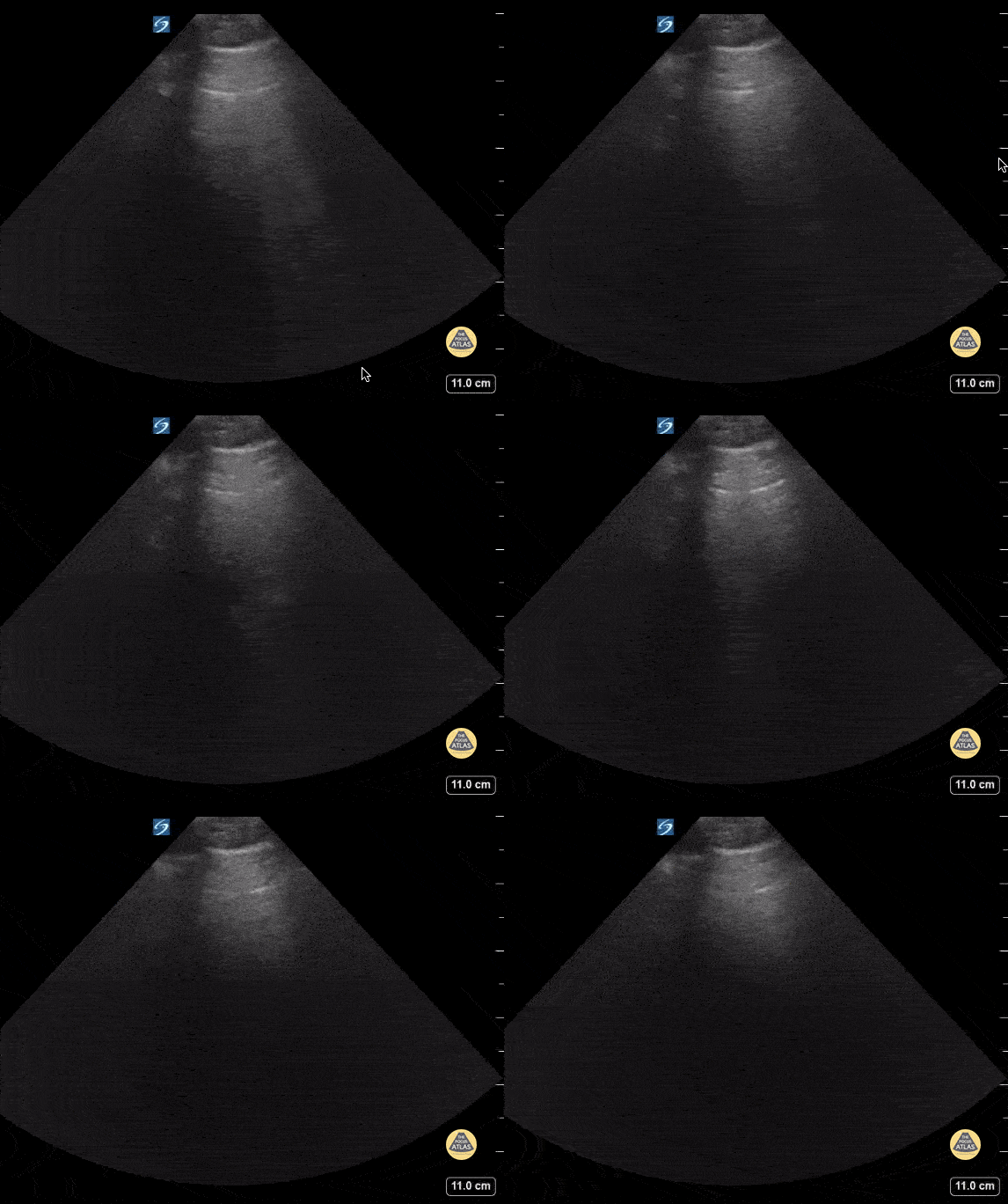
Pneumonia in a 4 Year Old
4 year old M with asthma who presents with respiratory distress and fever. + coronavirus (not COVID-19). POCUS shows focal B lines concerning for pneumonia, bacterial vs viral.
Contributor: Kathryn Pade, MD, Rady Children's Hospital San Diego
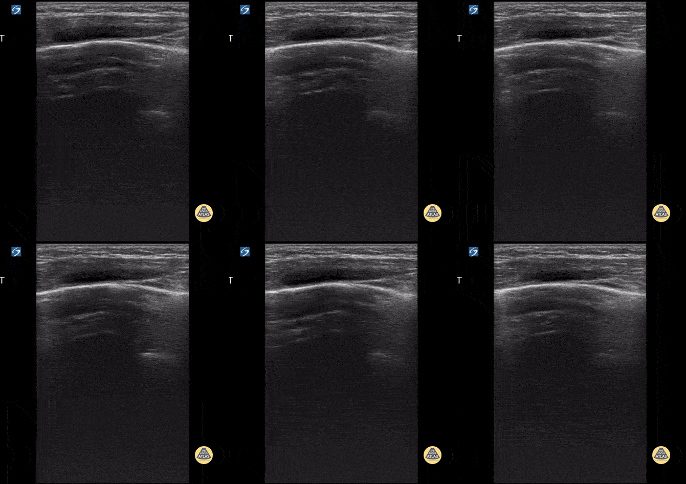
Normal Lung Sliding with Comet Tails
4 y/o with asthma who presents in respiratory distress. Using the linear probe, normal lung sliding is seen with comet tails.
Contributor: Kathryn Pade, MD, Rady Children's Hospital San Diego
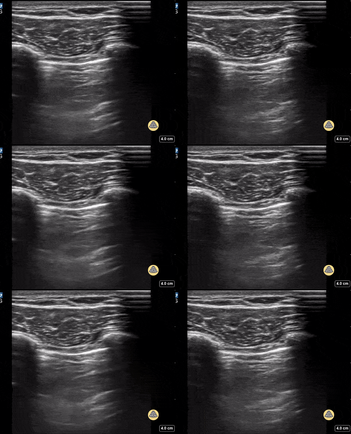
No Lung Sliding
Pt presented with shortness of breath of sudden onset. Found to have absence of lung sliding consistent with a large pneumothorax that required chest tube placement.
Contributor: Kathryn Pade, MD, Rady Children's Hospital San Diego
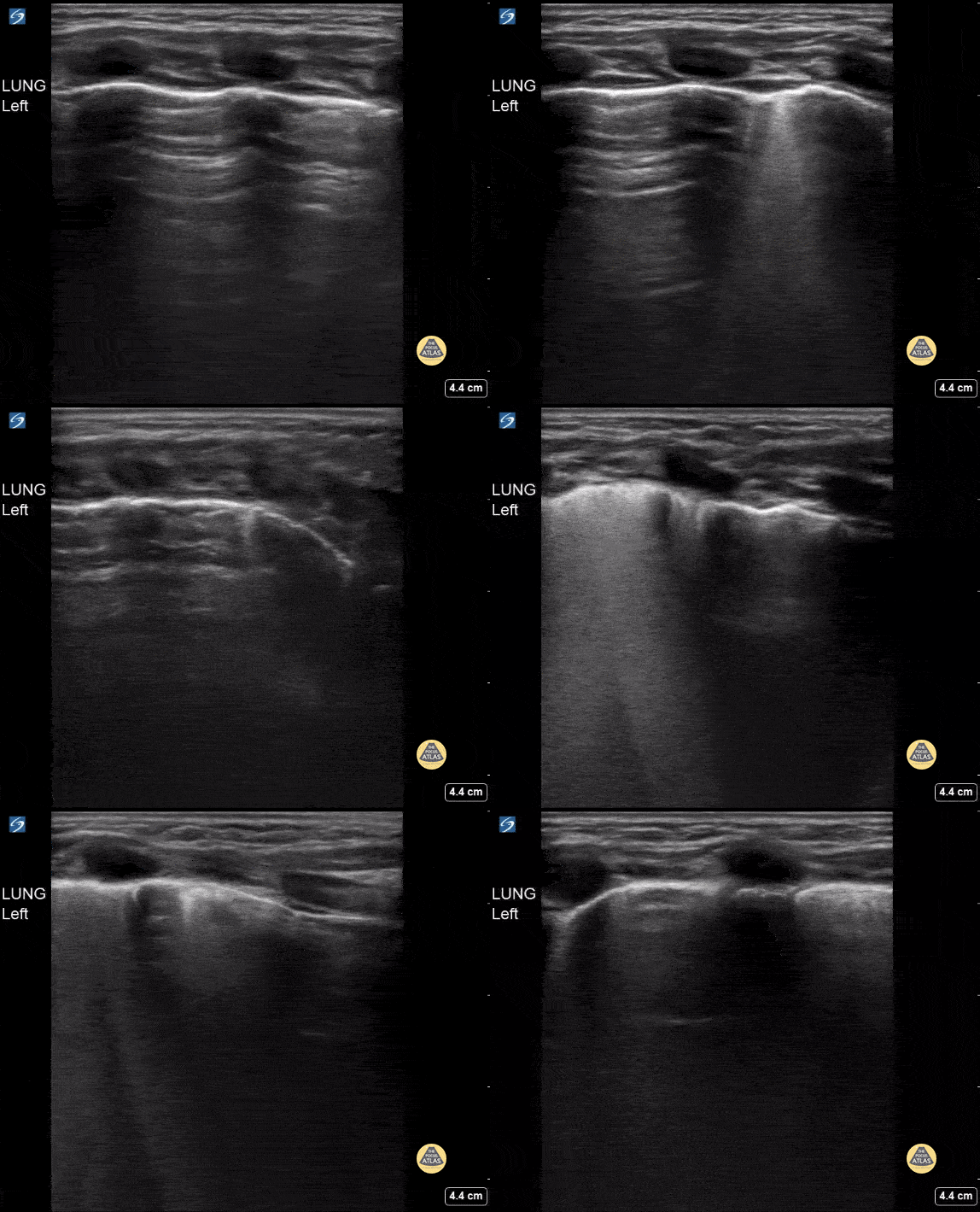
Bronchiolitis
9 month old with respiratory distress. Diffuse confluent B lines found on POCUS consistent with bronchiolitis.
Contributor: Kathryn Pade, MD, Rady Children's Hospital San Diego
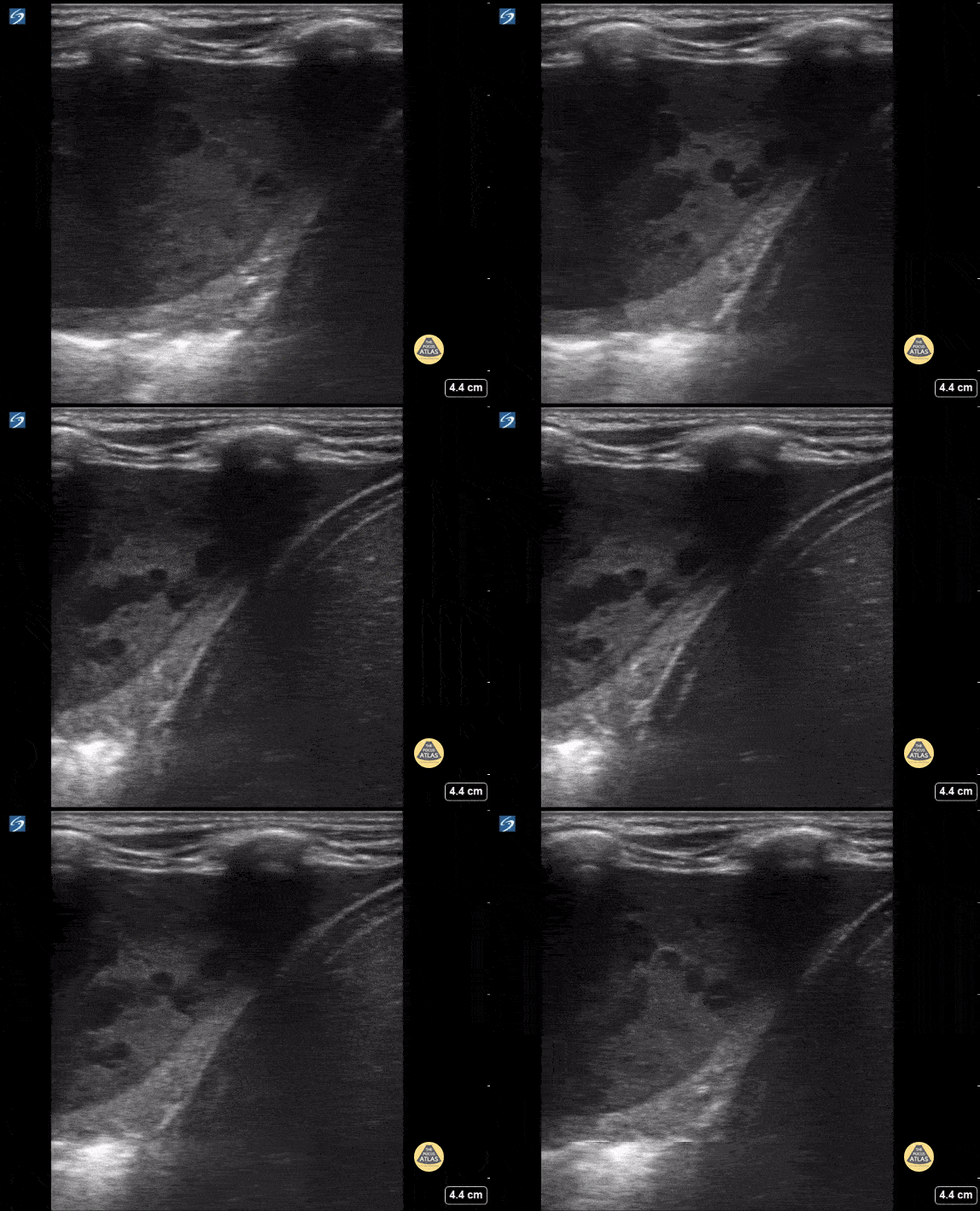
Empyema of the Lung
15 year old with complex PMH who presented with respiratory distress. CXR shows a large effusion. Bedside ultrasound showed an empyema.
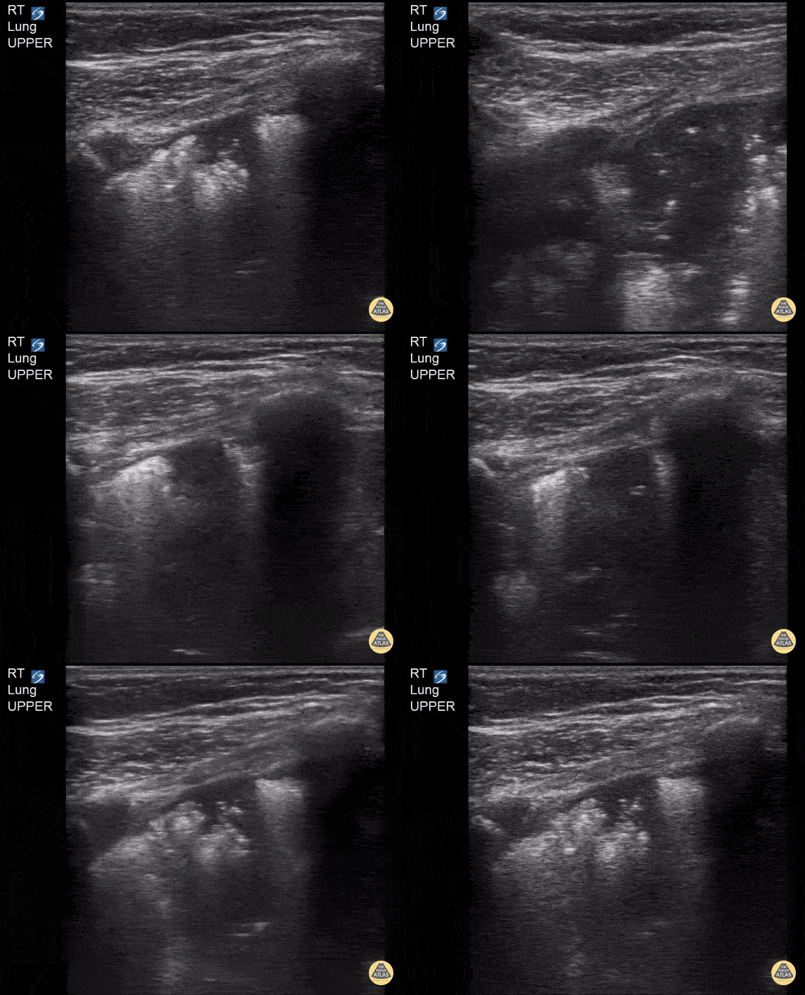
Subpleural Consolidation in 3 Year Old
3 y/o with respiratory distress. Found to have a large subpleural consolidation on CXR.
Contributor: Kathryn Pade, MD, Rady Children's Hospital San Diego

Atelectasis in Neonate
32 weeks premature. 4 weeks of life in the course of weaning from oxygen with respiratory deterioration requiring orotracheal intubation
Evidence of lung collapse with static bronchogram in the right upper lobe.
Contributor: Mg. Andres Silva Horna, Hospital Cayetano Heredia Piura-Peru
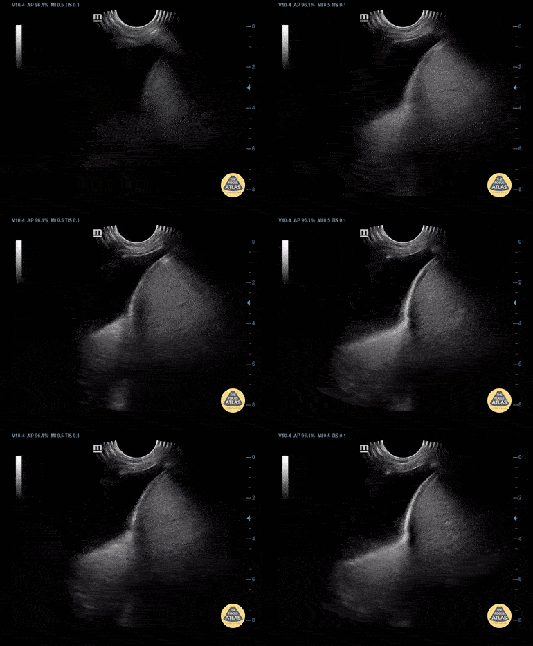
Pleural Effusion from Dengue
4-year-old boy with dengue who presents respiratory distress.
Pleural effusion with passive atelectasis (jellyfish sign) is visualized.
Contributor: Mg Andrés Fernando Silva Horna; Hospital Cayetano Heredia-Piura
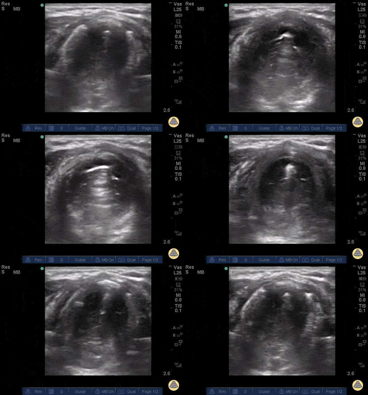
Tracheal Stenosis
The patient, presenting with stridor, is supine and the airway is seen from the anterior neck in transverse orientation. As the probe is fanned, the bright white line is seen to widen. This column of air moves with inspiration. At its narrowest it is only a few millimeters wide. Growth along the lateral tracheal walls has caused significant tracheal stenosis. In this case, US was used to determine the width of the tracheal column and determine that passage of an ETT would not be feasible. The patient was taken to the OR for an emergent surgical airway. Use of US to estimate tracheal diameter is a novel application.
Andrew Liteplo MD, RDMS - Massachusetts General Hospital
Chief, Division of Ultrasound in Emergency Medicine
Director, Emergency Ultrasound Fellowship
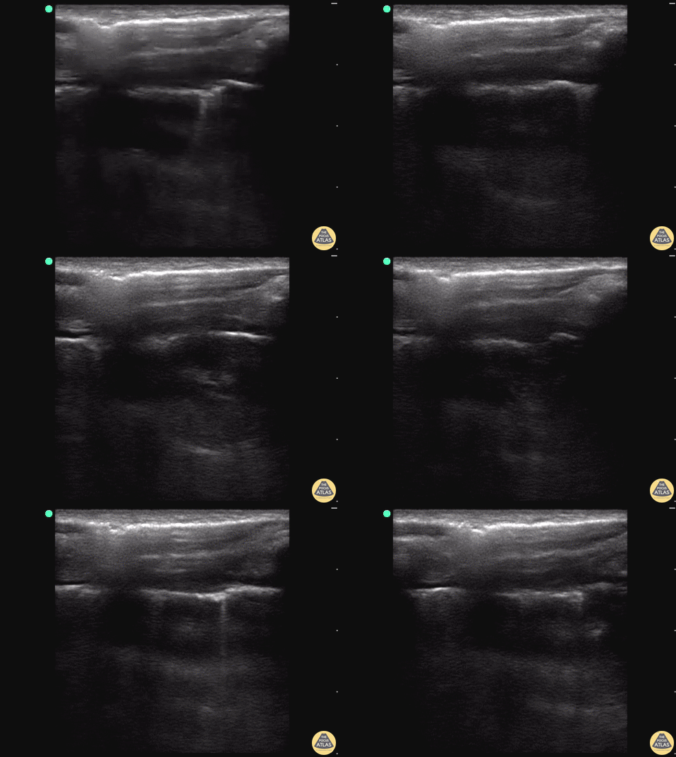
Pneumothorax with Lung Point
18 y/o M stabbed in the back presents to the trauma bay with left-sided chest pain and shortness of breath. E-FAST revealed decrease lung slide and a clear lung point.
While decreased lung slide is highly sensitive, it lacks specificity. Lung point indicates the transition point between normal pleura with normal lung sliding (on the left side of the image) and where there is air disrupting the pleural space with decreased lung sliding (on the right side of the image). Lung point is a highly specific finding indicating a pneumothorax.
Dr. Sathya Subramaniam, Pediatric EM Fellow - Kings County/SUNY Downstate
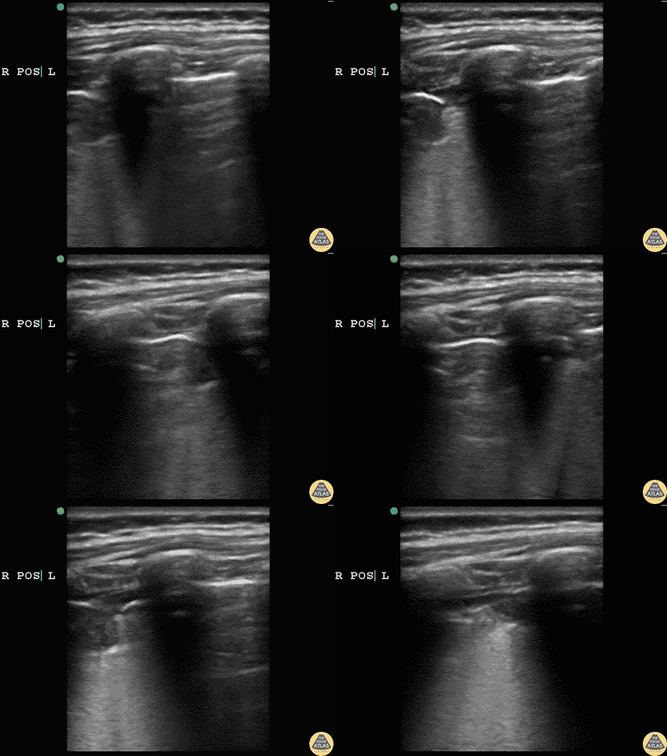
Hydrocarbon Ingestion with C-Lines
5 year old male that drank out of container with gasoline and started coughing and was breathing fast. On exam appeared tachypneic, with air entry bilaterally and subcostal retractions.
POCUS revealed bilateral subpleural consolidations, confirmed with CXR. This imaging finding is similar to findings seen in other consolidative processes. This was suggestive of an aspiration pneumonitis.
Dr. Sathya Subramaniam, Pediatric EM Fellow - Kings County/SUNY Downstate
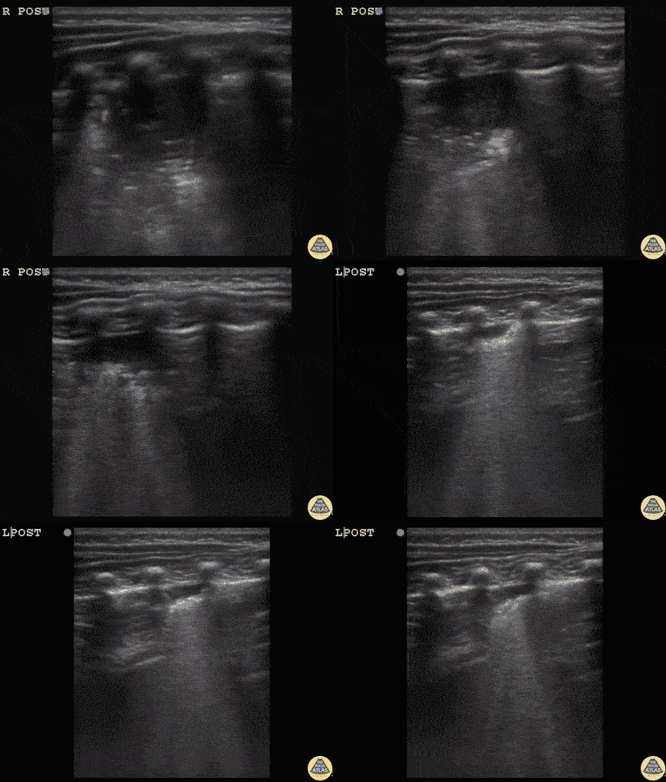
Infant Pneumonia with C-Lines
11 month old unvaccinated infant presenting with cough, fever and tachypnea starting today. Exam with crackles bilaterally in an infant with subcostal retractions and respiratory distress.
Right posterior lung with clear large consolidative process with C lines present.
Dr. Sathya Subramaniam, Pediatric EM Fellow - Kings County/SUNY Downstate
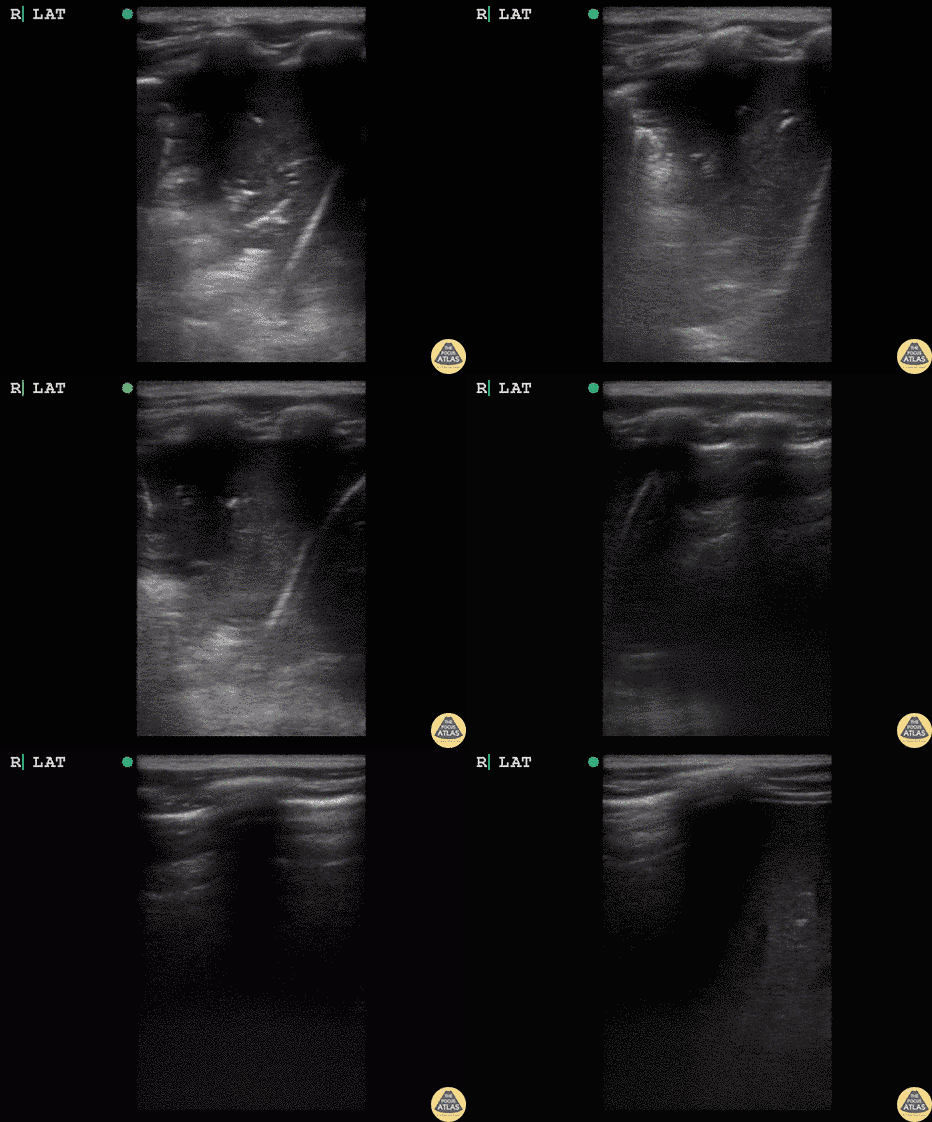
Lung Hepatization in Pneumonia
5 year old child with sickle cell disease. Coughing and fever for 3 days. On exam not ill appearing but decreased breath sounds over right lung. POCUS completed to evaluate for pneumonia.
Hepatization of the lung clearly demonstrates consolidative process concerning for pneumonia. The beginning of the image demonstrates hepatization in the lung field. The ultrasonographer then slides the probe inferiorly over normal lung past the diaphragm to the liver, demonstrating how similar lung hepatization can appear compared to the actual liver.
Dr. Sathya Subramaniam, Pediatric EM Fellow - Kings County/SUNY Downstate
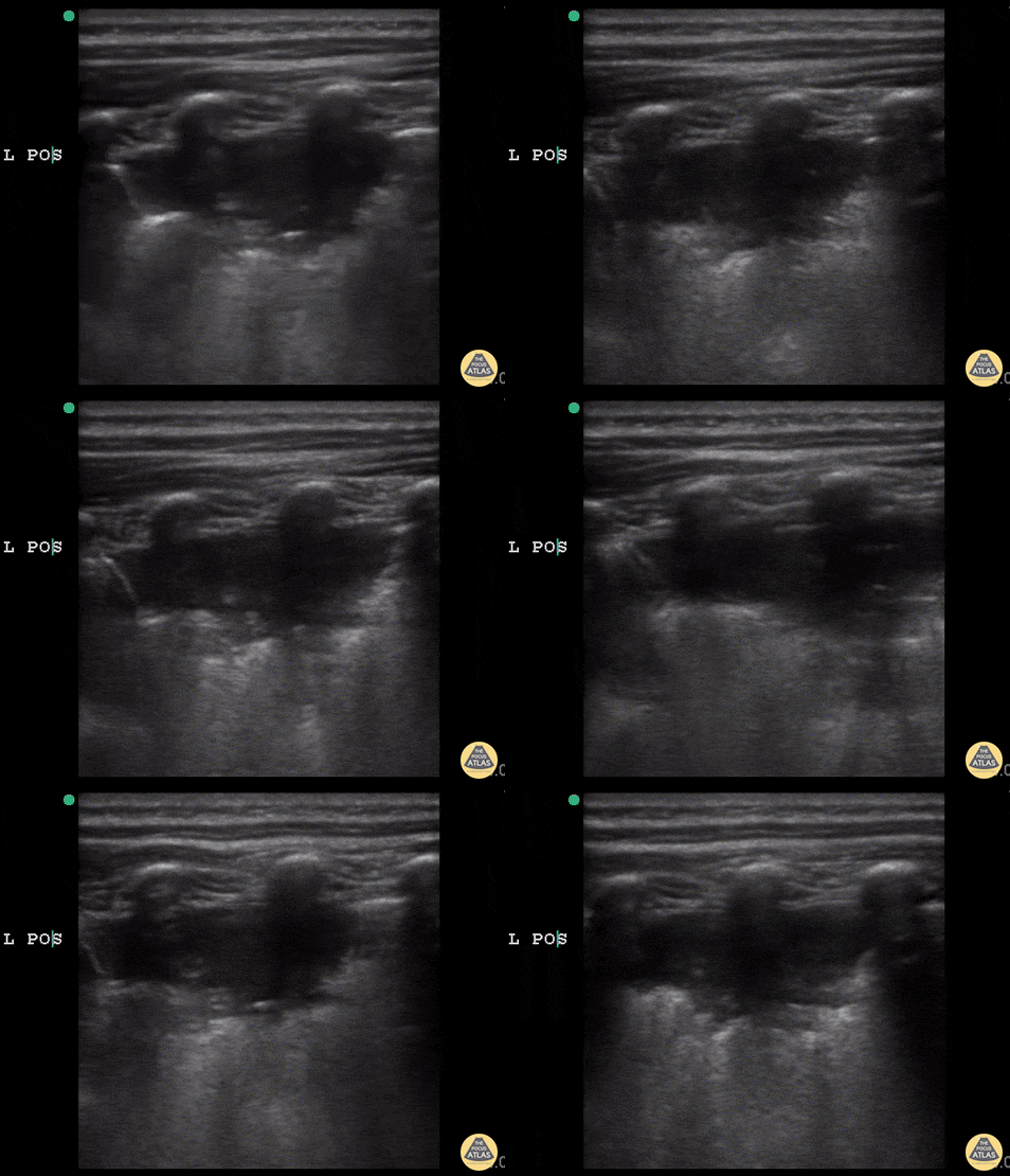
Acute Chest Syndrome
6 y/o sickle cell (HbSS) coughing with left-sided chest pain and 1 day of fever. Lungs without crackles, good air entry bilaterally.
A consolidative process is seen with a hypoechoic region with posterior enhancement greater than 1 cm in an area where normal A lines should be present. This is highly suggestive of acute chest syndrome given clinical features.
Dr. Sathya Subramaniam, Pediatric EM Fellow - Kings County/SUNY Downstate
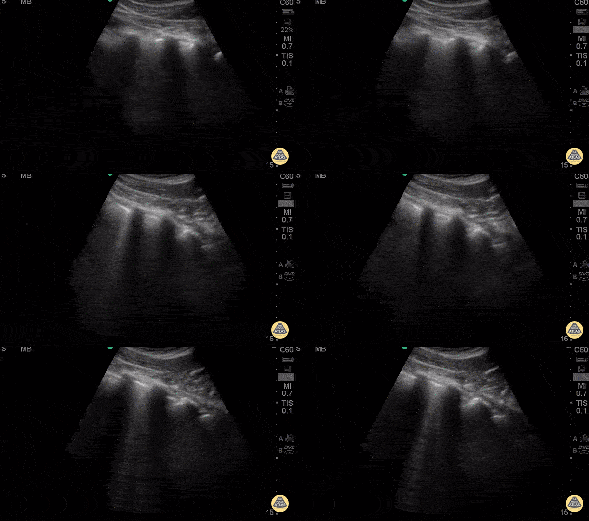
Focal B-lines - Pneumonia
2 years old male with hx of endotracheal intubation secondary to RSV infection presents with 2 days of fever, cough, rhinorrhea and nasal congestion. Denied nausea, emesis, diarrhea, chest pain, syncope, confusion, change in eating patterns and voiding patterns. POCUS demonstrates a focal area of B lines c/w pneumonia (likely viral).
Early PNA B lines: short path reverberation artifacts create by fluid filled alveoli. In the appropriate clinical scenario B lines and pleural consolidation suggest PNA.
Dr. Carolina Camacho Ruiz - Kings County Emergency Medicine
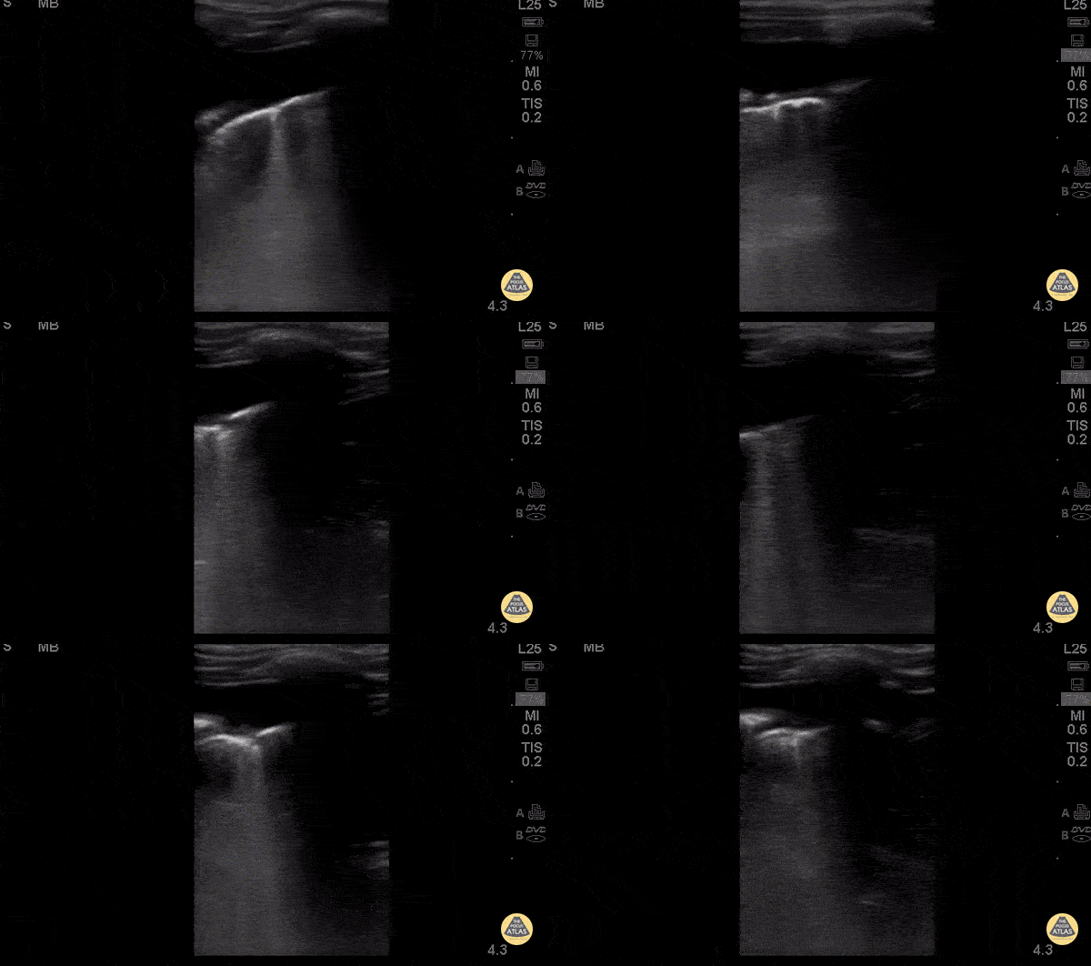
Pneumonia
3 y/o previously healthy UTD with vaccines. 5 days of cough and fevers. 3 days of abdominal pain, acutely worsening day of presentation with 1 episode of NBNB emesis.
Febrile tachypneic and hypoxic to 91% on RA.
CXR: white out of right lung.
POCUS: Right sided effusion associated with subpleural consolidation and focal b-lines on right lateral view.
Dr. Isaac Gordon - Kings County Pediatric Emergency Medicine

Hepatization of the Lung
A 15-month-old male presented with cough, fever, and tachypnea of 3-days duration. POCUS revealed findings of right lung consolidation, consistent with pneumonia referred to as hepatization of the lung. Seen here territories above and below the diaphragm show ultrasonographic findings resembling liver parenchyma.
Amar Singh, MD. Pediatrics specialist in Louisville, KY

COVID-19 Pneumonia
14 year-old female known to be SARS-CoV-2 positive presented with chest pain and shortness of breath. POCUS revealed findings consistent with COVID pneumonia including thickened pleura and presence of multiple B-lines.
Paul Khalil, MD. Assistant PEM POCUS director at University of Louisville/Norton Children’s
@khalil3paul

Dynamic Air Bronchograms
15 year-old male with cerebral palsy presents to the ED with hypoxia. Physical exam notable for left lung with decreased air movement on auscultation. POCUS demonstrates dynamic air bronchograms consistent with suspected diagnosis of pneumonia.
Paul Khalil, MD. Assistant PEM POCUS director at University of Louisville/Norton Children’s
@khalil3paul

Acute Chest Syndrome
A child with history of sickle cell presented with upper abdominal and back pain and was found to be tachypneic with low grade fever. Ultrasound of the left upper quadrant demonstrated basilar left lung consolidation. Subsequent chest x-ray was interpreted as infiltrate consistent with acute chest syndrome.
Michael Cover, @michaelc0ver

Pediatric Consolidation
A 10-year-old patient with no medical background is brought to the ED presenting a 2-day history of dry cough and right subcostal pain. There is neither fever nor shortness of breath. A lung ultrasound was performed following physical examination which prompted the discovery of a consolidation.
The probe is slid along 2 intercostal spaces revealing an oddly shaped structure with irregular edges that locates in between normal A-lines.
Dr. Felipe Urriola P.

Lung Atelectasis After Foreign Body Aspiration
2 yr old child with sudden respiratory distress with O2sat 70%. Child was intubated in prehospital setting by a HEMS physician and POCUS obtained en route to the hospital revealed a completely collapsed (atelectatic) left lung as seen in the clip; right lung was normal. In hospital an aspirated foreign body (a raisin) was removed a from the child’s left main bronchus. Child made a full recovery.
Victor Viersen
@victor_viersen
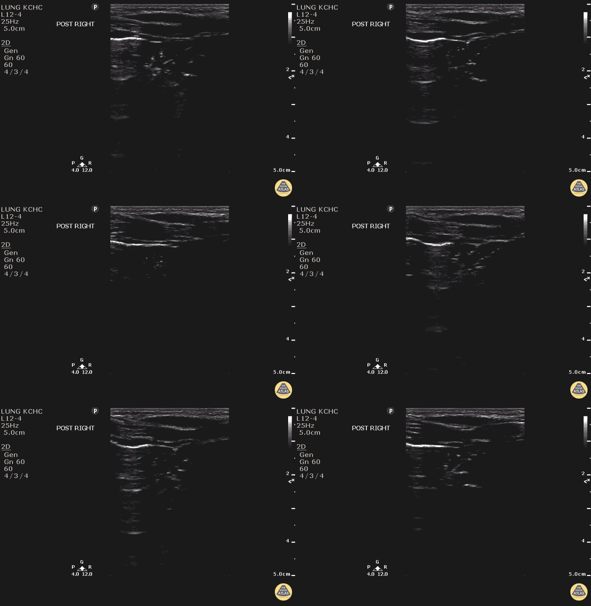
C-lines - Pneumonia
2 y/o M PMH RSV requiring intubation with 2 days of fevers, cough, rhinorrhea, congestion.
POCUS of right posterior lung demonstrates hyperechoic subpleural findings likely generated by the consolidated lung tissue known like C lines consistent with pneumonia. C lines are described as heterogenous hyperechoic irregularities below the pleural line. They are thought to arise from the pulling of the pleural by the consolidated lung tissue.
Shred sign is similar to C lines. Is an irregular line distinct from the pleural line due to consolidated lung tissues making contact with the aerated lung that is shredded and irregular.
Dr. Carolina Camacho and Dr. Michael Greisinger - Kings County Emergency Medicine


