![Radius/Ulnar fracture Long Axis (1/2)](https://images.squarespace-cdn.com/content/v1/58118909e3df282037abfad7/1732461824702-05AKJ49FMT4MC5GJWART/radius-ulna-long.gif)
Peds-MSK
![Radius/Ulnar fracture Long Axis (1/2)](https://images.squarespace-cdn.com/content/v1/58118909e3df282037abfad7/1732461824702-05AKJ49FMT4MC5GJWART/radius-ulna-long.gif)
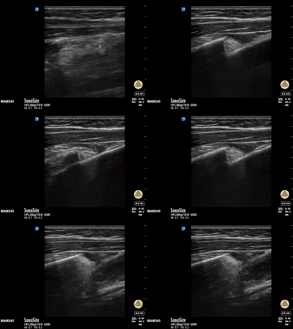
Radius/Ulnar fracture Long Axis (1/2)
11y female with L both bone forearm fx from fall during gym class. Long axis view with clear discontinuity of proximal and distal components of the shaft. Transverse view with transition from single bony cortex to overlap of fracture ends.
Matthew Moake, MD PhD
![Radius/ Ulna Fracture Transverse (1/2)](https://images.squarespace-cdn.com/content/v1/58118909e3df282037abfad7/1732461752674-Y322YESM0E611WLZZ8HT/radisu-ulna-short.gif)
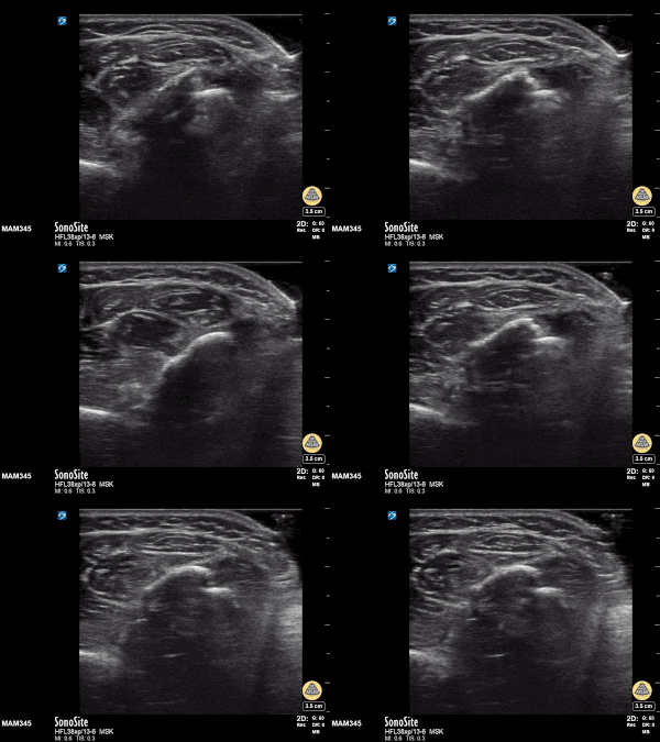
Radius/ Ulna Fracture Transverse (1/2)
11y female with L BB forearm fx from fall during gym class. Long axis view with clear discontinuity of proximal and distal components of the shaft. Transverse view with transition from single bony cortex to overlap of fracture ends.
Matthew Moake, MD PhD
![Humerus fracture- long axis (1/2)](https://images.squarespace-cdn.com/content/v1/58118909e3df282037abfad7/1732461298019-8DWQWN6O0M4KY9YIQX4Z/hum-long.gif)
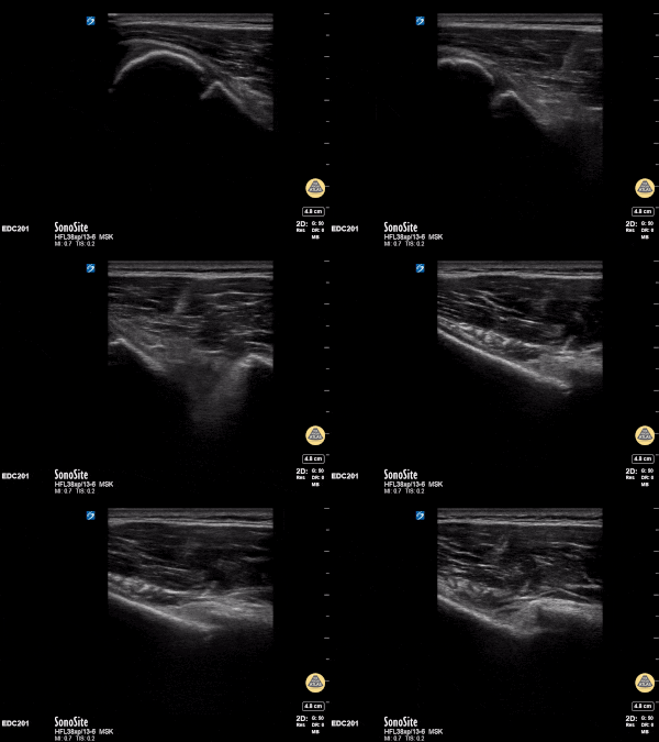
Humerus fracture- long axis (1/2)
12y male presenting with RUE pain after bike injury. POCUS demonstrating proximal humeral fracture. Long axis and short axis views of fracture.
Matthew Moake, MD PhD
![Humerus fracture- short axis (2/2)](https://images.squarespace-cdn.com/content/v1/58118909e3df282037abfad7/1732461174870-PJN8BCVBM3ORI66T1CJG/hum+short.gif)
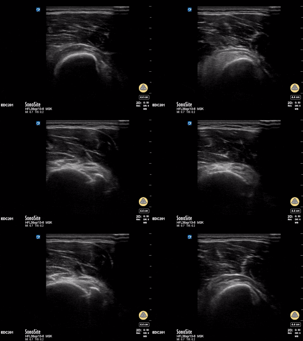
Humerus fracture- short axis (2/2)
12y male presenting with RUE pain after bike injury. POCUS demonstrating proximal humeral fracture. Long axis and short axis views of fracture.
Matthew Moake, MD PhD
![Distal Radius Buckle Fracture](https://images.squarespace-cdn.com/content/v1/58118909e3df282037abfad7/1727650853724-ORXDCGVJW28CRN2H9PC1/image-asset.gif)
Distal Radius Buckle Fracture
Clip shows longitudinal dorsal view of the radius with a "bump" in area of tenderness consistent with a torus or "buckle" fracture in a pediatric patient evaluated following a fall.
Miguel Agrait MD CAQ-SM, Eddie Rodriguez MD FPD-AEMUS
![Septic Elbow Effusion](https://images.squarespace-cdn.com/content/v1/58118909e3df282037abfad7/1664905809401-S4D9ROL7L3SOLA5LEYVH/image-asset.gif)
Septic Elbow Effusion
8 month old with arm swelling and fever. Longitudinal view of elbow with fluid collection displacing the posterior fat pad. Final diagnosis was septic arthritis of the elbow.
Contributor: Antonio Riera, MD, Yale University School of Medicine
![Soft Tissue Edema after Vaccination](https://images.squarespace-cdn.com/content/v1/58118909e3df282037abfad7/1664989607833-OAB8IWP9LC4RVPPQZN8J/image-asset.gif)
Soft Tissue Edema after Vaccination
Longitudinal suprapatellar evaluation with linear probe in a 23 month old with swelling to knee / lower leg after vaccination given in thigh. Note the impressive soft tissue edema. There is no cobblestoning pattern and no fluid collection layering under the quadriceps tendon / no signs of suprapatellar effusion. The patient had a dry arthrocentesis performed under procedural sedation by a consulting service.
Contributor: Antonio Riera, MD, Yale University School of Medicine
![Septic Knee Arthritis](https://images.squarespace-cdn.com/content/v1/58118909e3df282037abfad7/1664992034335-2NW6Z3BISKGDXIYGF6HR/image-asset.gif)
Septic Knee Arthritis
17 month old with knee effusion due to septic arthritis. Knee radiography was normal. Note the distal femur, physic and epiphysis seen on the lower part of the screen below the effusion and the hypoechoic non-ossified patella cartilage typically seen in this age.
Contributor: Antonio Riera, MD, Yale University School of Medicine
![Synovial Plica](https://images.squarespace-cdn.com/content/v1/58118909e3df282037abfad7/1664992471113-O8CPL5W328IF3Y7LWZ94/image-asset.gif)
Synovial Plica
11 year old with large knee effusion in the suprapatellar bursa. Note the visible synovial plica membrane
Contributor: Antonio Riera, MD Yale University School of Medicine
![Knee Effusion](https://images.squarespace-cdn.com/content/v1/58118909e3df282037abfad7/1664992740603-CXJIZ2T1PY9HYT5M921M/image-asset.gif)
Knee Effusion
10 year old with moderate size knee effusion in the suprapatellar bursa (seen deep to the quadriceps tendon). Etiology due to lyme arthritis.
Contributor: Antonio Riera, MD Yale University School of Medicine
![Knee Lipohemarthrosis-Case Series (1/3)](https://images.squarespace-cdn.com/content/v1/58118909e3df282037abfad7/1664993424509-ZHIPWIV01E4OQLR9FDGF/image-asset.gif)
Knee Lipohemarthrosis-Case Series (1/3)
15 year old female with patella fracture after a fall. Note the large suprapatellar effusion with discrete layering of the echogenic marrow/adipose above the acute hypoechoic blood. The lipohemarthrosis is visible in long and short axis (images 2 and 3 of case series).
Contributor: Antonio Riera, MD Yale University School of Medicine
![Knee Lipohemarthrosis-Case Series (2/3)](https://images.squarespace-cdn.com/content/v1/58118909e3df282037abfad7/1664993978199-XCBMGOYTXNA7ZU8FJMLS/image-asset.gif)
Knee Lipohemarthrosis-Case Series (2/3)
This is the short axis view of the case series.
15 year old female with patella fracture after a fall. Note the large suprapatellar effusion with discrete layering of the echogenic marrow/adipose above the acute hypoechoic blood. The lipohemarthrosis is visible in long and short axis.
Contributor: Antonio Riera, MD Yale University School of Medicine
![Knee Lipohemarthrosis-Case Series (3/3)](https://images.squarespace-cdn.com/content/v1/58118909e3df282037abfad7/1664994321489-DBMEORC2T4QVBVY8B2NS/image-asset.gif)
Knee Lipohemarthrosis-Case Series (3/3)
This is the long axis view of the case series.
15 year old female with patella fracture after a fall. Note the large suprapatellar effusion with discrete layering of the echogenic marrow/adipose above the acute hypoechoic blood. The lipohemarthrosis is visible in long and short axis.
Contributor: Antonio Riera, MD Yale University School of Medicine
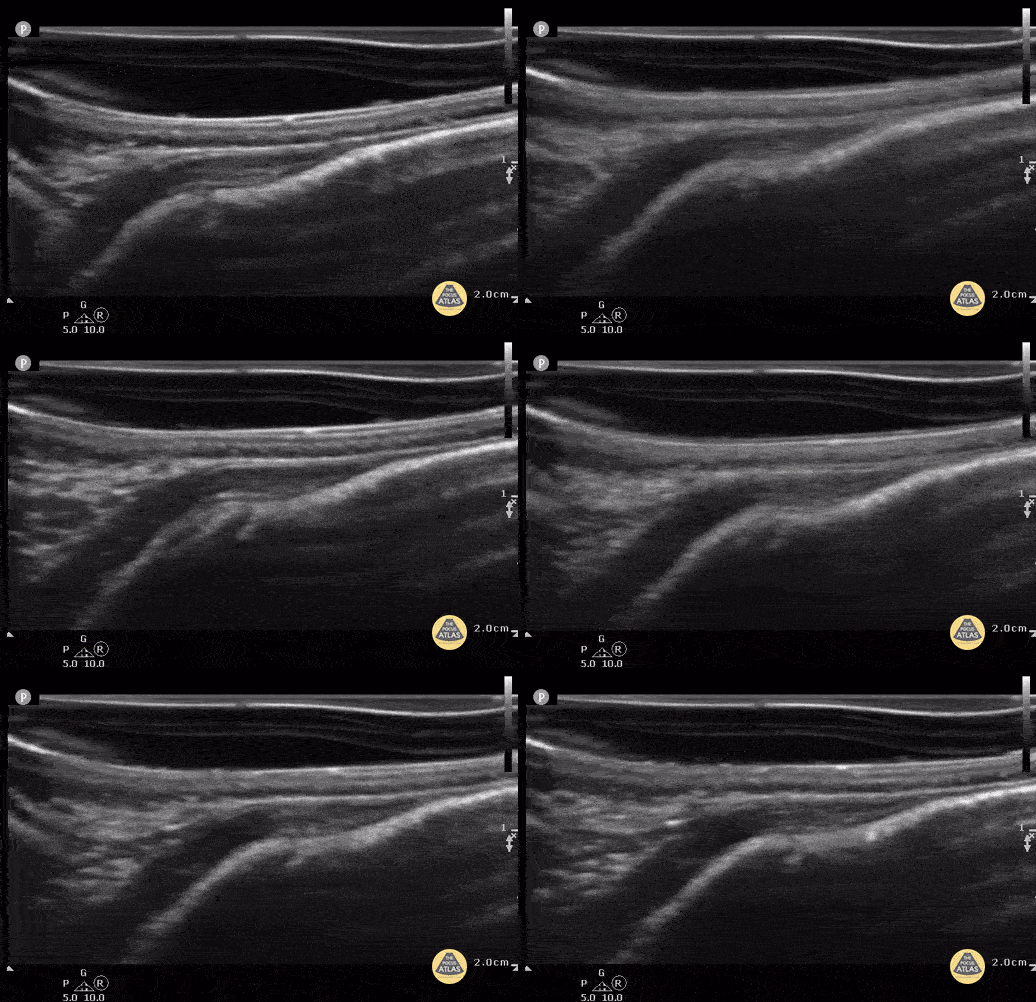
![Non-displaced parietal skull fracture (Case Series 1/2)](https://images.squarespace-cdn.com/content/v1/58118909e3df282037abfad7/1684791010445-Q26ZC59KPPTL2FPJ6BY9/ezgif.com-optimize.gif)
Non-displaced parietal skull fracture (Case Series 1/2)
4 week old with non displaced parietal fracture using water filled glove as stand off. (Case Series 1/2)
Contributor: Antonio Riera, MD
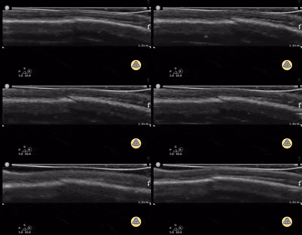
![Non-displaced parietal skull fracture (Case Series 2/2)](https://images.squarespace-cdn.com/content/v1/58118909e3df282037abfad7/1684791672380-UC2JJLDRF979QMWFRQW7/ezgif.com-optimize+%281%29.gif)
Non-displaced parietal skull fracture (Case Series 2/2)
same 4 week old with non-displaced parietal fracture after compression and displacement of fluid from the water filled glove. (Case Series 2/2)
Contributor: Antonio Riera, MD
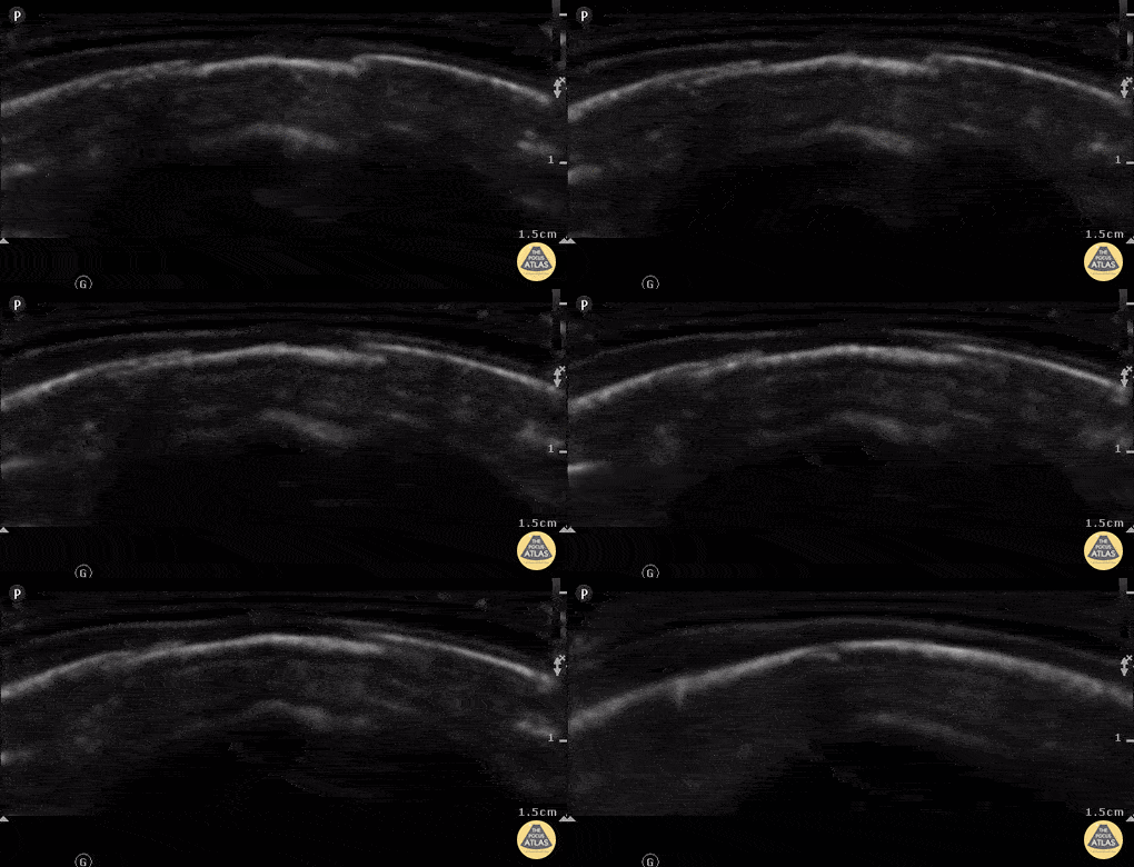
![Depressed parietal skull fracture](https://images.squarespace-cdn.com/content/v1/58118909e3df282037abfad7/1684792989643-IPZ6MNQ4EG0L51S13PDC/ezgif.com-optimize+%282%29.gif)
Depressed parietal skull fracture
7 week old with left parietal skull fracture. Note irregular edges on both sides and slightly depressed bony cortex.
Contributor: Antonio Riera, MD
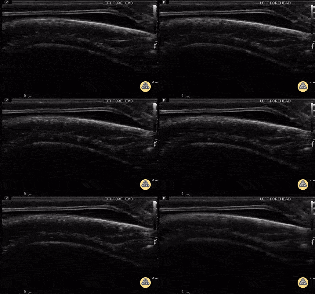
![Scalp Hematoma](https://images.squarespace-cdn.com/content/v1/58118909e3df282037abfad7/1684942270376-JBNV0CWWRSZ061GHKNC3/ezgif.com-optimize+%283%29.gif)
Scalp Hematoma
8 month old with scalp hematoma, no underlying skull fracture. The hematoma is denoted by the hypoechoic protuberance, superficial to the hyperechoic bone.
Contributor: Antonio Riera, MD
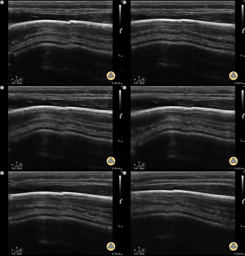
![Parietal Skull Fracture](https://images.squarespace-cdn.com/content/v1/58118909e3df282037abfad7/1684942637246-K0NWQZXHFPCG8MPLAK0N/ezgif.com-gif-maker+%281%29.gif)
Parietal Skull Fracture
9 month old with non-displaced parietal skull fracture. Note the diagonal jagged appearance of bony overlap.
Contributor: Antonio Riera, MD
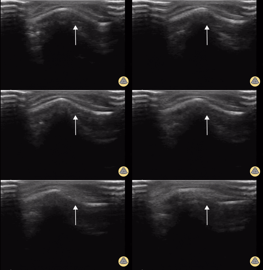
![Ping Pong Fracture](https://images.squarespace-cdn.com/content/v1/58118909e3df282037abfad7/1684942983776-ZBMMCMHPG2Z4VCDYIM52/ezgif.com-gif-maker+%282%29.gif)
Ping Pong Fracture
11 month old with depressed frontal ping pong fracture. These fractures occur when the bone is soft enough to indent rather than outright break.
Contributor: Antonio Riera, MD
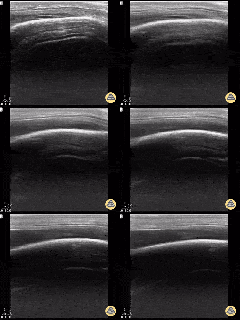
![Scalp Hematoma](https://images.squarespace-cdn.com/content/v1/58118909e3df282037abfad7/1684943326515-YNZ5W4RLOE26PXPI60YU/ezgif.com-optimize+%284%29.gif)
Scalp Hematoma
16 year old with large parietal hematoma and no underlying fracture. Bony cortex is intact.
Contributor: Antonio Riera, MD
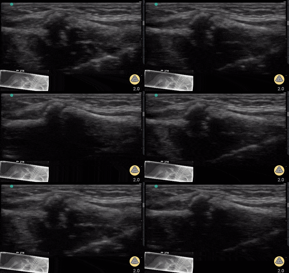
![Newborn Clavicle Fracture](https://images.squarespace-cdn.com/content/v1/58118909e3df282037abfad7/1684943933623-0SPGZI85RLWDVBC3HG48/ezgif.com-optimize+%285%29.gif)
Newborn Clavicle Fracture
2 week old with clavicle fracture from birth trauma. Note the rounded, bony protrusion with disruption of the cortex seen by ultrasound and callous formation seen on x-ray.
Contributor: Antonio Riera, MD
![Sprained Ankle](https://images.squarespace-cdn.com/content/v1/58118909e3df282037abfad7/1684944291855-WHMZQEZD746NGWJLPQ7T/ezgif.com-optimize+%286%29.gif)
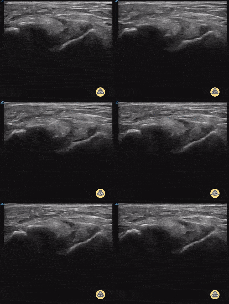
Sprained Ankle
17 yo male inverted his ankle playing basketball. POCUS shows partial tear of the ATFL.
Contributor: Paul Khalil, MD Nicklaus Children's Hospital @khalil3paul
![Skull Fracture](https://images.squarespace-cdn.com/content/v1/58118909e3df282037abfad7/1685646445325-D00QN3TC5Y3R0QPYITF2/ezgif.com-optimize+%287%29.gif)
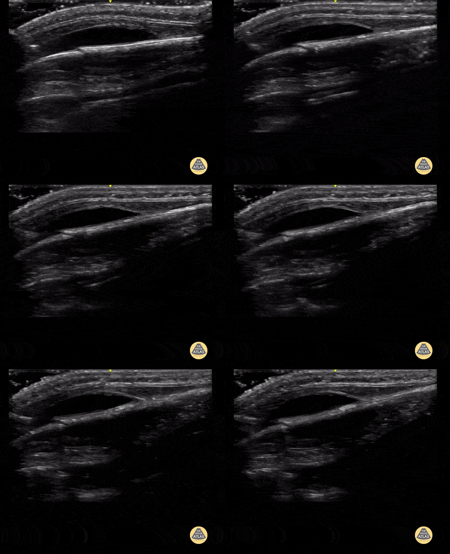
Skull Fracture
9 month-old presented after a 4 foot fall. There was a small frontal hematoma on exam without step off. POCUS was performed with a high frequency transducer over the area of hematoma, and demonstrates the skull as a hyperechoic linear structure, with nondisplaced discontinuity, indicative of a nondisplaced skull fracture. There is also a hypoechoic collection just anterior to the skull, suggestive of an associated hematoma.
Contributor: Allie Grither, MD, St Louis Children's Hospital (Washington University in St. Louis), @AGPemMD
![Tibia Fracture](https://images.squarespace-cdn.com/content/v1/58118909e3df282037abfad7/1685646923488-ISHAKIWEWJCY2Y0OPSLE/ezgif.com-optimize+%288%29.gif)
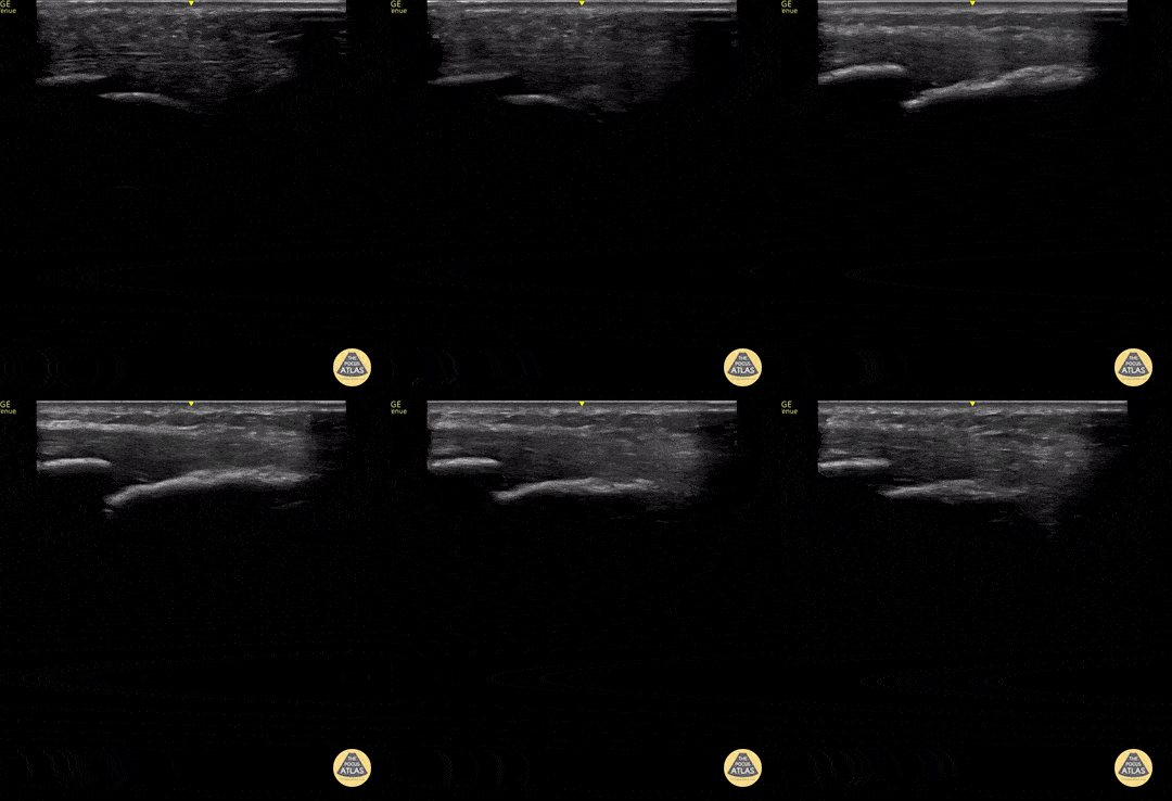
Tibia Fracture
12 year old came in with a fall from a scooter and right lower leg pain and swelling, just above the ankle. POCUS was performed with a high frequency transducer in longitudinal axis of the area of swelling, which demonstrates the tibia as a discontinuous, displaced, hyperechoic linear structure with an associated mixed echotexture collection just anterior to the discontinuity, suggestive of a displaced tibia fracture with hematoma.
Contributor: Allie Grither, MD, St. Louis Children's Hospital (Washington University in St. Louis), @AGPemMD
![Skull Fracture](https://images.squarespace-cdn.com/content/v1/58118909e3df282037abfad7/1685647722186-J2GKOMSVS10RIYI2GA7K/ezgif.com-optimize+%289%29.jpeg)
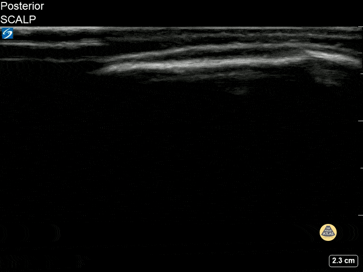
Skull Fracture
8 month old with fall off the bed with boggy mass palpated in the occiput.
Contributor: Kathryn Pade, MD, Rady Children's Hospital San Diego
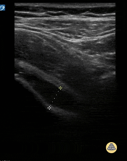
![Hip Effusion in 6yo 2/2 Transient Synovitis](https://images.squarespace-cdn.com/content/v1/58118909e3df282037abfad7/1685648160291-I9FQ2JF0GXDRUQKVDZFE/ezgif.com-optimize+%289%29.gif)
Hip Effusion in 6yo 2/2 Transient Synovitis
6 yo with acute onset limp. Afebrile. Decreased ROM of the left hip. POCUS showed a hip effusion consistent with the diagnosis of transient synovitis.
Contributor: Kathryn Pade, MD, Rady Children's Hospital San Diego
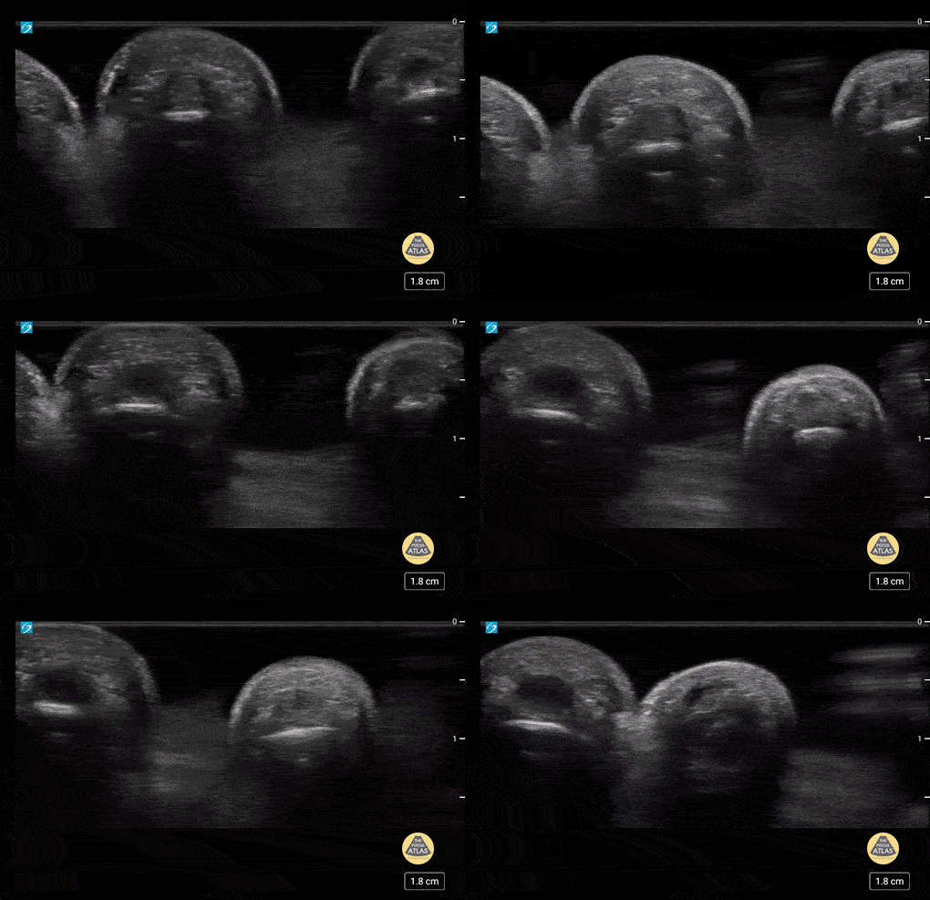
![Normal Fingers 1 of 2](https://images.squarespace-cdn.com/content/v1/58118909e3df282037abfad7/1685648672974-T7S4YRGE11R1UD1LTZWY/ezgif.com-optimize+%2810%29.gif)
Normal Fingers 1 of 2
5 yo with normal finger anatomy in a waterbath. Case series 1/2
Contributor: Paul Khalil, MD Nicklaus Children's Hospital @khalil3paul
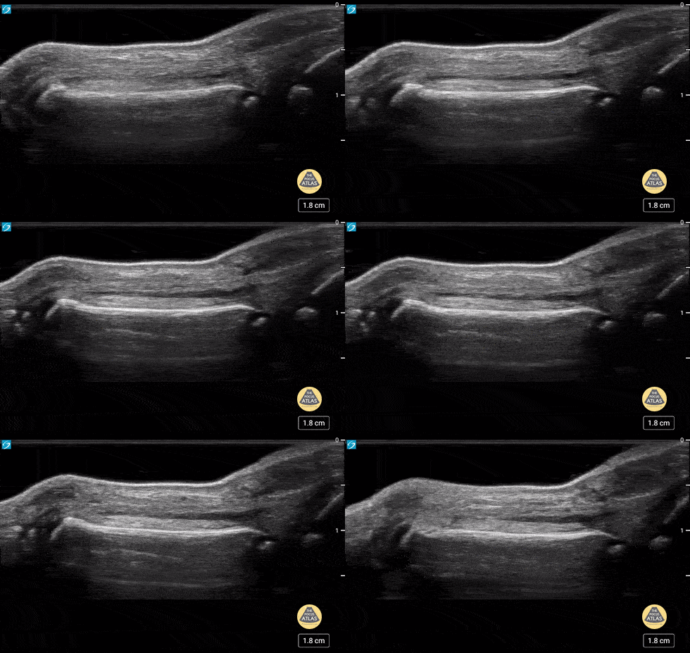
![Normal Fingers 2 of 2](https://images.squarespace-cdn.com/content/v1/58118909e3df282037abfad7/1685649027356-Z253K43HKBFLPG0MWJJR/ezgif.com-optimize+%2811%29.gif)
Normal Fingers 2 of 2
5 yo with normal finger anatomy in a waterbath. Case series 2 of 2
Contributor: Paul Khalil, MD Nicklaus Children's Hospital @khalil3paul
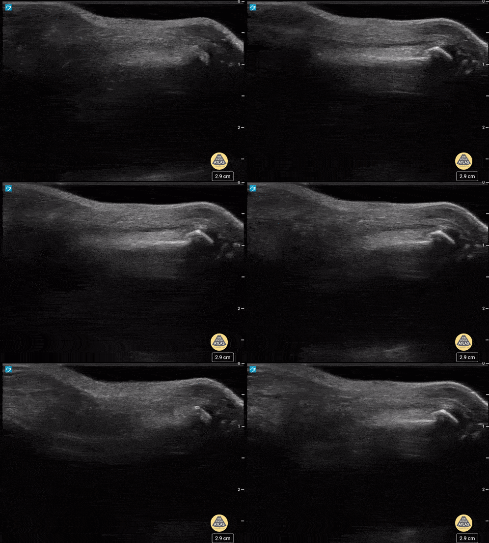
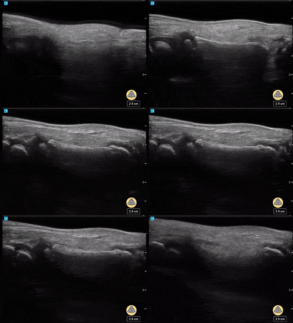
![Phalanx Fracture Reduction-Before](https://images.squarespace-cdn.com/content/v1/58118909e3df282037abfad7/1685649389817-KLW18Y7XFDCRIW4VX0PK/ezgif.com-optimize+%2812%29.gif)
Phalanx Fracture Reduction-Before
5 yo male with a phalanx fracture of the hand in a water bath before and after reduction. Case series 1 of 2
Contributor: Paul Khalil, MD Nicklaus Children's Hospital @khalil3paul
![Phalanx Fracture Reduction-After](https://images.squarespace-cdn.com/content/v1/58118909e3df282037abfad7/1685649649501-X3XT9WNQ02AW74HRF4LC/ezgif.com-optimize+%2813%29.gif)
Phalanx Fracture Reduction-After
5 yo male with a phalanx fracture of the hand in a water bath before and after reduction. Case series 2 of 2
Contributor: Paul Khalil, MD Nicklaus Children's Hospital @khalil3paul
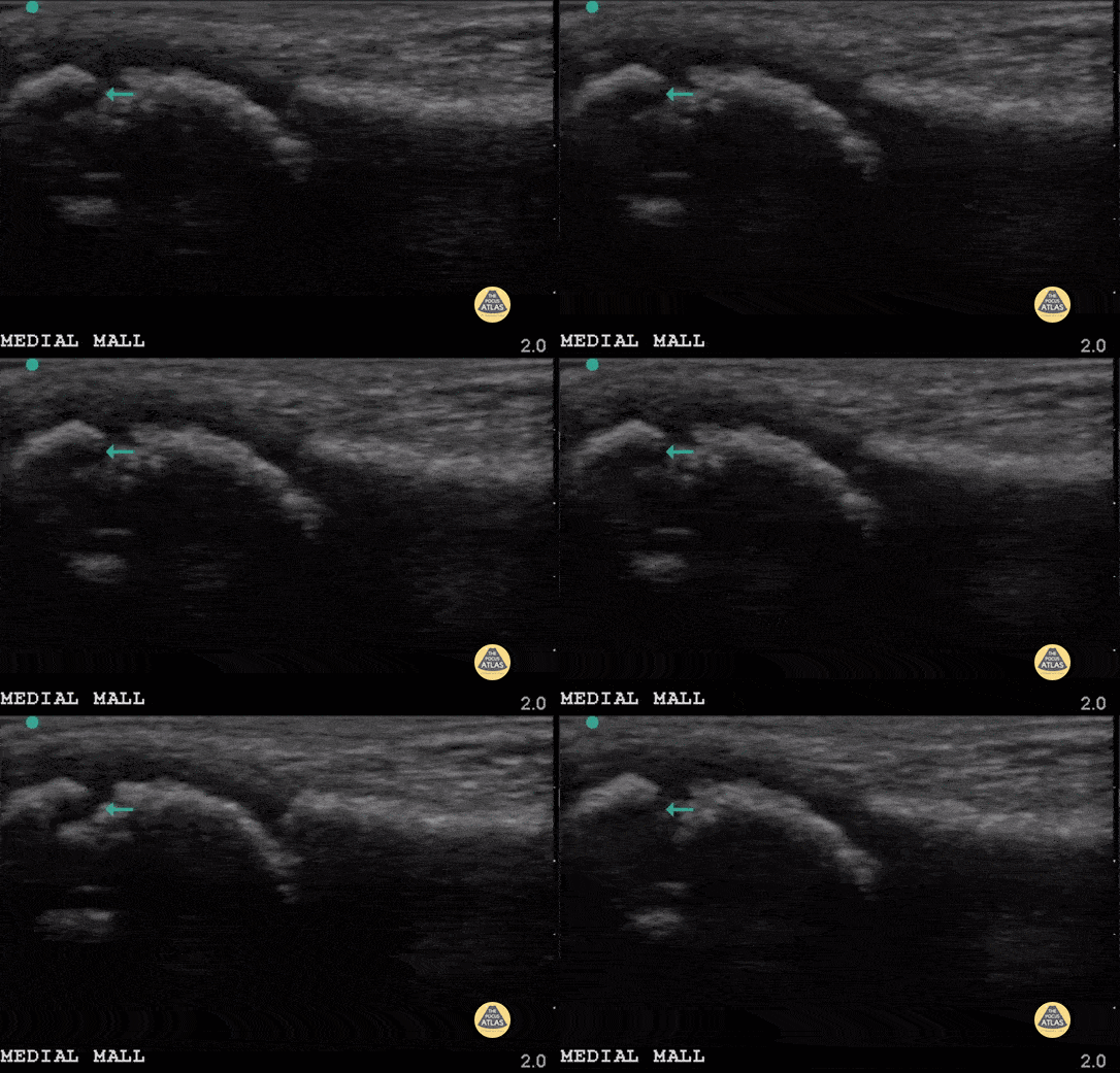
![Avulsion Fracture](https://images.squarespace-cdn.com/content/v1/58118909e3df282037abfad7/1685724011725-0868CXDQLG6KSQIRDI77/ezgif.com-optimize+%2814%29.gif)
Avulsion Fracture
7 year old with medial malleolus avulsion fracture of the distal tibia. The green arrow points to the gap where the avulsion piece (left of screen) is detached. Note a physis also visible (mid screen).
Contributor: Antonio Riera, MD
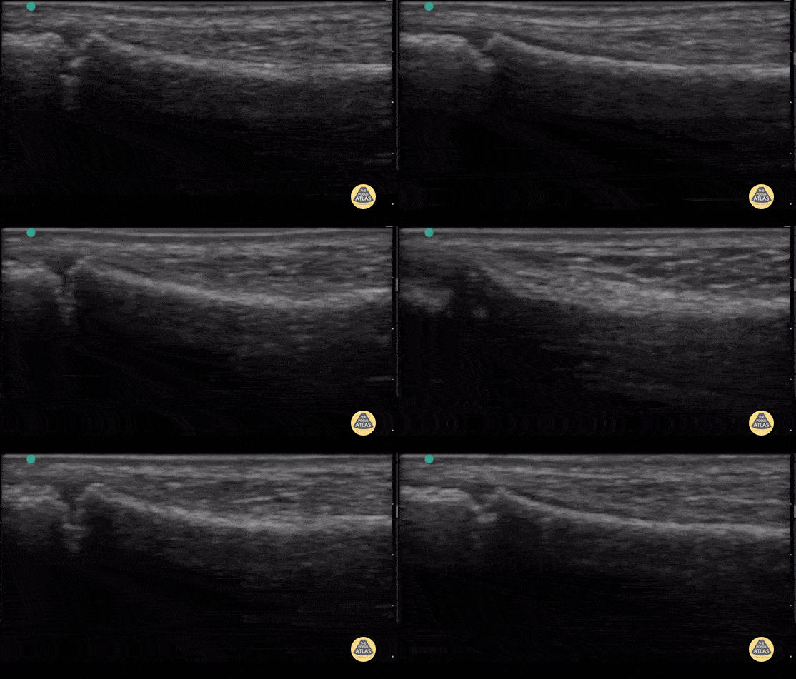
![Fibula with Normal Physis](https://images.squarespace-cdn.com/content/v1/58118909e3df282037abfad7/1685724366581-IV77XWPIOEU6W5T8TGQV/ezgif.com-optimize+%2815%29.gif)
Fibula with Normal Physis
7yo right fibular ultrasound with notable physis on left hand side of screen.
Contributor: Antonio Riera, MD
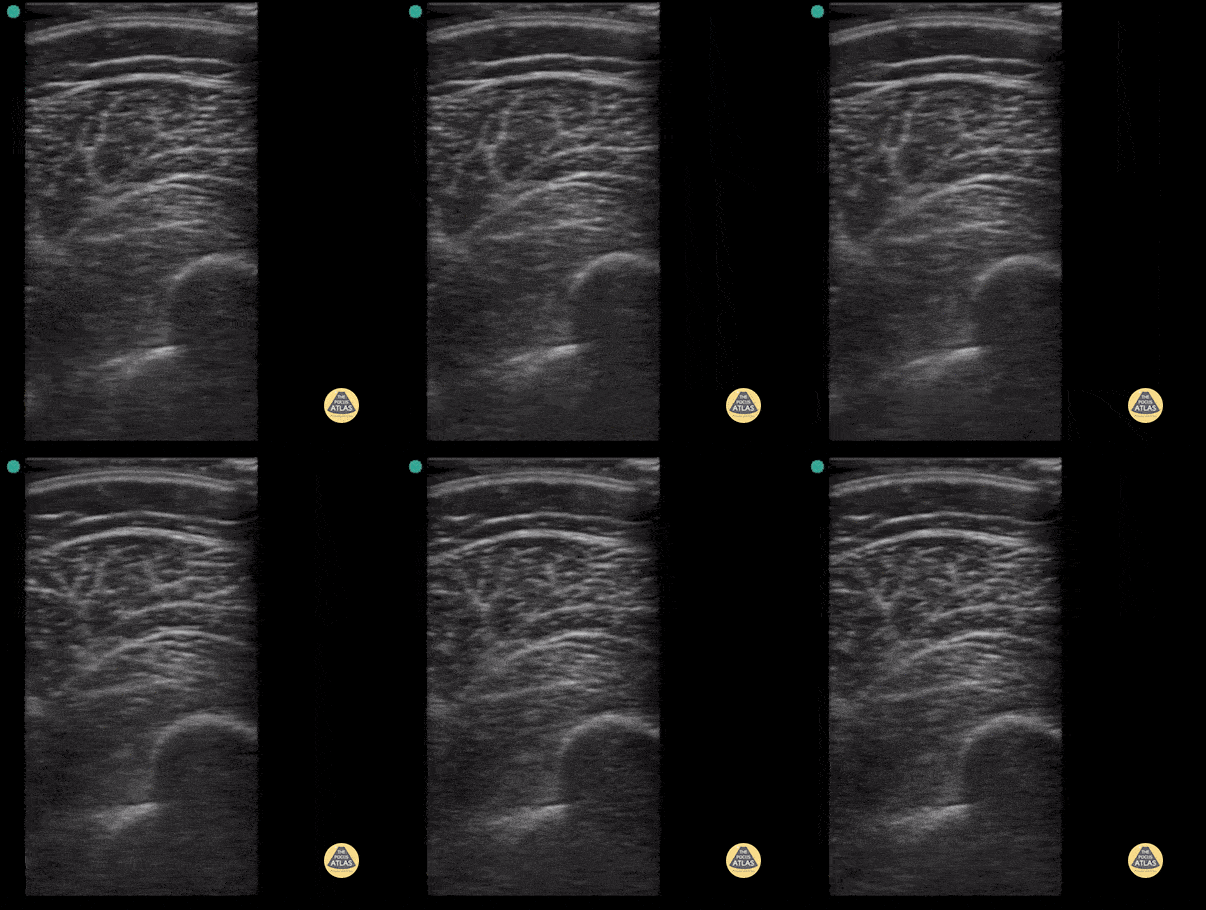
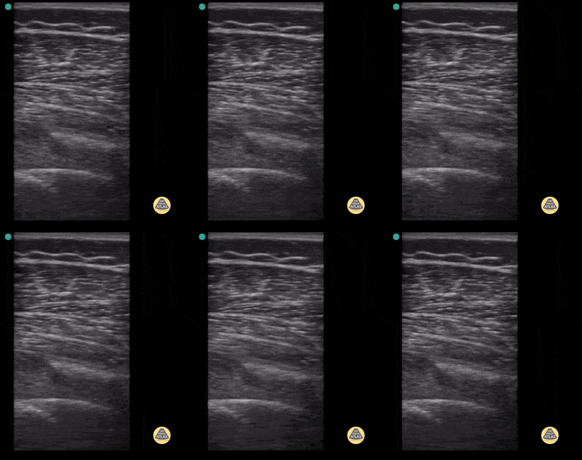
![Femur Fracture- Case Series, Short Axis](https://images.squarespace-cdn.com/content/v1/58118909e3df282037abfad7/1685725047655-I745I3PZ1ZC2R39K4X0Q/ezgif.com-optimize+%2816%29.gif)
Femur Fracture- Case Series, Short Axis
Short axis view of 9 year old male with spiral femur fracture with displacement. Note cortical disruption seen at about 5-6 cm in both long and short axis. Case series 1 of 2
Contributor: Antonio Riera, MD
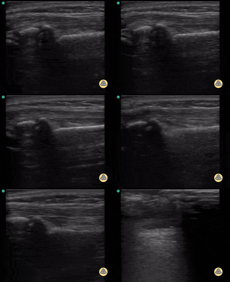
![Femur Fracture- Case Series, Long Axis](https://images.squarespace-cdn.com/content/v1/58118909e3df282037abfad7/1685725335805-LFB1IOHFB3T45VZQC1WB/ezgif.com-optimize+%2817%29.gif)
Femur Fracture- Case Series, Long Axis
Long axis view of 9 year old male with spiral femur fracture with displacement. Note cortical disruption seen at about 5-6 cm in both long and short axis. Case Series 2 of 2
Contributor: Antonio Riera, MD
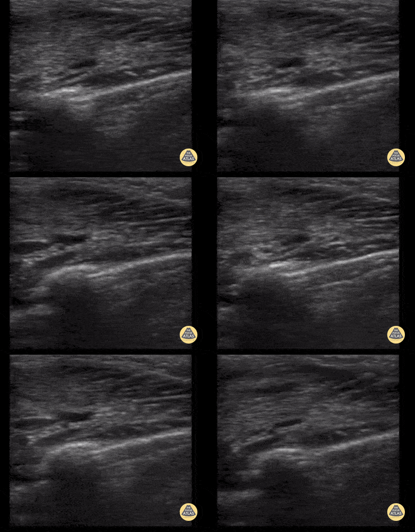
![Salter Harris II Fracture](https://images.squarespace-cdn.com/content/v1/58118909e3df282037abfad7/1685725944499-O3BY126X9L6XO9THO17J/ezgif.com-optimize+%2818%29.gif)
Salter Harris II Fracture
12 year old with SH-II fracture of distal radius. Not the abrupt metaphyseal angulation seen by ultrasound and physeal involvement.
Contributor: Antonio Riera, MD
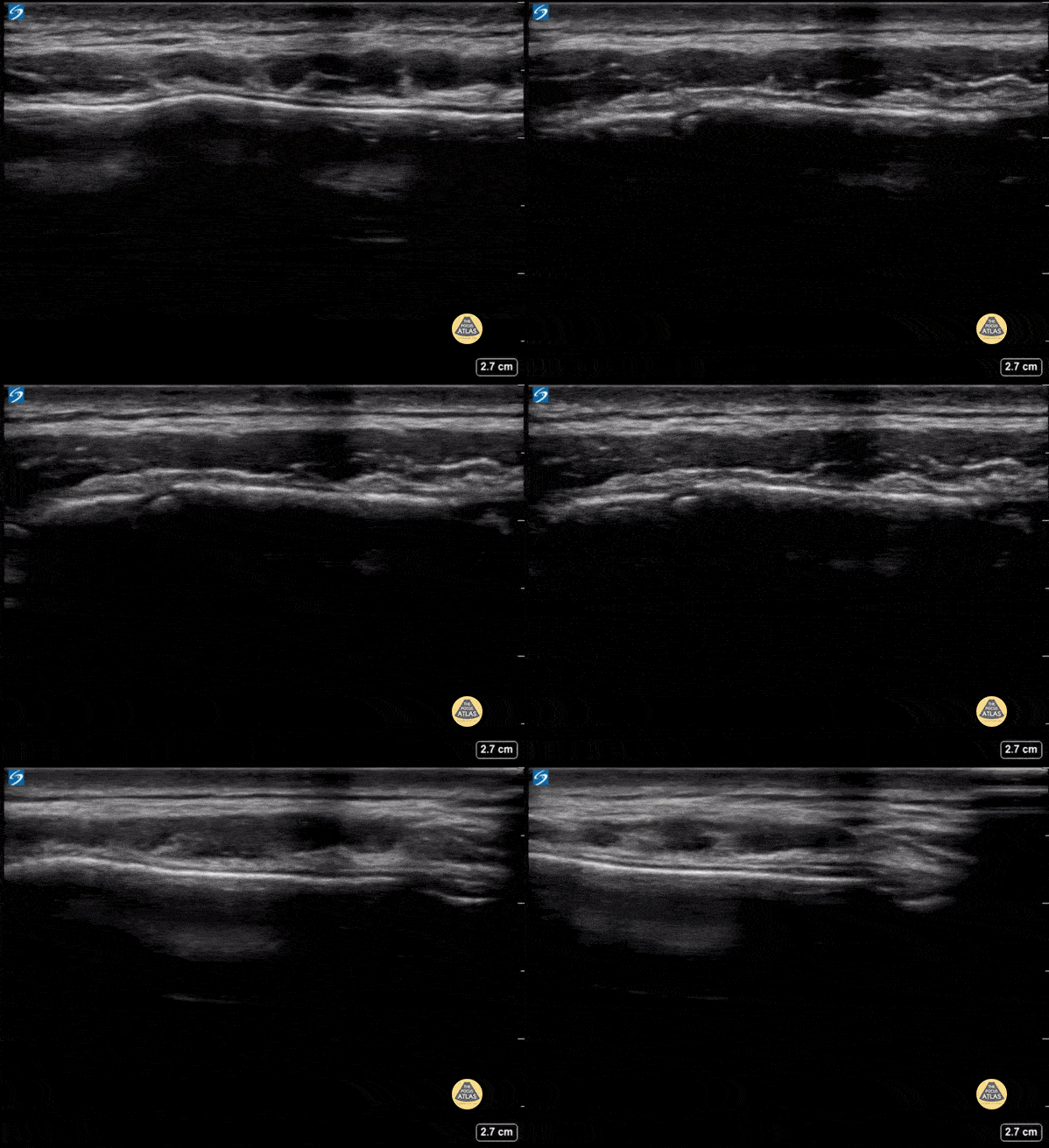
![Clavicle Fracture](https://images.squarespace-cdn.com/content/v1/58118909e3df282037abfad7/1685726205569-FMB3467N9I2QXYENMEXU/ezgif.com-optimize+%2819%29.gif)
Clavicle Fracture
29 month old with a mild, non-displaced clavicle fracture. Note the cortical disruption seen by ultrasound with an overlying small hematoma.
Contributor: Antonio Riera, MD
![Sternal Fracture](https://images.squarespace-cdn.com/content/v1/58118909e3df282037abfad7/1687969490597-0LR8JOK8M3CX1AW3UKD4/ezgif.com-optimize+%2840%29.gif)
Sternal Fracture
13 year old male who presented with sternal tenderness after a motor vehicle collision, found to have a step-off in the sternum on POCUS, indicating a sternal fracture. POCUS is more accurate than X-ray to identify sternal fracture.
Contributor: Zach Boivin, MD, @ZachBoivinMD
![Normal Hip Joint](https://images.squarespace-cdn.com/content/v1/58118909e3df282037abfad7/1687970363261-GC34DXSJFX5AJTN8G63H/ezgif.com-gif-maker+%284%29.gif)
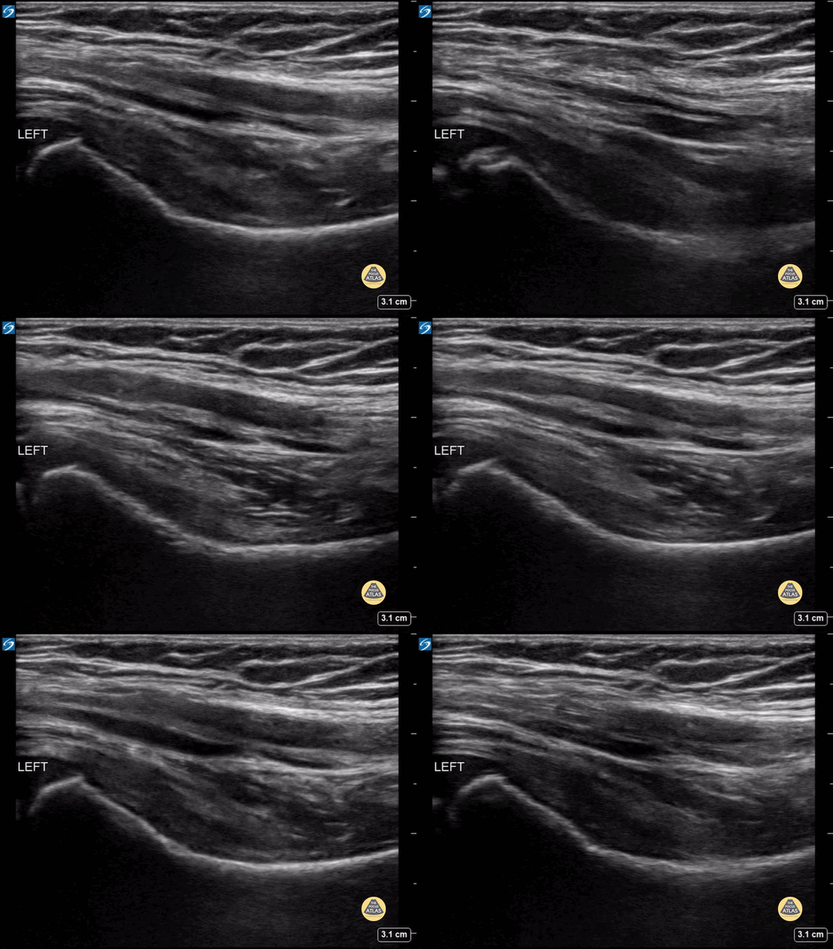
Normal Hip Joint
5 year old male presented with a left leg limp, assessment of the affected hip joint did not reveal a pathologic effusion.
Contributor: Zach Boivin, MD, @ZachBoivinMD
![Nursemaid's Elbow](https://images.squarespace-cdn.com/content/v1/58118909e3df282037abfad7/1609961854525-2SCIMYE89Y1KVLJOM724/image-asset.gif)
Nursemaid's Elbow
Nursemaid's pre and post reduction.
Nathan Jia, Orthopedic Resident
![Shoulder Relocation](https://images.squarespace-cdn.com/content/v1/58118909e3df282037abfad7/1616093328575-6W3O2OC7QUW0M7480BBT/image-asset.gif)
Shoulder Relocation
A 9-year-old female presented with left shoulder pain. She has a history of multiple dislocations and, as seen here on POCUS, is able to reduce the dislocation herself.
Julie Klensch, PEM Fellow & Paul Khalil, MD
University of Louisville/Norton Children's Hospital
![Ruptured Achilles Tendon](https://images.squarespace-cdn.com/content/v1/58118909e3df282037abfad7/1601048210657-RVGMTUGC6WGSOR5CTBJ9/image-asset.gif)
Ruptured Achilles Tendon
16-year-old male presented with acute onset sharp pain to his LE and inability to bare weight after having landed oddly while playing basketball. POCUS revealed a near-complete disruption of his Achilles Tendon.
Paul Khalil, MD. Assistant PEM POCUS director at University of Louisville/Norton Children’s
@Khalil3Paul
![Mildly Displaced Clavicle Fracture](https://images.squarespace-cdn.com/content/v1/58118909e3df282037abfad7/1599260703664-0IHNPMY5BDHC7NVH941G/image-asset.gif)
Mildly Displaced Clavicle Fracture
A 15 year old wrestler landed on his right shoulder. POCUS was performed over point of maximal pain demonstrating cortical displacement consistent with a clavicle fracture.
Paul Khalil, MD @Khalil3Paul
Assistant PEM POCUS Director at University of Louisville/Norton Children’s
![Sternal Fracture](https://images.squarespace-cdn.com/content/v1/58118909e3df282037abfad7/1579283346380-YX68Q1OH1ELOXXHDFXWI/image-asset.gif)
Sternal Fracture
An 8 year old female presented with chest pain after a fall out of a bouncy house at her neighborhood block party. She has notable bony tenderness to the anterior chest wall over the sternum. POCUS revealed normal lung slide, but on evaluation of the sternum, a fracture was noted. In this clip the fracture is seen on the right as cortical disruption with surrounding trace hypoechoic hematoma formation. On the left side of the screen a normal growth plate is noted.
Image courtesy of Dr. Paul Khalil
Twitter: @khalil3paul
![Normal Ankle](https://images.squarespace-cdn.com/content/v1/58118909e3df282037abfad7/1693606638154-K2Z6Q28HZDHRXJ4K2S4S/ezgif.com-gif-maker.gif)
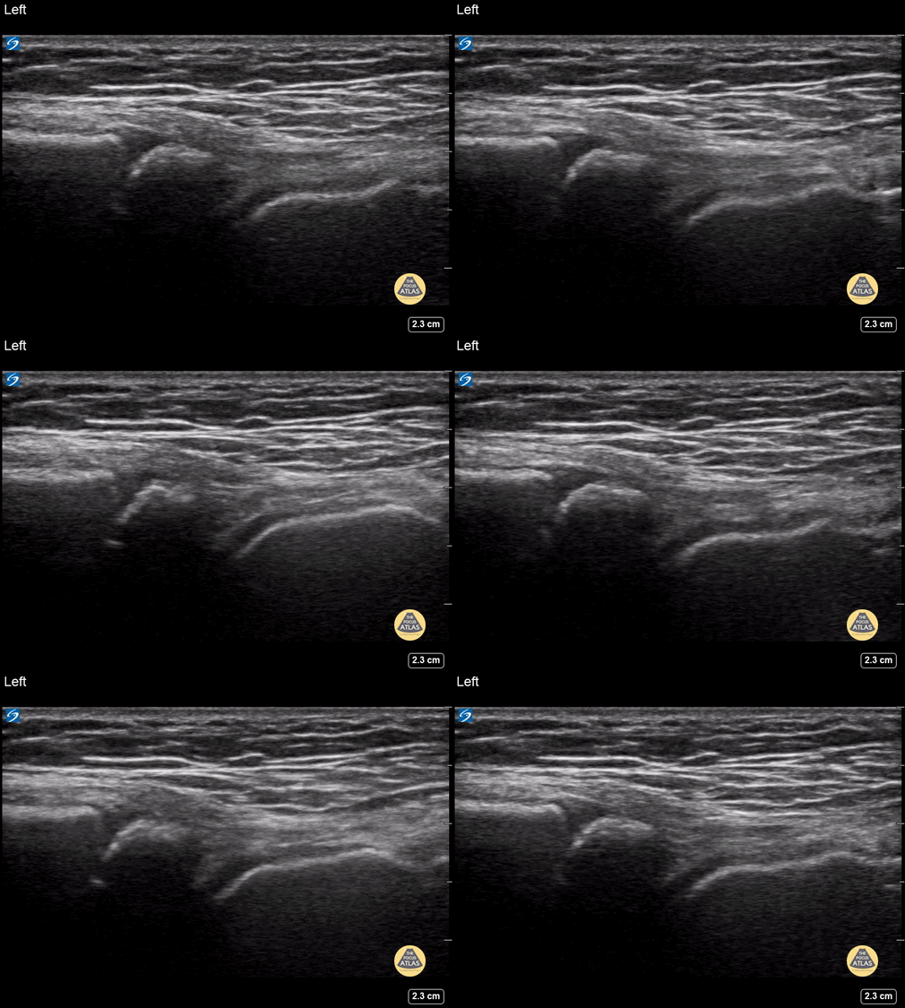
Normal Ankle
5y female presenting with swelling, redness, and limp to the R foot. Contralateral normal side imaged for comparison. The normal tibiotalar joint in the long axis is seen here. Since the patient skeletally immature, the distal tibial physis can also be seen.
Contributor: Matthew Moake, MD PhD
![Radius/Ulnar fracture Long Axis (1/2)](https://images.squarespace-cdn.com/content/v1/58118909e3df282037abfad7/1732461824702-05AKJ49FMT4MC5GJWART/radius-ulna-long.gif)
![Radius/ Ulna Fracture Transverse (1/2)](https://images.squarespace-cdn.com/content/v1/58118909e3df282037abfad7/1732461752674-Y322YESM0E611WLZZ8HT/radisu-ulna-short.gif)
![Humerus fracture- long axis (1/2)](https://images.squarespace-cdn.com/content/v1/58118909e3df282037abfad7/1732461298019-8DWQWN6O0M4KY9YIQX4Z/hum-long.gif)
![Humerus fracture- short axis (2/2)](https://images.squarespace-cdn.com/content/v1/58118909e3df282037abfad7/1732461174870-PJN8BCVBM3ORI66T1CJG/hum+short.gif)
![Distal Radius Buckle Fracture](https://images.squarespace-cdn.com/content/v1/58118909e3df282037abfad7/1727650853724-ORXDCGVJW28CRN2H9PC1/image-asset.gif)
![Septic Elbow Effusion](https://images.squarespace-cdn.com/content/v1/58118909e3df282037abfad7/1664905809401-S4D9ROL7L3SOLA5LEYVH/image-asset.gif)
![Soft Tissue Edema after Vaccination](https://images.squarespace-cdn.com/content/v1/58118909e3df282037abfad7/1664989607833-OAB8IWP9LC4RVPPQZN8J/image-asset.gif)
![Septic Knee Arthritis](https://images.squarespace-cdn.com/content/v1/58118909e3df282037abfad7/1664992034335-2NW6Z3BISKGDXIYGF6HR/image-asset.gif)
![Synovial Plica](https://images.squarespace-cdn.com/content/v1/58118909e3df282037abfad7/1664992471113-O8CPL5W328IF3Y7LWZ94/image-asset.gif)
![Knee Effusion](https://images.squarespace-cdn.com/content/v1/58118909e3df282037abfad7/1664992740603-CXJIZ2T1PY9HYT5M921M/image-asset.gif)
![Knee Lipohemarthrosis-Case Series (1/3)](https://images.squarespace-cdn.com/content/v1/58118909e3df282037abfad7/1664993424509-ZHIPWIV01E4OQLR9FDGF/image-asset.gif)
![Knee Lipohemarthrosis-Case Series (2/3)](https://images.squarespace-cdn.com/content/v1/58118909e3df282037abfad7/1664993978199-XCBMGOYTXNA7ZU8FJMLS/image-asset.gif)
![Knee Lipohemarthrosis-Case Series (3/3)](https://images.squarespace-cdn.com/content/v1/58118909e3df282037abfad7/1664994321489-DBMEORC2T4QVBVY8B2NS/image-asset.gif)
![Non-displaced parietal skull fracture (Case Series 1/2)](https://images.squarespace-cdn.com/content/v1/58118909e3df282037abfad7/1684791010445-Q26ZC59KPPTL2FPJ6BY9/ezgif.com-optimize.gif)
![Non-displaced parietal skull fracture (Case Series 2/2)](https://images.squarespace-cdn.com/content/v1/58118909e3df282037abfad7/1684791672380-UC2JJLDRF979QMWFRQW7/ezgif.com-optimize+%281%29.gif)
![Depressed parietal skull fracture](https://images.squarespace-cdn.com/content/v1/58118909e3df282037abfad7/1684792989643-IPZ6MNQ4EG0L51S13PDC/ezgif.com-optimize+%282%29.gif)
![Scalp Hematoma](https://images.squarespace-cdn.com/content/v1/58118909e3df282037abfad7/1684942270376-JBNV0CWWRSZ061GHKNC3/ezgif.com-optimize+%283%29.gif)
![Parietal Skull Fracture](https://images.squarespace-cdn.com/content/v1/58118909e3df282037abfad7/1684942637246-K0NWQZXHFPCG8MPLAK0N/ezgif.com-gif-maker+%281%29.gif)
![Ping Pong Fracture](https://images.squarespace-cdn.com/content/v1/58118909e3df282037abfad7/1684942983776-ZBMMCMHPG2Z4VCDYIM52/ezgif.com-gif-maker+%282%29.gif)
![Scalp Hematoma](https://images.squarespace-cdn.com/content/v1/58118909e3df282037abfad7/1684943326515-YNZ5W4RLOE26PXPI60YU/ezgif.com-optimize+%284%29.gif)
![Newborn Clavicle Fracture](https://images.squarespace-cdn.com/content/v1/58118909e3df282037abfad7/1684943933623-0SPGZI85RLWDVBC3HG48/ezgif.com-optimize+%285%29.gif)
![Sprained Ankle](https://images.squarespace-cdn.com/content/v1/58118909e3df282037abfad7/1684944291855-WHMZQEZD746NGWJLPQ7T/ezgif.com-optimize+%286%29.gif)
![Skull Fracture](https://images.squarespace-cdn.com/content/v1/58118909e3df282037abfad7/1685646445325-D00QN3TC5Y3R0QPYITF2/ezgif.com-optimize+%287%29.gif)
![Tibia Fracture](https://images.squarespace-cdn.com/content/v1/58118909e3df282037abfad7/1685646923488-ISHAKIWEWJCY2Y0OPSLE/ezgif.com-optimize+%288%29.gif)
![Skull Fracture](https://images.squarespace-cdn.com/content/v1/58118909e3df282037abfad7/1685647722186-J2GKOMSVS10RIYI2GA7K/ezgif.com-optimize+%289%29.jpeg)
![Hip Effusion in 6yo 2/2 Transient Synovitis](https://images.squarespace-cdn.com/content/v1/58118909e3df282037abfad7/1685648160291-I9FQ2JF0GXDRUQKVDZFE/ezgif.com-optimize+%289%29.gif)
![Normal Fingers 1 of 2](https://images.squarespace-cdn.com/content/v1/58118909e3df282037abfad7/1685648672974-T7S4YRGE11R1UD1LTZWY/ezgif.com-optimize+%2810%29.gif)
![Normal Fingers 2 of 2](https://images.squarespace-cdn.com/content/v1/58118909e3df282037abfad7/1685649027356-Z253K43HKBFLPG0MWJJR/ezgif.com-optimize+%2811%29.gif)
![Phalanx Fracture Reduction-Before](https://images.squarespace-cdn.com/content/v1/58118909e3df282037abfad7/1685649389817-KLW18Y7XFDCRIW4VX0PK/ezgif.com-optimize+%2812%29.gif)
![Phalanx Fracture Reduction-After](https://images.squarespace-cdn.com/content/v1/58118909e3df282037abfad7/1685649649501-X3XT9WNQ02AW74HRF4LC/ezgif.com-optimize+%2813%29.gif)
![Avulsion Fracture](https://images.squarespace-cdn.com/content/v1/58118909e3df282037abfad7/1685724011725-0868CXDQLG6KSQIRDI77/ezgif.com-optimize+%2814%29.gif)
![Fibula with Normal Physis](https://images.squarespace-cdn.com/content/v1/58118909e3df282037abfad7/1685724366581-IV77XWPIOEU6W5T8TGQV/ezgif.com-optimize+%2815%29.gif)
![Femur Fracture- Case Series, Short Axis](https://images.squarespace-cdn.com/content/v1/58118909e3df282037abfad7/1685725047655-I745I3PZ1ZC2R39K4X0Q/ezgif.com-optimize+%2816%29.gif)
![Femur Fracture- Case Series, Long Axis](https://images.squarespace-cdn.com/content/v1/58118909e3df282037abfad7/1685725335805-LFB1IOHFB3T45VZQC1WB/ezgif.com-optimize+%2817%29.gif)
![Salter Harris II Fracture](https://images.squarespace-cdn.com/content/v1/58118909e3df282037abfad7/1685725944499-O3BY126X9L6XO9THO17J/ezgif.com-optimize+%2818%29.gif)
![Clavicle Fracture](https://images.squarespace-cdn.com/content/v1/58118909e3df282037abfad7/1685726205569-FMB3467N9I2QXYENMEXU/ezgif.com-optimize+%2819%29.gif)
![Sternal Fracture](https://images.squarespace-cdn.com/content/v1/58118909e3df282037abfad7/1687969490597-0LR8JOK8M3CX1AW3UKD4/ezgif.com-optimize+%2840%29.gif)
![Normal Hip Joint](https://images.squarespace-cdn.com/content/v1/58118909e3df282037abfad7/1687970363261-GC34DXSJFX5AJTN8G63H/ezgif.com-gif-maker+%284%29.gif)
![Nursemaid's Elbow](https://images.squarespace-cdn.com/content/v1/58118909e3df282037abfad7/1609961854525-2SCIMYE89Y1KVLJOM724/image-asset.gif)
![Shoulder Relocation](https://images.squarespace-cdn.com/content/v1/58118909e3df282037abfad7/1616093328575-6W3O2OC7QUW0M7480BBT/image-asset.gif)
![Ruptured Achilles Tendon](https://images.squarespace-cdn.com/content/v1/58118909e3df282037abfad7/1601048210657-RVGMTUGC6WGSOR5CTBJ9/image-asset.gif)
![Mildly Displaced Clavicle Fracture](https://images.squarespace-cdn.com/content/v1/58118909e3df282037abfad7/1599260703664-0IHNPMY5BDHC7NVH941G/image-asset.gif)
![Sternal Fracture](https://images.squarespace-cdn.com/content/v1/58118909e3df282037abfad7/1579283346380-YX68Q1OH1ELOXXHDFXWI/image-asset.gif)
![Normal Ankle](https://images.squarespace-cdn.com/content/v1/58118909e3df282037abfad7/1693606638154-K2Z6Q28HZDHRXJ4K2S4S/ezgif.com-gif-maker.gif)