
Renal/GU

Horseshoe Kidney Transverse
Horseshoe kidney. No kidney was identified in either flank area, this anterior abdominal view shows one large kidney that is anterior to the Aorta.
Contributed by: Marion Memmott, DO; Michael Bernard, DO; Central Michigan University Emergency Medicine Residency

Horseshoe Kidney Longitudinal
Horseshoe kidney. No kidney was identified in either flank area, this anterior abdominal view shows one large kidney that is anterior to the Aorta.
Marion Memmott, DO; Michael Bernard, DO; Central Michigan University Emergency Medicine Residency

Prostate Carcinoma
Prominent prostate on ultrasound in a 72 year-old-male with known prostate carcinoma and recent PET scan with confirmation. No known masses or metastasis. This is seen in both transverse and sagittal views.
Lindsay Davis, DO, MPH, PGY3; Matthew Welch, MS4; Alex Schlangen, DO, PGY1

Bladder Diverticulum 2
A bladder diverticulum is demonstrated deep to the bladder as the ultrasound probe is fanned.
Contributed by: Brittany Garza, DO; Saleem Nasseh, MD; Sadie Ellerson, MS4

Moderate Hydronephrosis from Nephrolithiasis
Here is an example of hydronephrosis in the presence of nephrolithasis. This patient presented to the emergency department complaining of new right flank pain after waking up in the morning. She was seen previously for urinary complaints, diagnosed with UTI and started on antibiotic regiment that had been altered twice due to concerns of sub-optimal coverage. She demonstrated right CVA tenderness on physical exam, raising suspicion for pyelonephritis as well as hydronephrosis. Subsequent point of care ultrasound revealed moderate hydronephrosis. CT was obtained to further evaluate and confirmed the presence of an obstructing kidney stone. Note presence of hydronephrosis as the probe is fanned/tilted through the kidney, highlighting the importance of scanning through the entire organ.
Dr. Carlo Zamora, DO, PGY-1
Riverside Regional Medical Center Emergency Medicine Residency (Newport News, VA)

UVJ stone with ureteral dilation
A young female presented with acute RLQ abdominal pain and found to have a 5mm right ureter stone at the UVJ with ureteral dilation. The patient also had moderate hydronephrosis.
Matthew Petruso, DO; Harpreet Grewal, MD; Eric Versluis, MS4; Central Michigan University: Emergency Medicine Residency.

Large Renal Cysts
83 year old male presented to the ED for right flank pain.
The renal ultrasound showed two right renal cysts. The superior renal pole cyst measuring 12 x 13cm. The inferior pole cyst measuring 4 x 5cm.
Laura Schroeder, MS, MD; Syeda Zehra, MD; Conner Shea (Medical Student) Central Michigan University Emergency Medicine Residency

Right Sided Emphysematous Pyelonephritis
60 year old female with poorly controlled diabetes. Initial CT showing R-sided pyelonephritis, US performed 12 hours later demonstrated development of emphysematous pyelonephritis noted by the hyperechoic areas with dirty shadowing seen within the renal parenchyma (indicative of gas). This was later confirmed on repeat CT imaging.
Contributor: Shane Solger, MD
Kings County/SUNY Downstate EM/IM

Bladder Diverticula
82 year old male with history of subarachnoid bleed presented for evaluation after a fall. During a EFAST exam, bladder US revealed multiple, anechoic, cystic-appearing structures consistent with bladder diverticula (confirmed by CT).
A bladder diverticulum is a protrusion of the mucosa through the muscular wall of the bladder and can be congenital or acquired. Acquired diverticula are more common, with bladder outlet obstruction being the most common cause. Urology follow up is recommended to confirm the cause and screen for other concerning etiologies or complications including carcinoma, recurrent UTIs and bladder calculi.
Contributors: Michael Huber, 4th year Medical Student; Chad Bambrick, MD; Ana Camagay, MD
Central Michigan University College of Medicine

Large Simple Renal Cyst
Large Renal Cyst seen coming off of the kidney in short axis.
Dax Spencer

Moderate-Severe Hydronephrosis
This is the right renal ultrasound of a 20 year old male with a past medical history of recurrent obstructing nephrolithiasis. There is a moderate to severe hydronephrosis present. with what appears to be some thinning of the renal cortex.
Brittany Ladson, DO; Sara Schumbach, MD

Two Stones, One Image
Ultrasound clip demonstrating both cholelithiasis as well as obstructing stone in the ureter!
Image courtesy of Robert Jones DO, FACEP @RJonesSonoEM
Director, Emergency Ultrasound; MetroHealth Medical Center; Professor, Case Western Reserve Medical School, Cleveland, OH
View his original post here.

Penile Prosthesis Reservoir
60s M presented to the ED sent in by PMD for evaluation of elevated BUN and Creatinine. POCUS performed with transverse view of the urinary bladder demonstrating discrete cystic structure on anatomic right with second circular, hyperechoic structure visualized. Structures are consistent with penile prosthesis fluid reservoir abutting the right anterior bladder. Findings confirmed on chart review of previous urinary bladder imaging.
Hydraulic inflatable prostheses consist of two or three piece types. The three piece types (as pictured) consist of a pair of cylinders placed in each corpus cavernosum, a pump placed in the scrotum, and a reservoir placed adjacent to the bladder (1).
Yahyavi-Firouz-Abadi N, Menias C, Bhalla S, Siegel C, Gayer G, Katz D. Imaging of Cosmetic Plastic Procedures and Implants in the Body and Their Potential Complications. AJR Am J Roentgenol. 2015;204(4):707-15. doi:10.2214/AJR.14.13516 - Pubmed
Contributors: Joshua Taubman MD, Resident Physician - University at Buffalo Emergency Medicine, @UBEMSono

Renal Cell Carcinoma At Inferior Pole
This patient presented with hematuria and flank pain. In this scan, you can see a visible circular mass along the inferior pole of the kidney with further testing later confirming presence of a renal cell carcinoma.
Image courtesy of Robert Jones DO, FACEP @RJonesSonoEM
Director, Emergency Ultrasound; MetroHealth Medical Center; Professor, Case Western Reserve Medical School, Cleveland, OH
View his original post here

High Grade Pyelonephritis With Urothelial Thickening
Although most cases of uncomplicated pyelonephritis will appear normal on ultrasound, urothelial thickening might be observed. It should be noted that this can also be observed in different clinical contexts including urolithiasis, stents and malignancy.
Image courtesy of Robert Jones DO, FACEP @RJonesSonoEM
Director, Emergency Ultrasound; MetroHealth Medical Center; Professor, Case Western Reserve Medical School, Cleveland, OH
View his original post here

Renal Abscess
This clip demonstrates a renal abscess seen along the inferior pole of the kidney. Notice the circular shape with internal echoes and a hyperechoic rim demonstrated in both long and short axis.
Image courtesy of Robert Jones DO, FACEP @RJonesSonoEM
Director, Emergency Ultrasound; MetroHealth Medical Center; Professor, Case Western Reserve Medical School, Cleveland, OH
View his original post here

Air Within Bladder
This sagittal image of the bladder demonstrates air within the bladder. This could be from emphysematous cystitis but could also be iatrogenic due to recent foley placement. Also note the presence of layered echoes in the bladder due to an infectious process.
Image courtesy of Robert Jones DO, FACEP @RJonesSonoEM
Director, Emergency Ultrasound; MetroHealth Medical Center; Professor, Case Western Reserve Medical School, Cleveland, OH
View his original post here

Medullary Nephrocalcinosis
This clip demonstrates medullary nephrocalcinosis. Note the increased echogenicity of the renal pyramids due to calcium deposition.
Image courtesy of Robert Jones DO, FACEP @RJonesSonoEM
Director, Emergency Ultrasound; MetroHealth Medical Center; Professor, Case Western Reserve Medical School, Cleveland, OH
View his original post here

Pyonephrosis in Setting of Proximal Ureteral Stone
This patient presented with fever and flank pain with a known history of medullary nephrocalcinosis. Here we can see internal echoes within the dilated collecting system consistent with pyonephrosis. A large stone can be seen in the proximal ureter as a hyperechoic line with posterior shadowing.
Image courtesy of Robert Jones DO, FACEP @RJonesSonoEM
Director, Emergency Ultrasound; MetroHealth Medical Center; Professor, Case Western Reserve Medical School, Cleveland, OH
View his original post here

Emphysematous Pyelonephritis
This clip demonstrates air within the parenchyma of the kidney consistent with emphysematous pyelonephritis. This is different than emphysematous pyelitis in which air is only seen in the collection system.
Image courtesy of Robert Jones DO, FACEP @RJonesSonoEM
Director, Emergency Ultrasound; MetroHealth Medical Center; Professor, Case Western Reserve Medical School, Cleveland, OH
View his original post here

Acute Focal Nephritis
Right presented with right flank pain. A known ureterovesical junction stone is present. But also notice the focal hyperechoic area at the superior pole of the kidney. This patient was diagnosised with acute focal nephritis. This is on spectrum somewhere between acute pyelonephritis and renal abscess.
Image courtesy of Robert Jones DO, FACEP @RJonesSonoEM
Director, Emergency Ultrasound; MetroHealth Medical Center; Professor, Case Western Reserve Medical School, Cleveland, OH
View his original post here

Bladder cancer
Older male presented with right flank pain and hematuria. Bladder ultrasound revealed mass in bladder with internal color flow. Subsequent CT imaging performed and patient eventually diagnosed with bladder cancer.
Image courtesy of Robert Jones DO, FACEP @RJonesSonoEM
Director, Emergency Ultrasound; MetroHealth Medical Center; Professor, Case Western Reserve Medical School, Cleveland, OH
View his original post here

Megaureter
This patient presented with left flank pain. The large anechoic structure had no color flow when doppler was applied. This was a megaureter which can result from VUR, bladder outlet obstruction, idiopathic. In this patient it was known and due to VUR.
Image courtesy of Robert Jones DO, FACEP @RJonesSonoEM
Director, Emergency Ultrasound; MetroHealth Medical Center; Professor, Case Western Reserve Medical School, Cleveland, OH
View his original post here

Atypical Cause of Hydronephrosis
Patient with a history of hypertension presented with left flank pain and microhematuria. Kidney was scanned and revealed hydronephrosis without presence of a stone. Ultimately MRI was obtained and received a final diagnosis of idiopathic retroperitoneal fibrosis which was causing external compression of ureter.
Image courtesy of Robert Jones DO, FACEP @RJonesSonoEM
Director, Emergency Ultrasound; MetroHealth Medical Center; Professor, Case Western Reserve Medical School, Cleveland, OH
View his original post here

Adrenal Metastasis
Patient presented with right flank pain and reported pain was similar to kidney stones. Large solid mass is observed and received a final diagnosis of adrenal metastasis after further imaging.
Image courtesy of Robert Jones DO, FACEP @RJonesSonoEM
Director, Emergency Ultrasound; MetroHealth Medical Center; Professor, Case Western Reserve Medical School, Cleveland, OH
View his original post here

Severe Hydronephrosis vs Cyst?
This is a long axis image of severe hydronephrosis that mimics a cystic kidney.
Image courtesy of Robert Jones DO, FACEP @RJonesSonoEM
Director, Emergency Ultrasound; MetroHealth Medical Center; Professor, Case Western Reserve Medical School, Cleveland, OH
View his original post here

Perinephric Abscess
Patient was admitted to ICU for DKA + pyelonephritis + E. Coli bacteremia. She was treated in the ICU and downgraded to the medicine floors, after tx for septic shock, but had persistent leukocytosis and intermittent LUQ px. Repeat CT showed 2.2 x 3.5 x 3.9 cm perinephric abscess, with the following images taken after identification on CT. 8cc of purulent drainage was subsequently drained via IR.
Shane Solger, MD
King's County/SUNY Downstate

Ureterocele
34-year-old female with a history of type 1 diabetes and recurrent urinary tract infections presenting to the emergency department with complaints of left flank pain and dysuria for two days. CT scan showed a 5.4 x 5.8mm obstructive calculus in the left lower ureter and moderate left hydronephrosis. POCUS additionally noted an anechoic, cystic structure projecting into the bladder, consistent with a ureterocele. Shown here is the transverse bladder scan image.
Ibrahim Baida, MS4, @ibbaida
Ian Keck, PGY-3
Dax Spencer, PGY-1
Central Michigan University College of Medicine

Urinoma
Longitudinal view of the kidney reveals an anechoic space adjacent to the kidney, likely due to a urinoma secondary to a calyx rupture.
Image courtesy of Robert Jones DO, FACEP @RJonesSonoEM
Director, Emergency Ultrasound; MetroHealth Medical Center; Professor, Case Western Reserve Medical School, Cleveland, OH
View his original post here

Bladder Hematoma
Sagittal view of the bladder shows a post-procedural hematoma within the bladder wall obstructing one of the ureteric openings.
Image courtesy of Robert Jones DO, FACEP @RJonesSonoEM
Director, Emergency Ultrasound; MetroHealth Medical Center; Professor, Case Western Reserve Medical School, Cleveland, OH
View his original post here

Renal Abscess
Longitudinal view of the kidney representative of a renal abscess. Note the heterogenous cystic structure within the renal cortex.
Image courtesy of Robert Jones DO, FACEP @RJonesSonoEM
Director, Emergency Ultrasound; MetroHealth Medical Center; Professor, Case Western Reserve Medical School, Cleveland, OH
View his original post here

UVJ Stone
Patient with flank pain was found to have a 3mm unilateral obstructing ureteric stone without hydronephrosis.
Image courtesy of Robert Jones DO, FACEP @RJonesSonoEM
Director, Emergency Ultrasound; MetroHealth Medical Center; Professor, Case Western Reserve Medical School, Cleveland, OH
View his original post here

Renal Cell Carcinoma
Renal parenchyma showed a dysmorphic appearance with the presence of mild ascites in this patient with flank pain and hematuria. Patient was later diagnosed with renal cell carcinoma.
Image courtesy of Robert Jones DO, FACEP @RJonesSonoEM
Director, Emergency Ultrasound; MetroHealth Medical Center; Professor, Case Western Reserve Medical School, Cleveland, OH
View his original post here

Ureteral Erosion
A patient with a history of ureteral stent placement presented with flank pain. POCUS revealed no hydronephrosis but shows signs of free retroperitoneal fluid likely due to erosion of the ureter by the stent.
Image courtesy of Robert Jones DO, FACEP @RJonesSonoEM
Director, Emergency Ultrasound; MetroHealth Medical Center; Professor, Case Western Reserve Medical School, Cleveland, OH
View his original post here

Testicular Microlithiasis
Testicular ultrasound revealed microcalcifications diffusely within the testes presenting with testicular pain. These findings can be associated with germ cell tumors.
Image courtesy of Robert Jones DO, FACEP @RJonesSonoEM
Director, Emergency Ultrasound; MetroHealth Medical Center; Professor, Case Western Reserve Medical School, Cleveland, OH
View his original post here

Medullary Pyramids
A patient was being evaluated for flank pain. This clip shows findings of normal but prominent medullary pyramids as indicated by how they taper down toward the renal pelvis.
Image courtesy of Robert Jones DO, FACEP @RJonesSonoEM
Director, Emergency Ultrasound; MetroHealth Medical Center; Professor, Case Western Reserve Medical School, Cleveland, OH
View his original post here

Horseshoe Kidney
POCUS was used for evaluation of right flank pain with suspected hydronephrosis. A transabdominal view revealed obstructive uropathy due to a horseshoe kidney.
Image courtesy of Robert Jones DO, FACEP @RJonesSonoEM
Director, Emergency Ultrasound; MetroHealth Medical Center; Professor, Case Western Reserve Medical School, Cleveland, OH
View his original post here

Ureteral Stone during Pregnancy
A pregnant female of 8 week gestation presented to the ED with sudden severe right adnexal pain with stable vitals. Ultrasound revealed a viable IUP and a stone within the right distal ureter.
Image courtesy of Robert Jones DO, FACEP @RJonesSonoEM
Director, Emergency Ultrasound; MetroHealth Medical Center; Professor, Case Western Reserve Medical School, Cleveland, OH
View his original post here

Pelvic Kidney
A G1P0 female presented to the ED with vaginal bleeding, abdominal pain, and stable vital signs. Examination reveals a mass palpable supero-lateral to the uterus on the right. Ultrasound revealed a pelvic kidney.
Image courtesy of Robert Jones DO, FACEP @RJonesSonoEM
Director, Emergency Ultrasound; MetroHealth Medical Center; Professor, Case Western Reserve Medical School, Cleveland, OH
View his original post here

Urinoma
A patient presented with right flank pain. Ultrasound of the kidney revealed hydronephrosis with surrounding urinoma due to a suspected minor calyx rupture.
Image courtesy of Robert Jones DO, FACEP @RJonesSonoEM
Director, Emergency Ultrasound; MetroHealth Medical Center; Professor, Case Western Reserve Medical School, Cleveland, OH
View his original post here

Misplaced Foley
An elderly male presented with complaints of suprapubic pain. He recently had his foley catheter changed, which is draining but with significant pain. Sagittal view of the pelvis revealed the foley balloon inflated within the prostatic urethra.
Image courtesy of Robert Jones DO, FACEP @RJonesSonoEM
Director, Emergency Ultrasound; MetroHealth Medical Center; Professor, Case Western Reserve Medical School, Cleveland, OH
View his original post here

Urothelial Thickening
A young female presented to the ED with flank pain and a fever. Ultrasound revealed the urothelial lining of the renal pelvis was >2mm suggestive of a recent underlying obstructive process.
Image courtesy of Robert Jones DO, FACEP @RJonesSonoEM
Director, Emergency Ultrasound; MetroHealth Medical Center; Professor, Case Western Reserve Medical School, Cleveland, OH
View his original post here

Bladder Mass
An elderly male with a history of atrial fibrillation, hypertension, and COPD presented for evaluation of lower abdominal pain. He denied associated fever, chills, nausea, vomiting, dysuria, increased urinary frequency, hematuria, or incontinence. Also denied history of smoking or exposure to aniline dyes. POCUS was notable for the presence of a hyperechoic mass along the posterior wall of his urinary bladder. This prompted additional image acquisition; CT confirmed the presence of a 2.8 x 1.6 x 3.6 cm bladder mass warranting additional work-up. This serves as an example of how POCUS can help localize intra-abdominal areas of interest, particularly in the setting of a vague HPI.
Thomas G. Weiss, MS4; Matthew French, PGY3
Central Michigan University College of Medicine

Ureteropelvic Junction Stone
A young patient presented to the ED with abdominal and back pain. POCUS revealed a hyperechoic stone with acoustic shadowing within the right ureteropelvic junction.
Image courtesy of Robert Jones DO, FACEP @RJonesSonoEM
Director, Emergency Ultrasound; MetroHealth Medical Center; Professor, Case Western Reserve Medical School, Cleveland, OH
View his original post here

Xanthogranulomatous Pyelonephritis
A female presented to the ED with right flank pain, fever, leukocytosis, soft vitals, and dirty urine. Urine culture grew Proteus mirabilis. Bedside ultrasound revealed a bear claw sign with perinephric abscess indicative of xanthogranulomatous pyelonephritis.
Image courtesy of Robert Jones DO, FACEP @RJonesSonoEM
Director, Emergency Ultrasound; MetroHealth Medical Center; Professor, Case Western Reserve Medical School, Cleveland, OH
View his original post here

Right Distal Ureterovesicular Junction Stone
Transabdominal ultrasound showing right-sided twinkle artifact with strong left-sided urine jet indicative of right-sided UVJ stone obstruction.
Image courtesy of Robert Jones DO, FACEP @RJonesSonoEM
Director, Emergency Ultrasound; MetroHealth Medical Center; Professor, Case Western Reserve Medical School, Cleveland, OH
View his original post here

Incidental Polycystic Kidney Disease
A 12 year old male presented after a MVC. A FAST exam was performed demonstrating incidental finding of Polycystic Kidney Disease (PCKD).
Paul Khalil, MD @Khalil3Paul
Assistant PEM POCUS director at University of Louisville/Norton Children’s
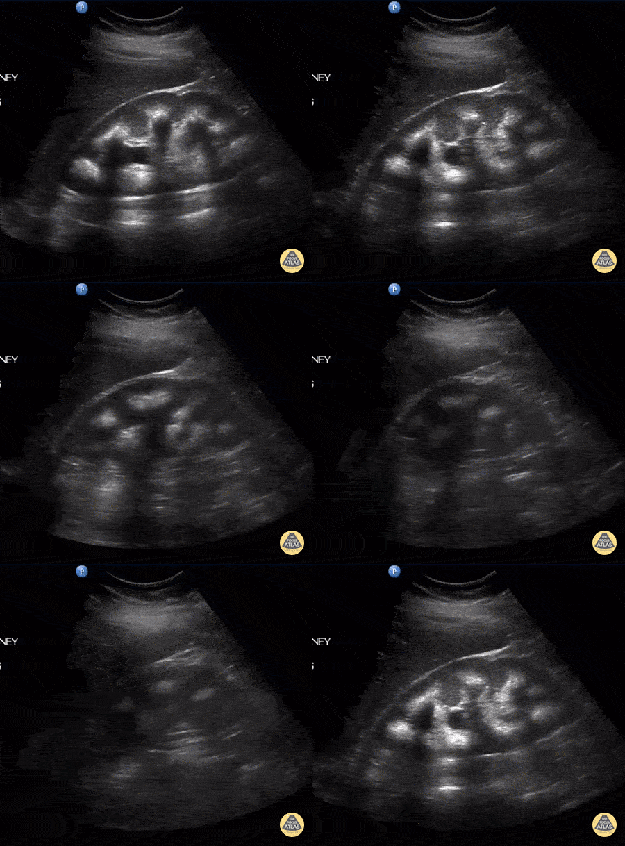
Medullary Nephrocalcinosis with Moderate Hydronephrosis
A female patient presented with right flank pain. A longitudinal view of the right kidney indicates medullary nephrocalcinosis with posterior shadowing and moderate hydronephrosis. Note the posterior shadowing partially masks the hydronephrosis. Further ultrasound found a distal ureteral stone.
Image courtesy of Robert Jones DO, FACEP @RJonesSonoEM
Director, Emergency Ultrasound; MetroHealth Medical Center; Professor, Case Western Reserve Medical School, Cleveland, OH
View his original post here
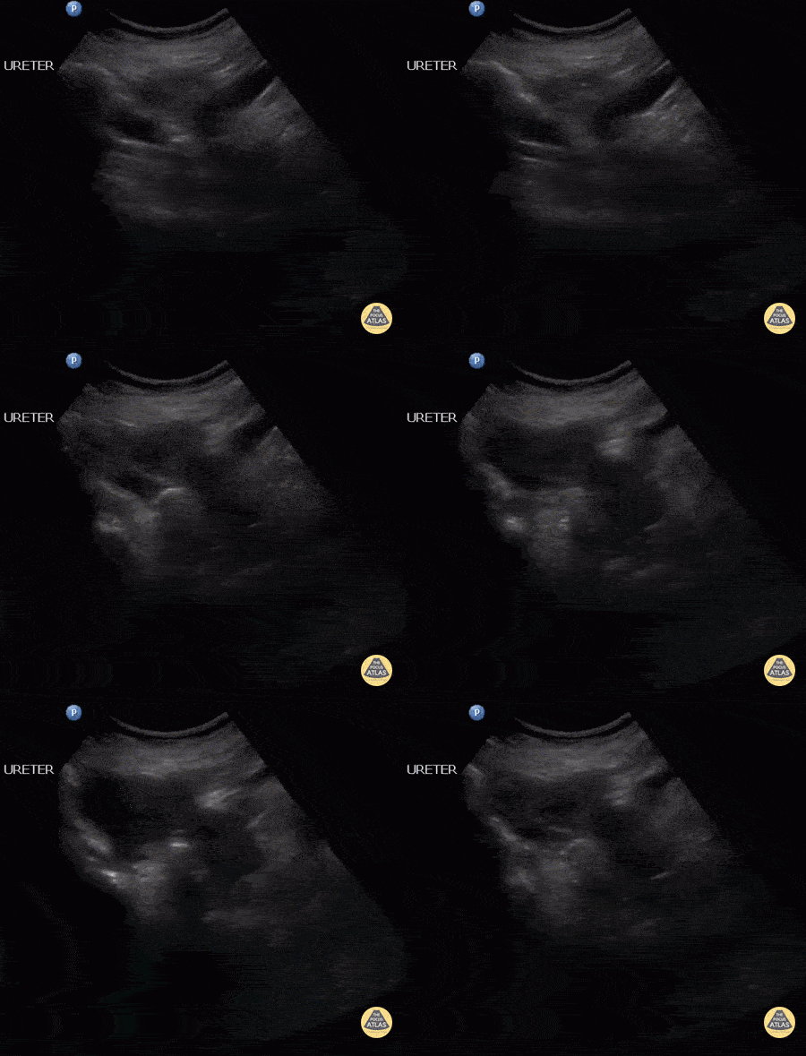
Distal Ureteral Stone
A female patient presented with right flank pain. A longitudinal view of the lower ureter reveals a distal ureteral stone. Further ultrasound of the right kidney depicted medullary nephrocalcinosis.
Image courtesy of Robert Jones DO, FACEP @RJonesSonoEM
Director, Emergency Ultrasound; MetroHealth Medical Center; Professor, Case Western Reserve Medical School, Cleveland, OH
View his original post here

Ureteral Stones at Ureterovesical Junction
Multiple ureteral stones visualized at UVJ with notable ureteral peristalsis.
Image courtesy of Robert Jones DO, FACEP @RJonesSonoEM
Director, Emergency Ultrasound; MetroHealth Medical Center; Professor, Case Western Reserve Medical School, Cleveland, OH
View his original post here

Bear Paw Sign
A 43-year-old female presented to the ED reporting fever and left-sided flank and low back pain. HPI was notable for recurrent urinary tract infections. POCUS performed on the Left Upper Quadrant revealed severe hydronephrosis, with hypdronephrotic collections in the region of the calyces resembling the outline of a bear’s paw (referred to as “bear paw sign”). Subsequent abdominal CT confirmed severe hydronephrosis secondary to stenosis of the ureteroplevic junction (UPJ).
Josiane Almeida

Simple Renal Cyst
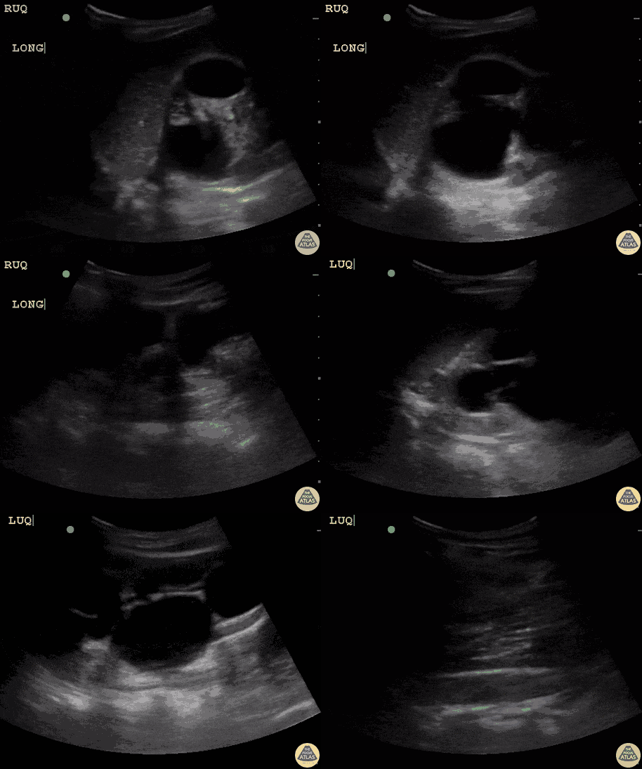
A 84-year-old female presented to the ED in decompensated heart failure. During the POCUS of her RUQ we incidentally identified this simple renal cyst; also note a single calculi within her gallbladder.
Josiane Almeida, Emergency Physician Department of Marilia Clinic Hospital, Sao Paulo- Brazil

Diverticuli of Urinary Bladder
A middle aged male presented with lower abdominal pain and difficulty urinating. He also reported incomplete bladder emptying. POCUS demonstrated multiple bladder diverticuli that were subsequently confirmed on CT pelvis.
Acquired bladder diverticula are often secondary to bladder outlet obstruction that may be related to an enlarged prostate, urethral stricture, or neurologic disease.
Lydia Mansour, DO, PGY1 & Sohaib Mandoorah, MD, PGY3 Central Michigan University Emergency Medicine Residents

Hydronephrosis
POCUS evidence of severe right renal hydronephrosis, as identified in a patient who had an ipsilateral 2.5cm mid-ureteral calculus.
Aaron Inouye, PA-C, North Canyon Medical Center
@PAintheED
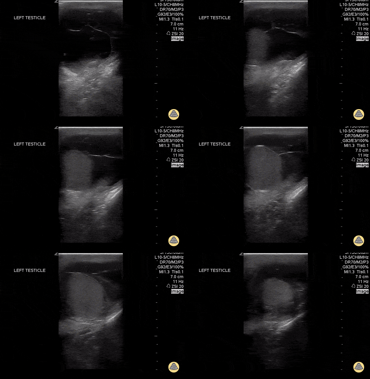
Septated Hydrocele
A 30 year old sexually active male with no previous medical history presented to the emergency department with testicular pain. POC US demonstrates a septated hydrocele.
Mario Corro, MD, PGY-3 Staten Island University Hospital
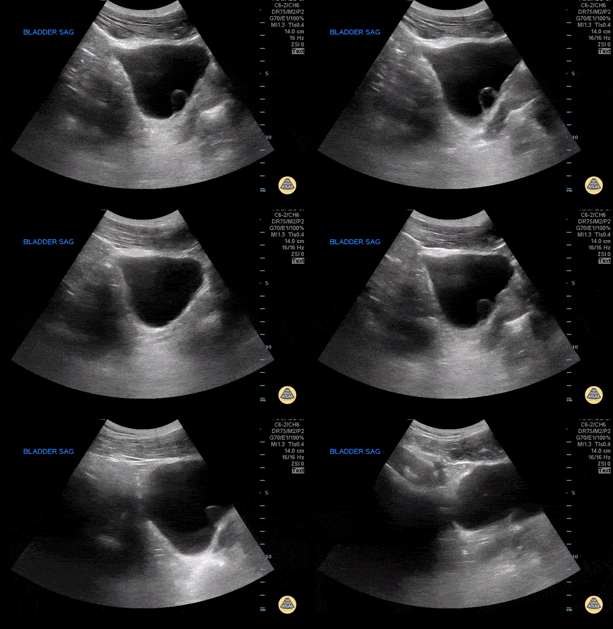
Ureterocele
A 27 year old female with no significant past medical history, presented to the emergency department for dysuria. POCUS demonstrated a ureterocele seen projecting into the bladder.
Mario Corro, MD, PGY-3 Staten Island University Hospital
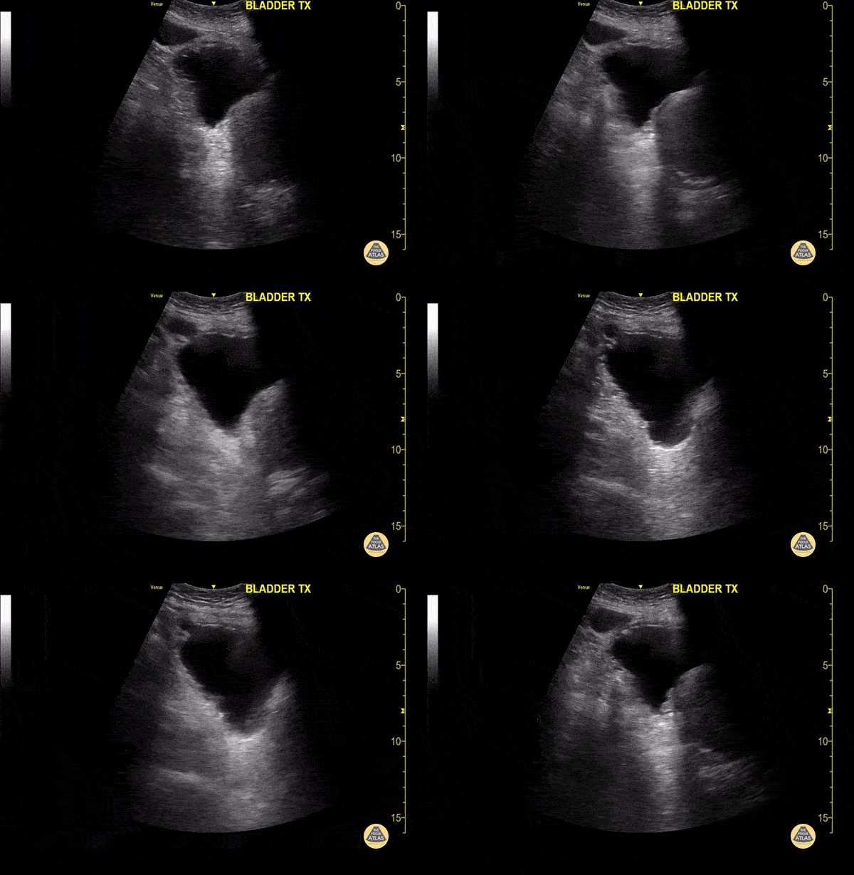
Anterior Bladder Diverticulum
A 60 year old male presented to the emergency department for evaluation of hematuria. POCUS demonstrated a diverticulum extending from the anterior bladder surface.
Mario Corro, MD, PGY-3, Staten Island University Hospital

Epididymitis with Complex Hydrocele
This image demonstrates fluid filling the scrotal sac with multiple thin septations consistent with a complex hydrocele. In the setting of epididymitis, a pyocele should be considered.
Image courtesy of Aventura Ultrasound
See Original Post Via their Twiiter: @AventuraEUS

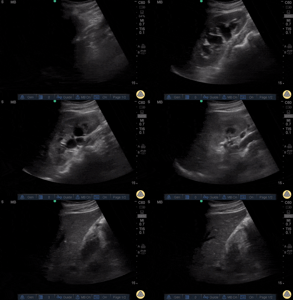
Normal Kidney
Sukh Singh, MD
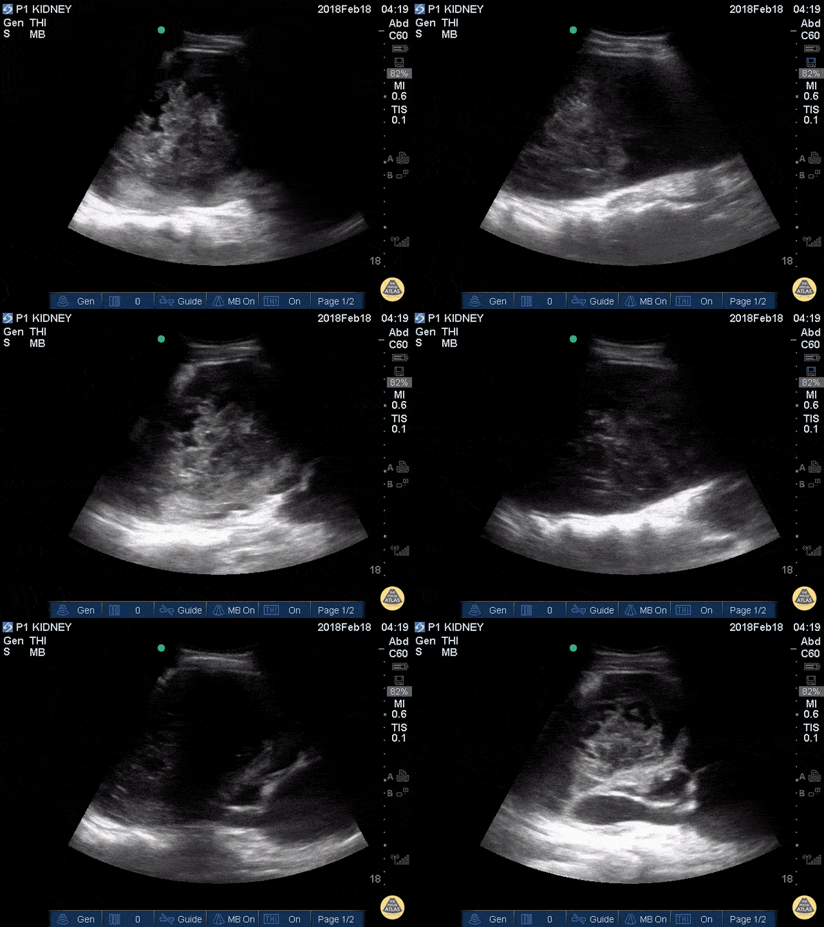
Traumatic Kidney Hematoma
19M w/ hx “fell down stairs 1 week ago,” won’t provide additional details. Admitted to an outside hospital for renal laceration and subcapsular hematoma, here in ED today wondering if hematoma still present, asymptomatic; normal vitals.
Greg Powell, MD
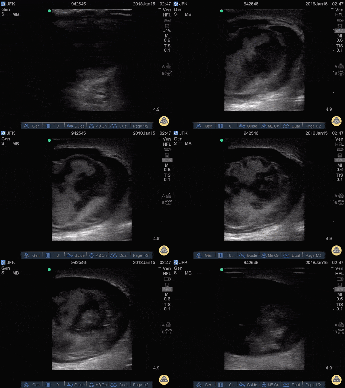
Testicular Rupture
27 y/o M presented wit scrotal pain upon awakening after a night of heavy drinking. He could not recall any trauma. Physical exam revealed a tender, swollen, and ecchymotic scrotum. POCUS demonstrates hematocele, as well as (1) disruption of the tunica albuginea, (2) contour abnormality of the testis, and (3) heterogeneous echotexture of testicular parenchyma. These three findings collectively are highly sensitive and specific for testicular rupture1-3, and warrant urgent surgical exploration.
Elizabeth Hanson, MD - EM resident, Kings County/SUNY Downstate
1. Deurdulian C, Mittelstaedt CA, Chong WK, Fielding JR. US of acute scrotal trauma: optimal technique, imaging findings, and management. RadioGraphics2007; 27: 357–369.
2. BuckleyJC, McAninch JW. Use of ultrasonography for the diagnosis of testicular injuries in blunt scrotal trauma. J Urol2006; 175: 175–178.
3. Micalle FM, Ahmad I, Ramesh N, Hurley M, McInerney D. Ultrasound features of blunt testicular injury. Injury2001; 32: 23–26.

Bladder Diverticulum
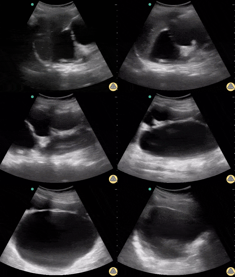
78 yo M h/o BPH s/p TURP 1yr ago presents c/o difficulty voiding x2 wks. US of bladder revealed 2 large fluid filled (anechoic) structures w/ a communicating tract and bidirectional flow on color Doppler. The superficial structure is the urinary bladder while the deep structure is a large bladder diverticulum. Pt didn't have any previously documented hx of a bladder diverticulum.
A bladder diverticulum is a rare congenital or acquired defect consisting of a protrusion of the mucosa through the bladder musculature. The most common acquired cause is bladder outlet obstruction 2/2 to BPH. Pts w/ a new diagnosis in the ED should be referred for urology follow up. These pts are at high risk of UTIs and bladder calculi due to urinary stasis from incomplete emptying of the diverticulum. This can even occur in pts w/ a Foley, as the catheter may not drain the diverticulum. In fact, having a chronic indwelling catheter is a rare cause of bladder diverticula. Pts w/ hematuria, lower urinary tract symptoms, recurrent UTIs, or bladder calculi may require diverticulectomy.
Drs. Justin Berkowitz, Adrian Aurrecoechea, and Catherine Bon
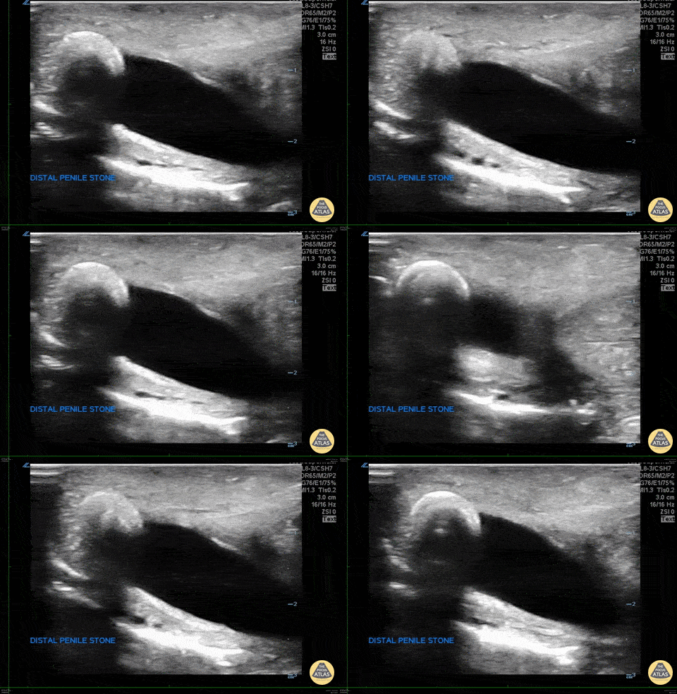
Penile Urethral Calculus
WCUME 2017 Submission and WINNER for "Creative Caption" Category
"Stone in the Sword"
The patient presented with penile pain and blood in urine. POCUS demonstrates a calculus obstructing the distal urethra.
Inna Shniter, MD - UCI Ultrasound Fellow

Perinephric Hematoma
This is a longitudinal view of the right kidney in a patient who presented with sudden, severe right flank pain. There was no history of trauma. No gross hematuria. The patient’s pain was difficult to control with analgesics and bedside ultrasound revealed apparent spontaneous perinephric hematoma.
Therese Mead, DO
Emergency Physician
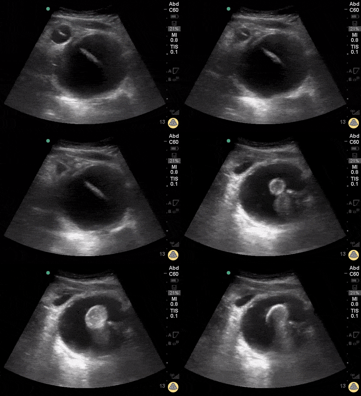
Difficult Foley: Bladder Diverticulum
WCUME 2017 Submission for "Novel Indication"
A urinary catheter is not draining appropriately and bedside ultrasound reveals inflated balloon caught within a bladder diverticulum. Under dynamic ultrasound guidance, the balloon is deflated, the catheter withdrawn into the bladder lumen, and then reinflated in the appropriate position.
Sam Langberg, MD - Ochsner Medical Center, New Orleans, LA
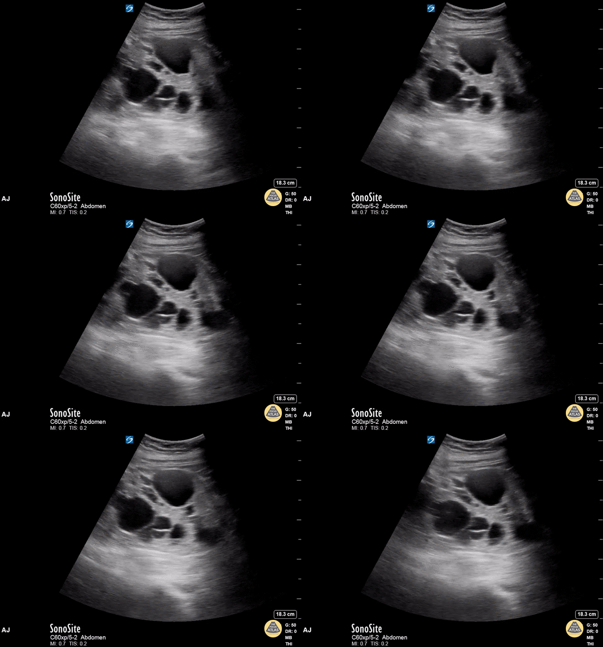
Polycystic Kidney Disease
This middle-aged adult female presented to the emergency department with abdominal pain. The patient reported history of polycystic kidney disease. Bedside renal ultrasound revealed multiple renal cysts in both the cortex and medullary areas of the kidney, consistent with her history. Stones and hydronephrosis would be difficult to detect in the setting of polycystic kidney disease.
Ahmad Jaber, MBBS PGY3
Resident Physician, Central Michigan University Emergency Medicine Residency

Ureterovesical Junction Nephrolithiasis
This is a 58 year old man that presented with first episode of severe LLQ pain and vomiting. The differentials were diverticulitis vs nephro/urolithiasis. POCUS was performed obtaining images of left and right kidneys, bladder and aorta. The image shows a 7mm stone seen with shadowing at the L UVJ.
Maria Perez; Emergency Registrar; St Vincent’s Hospital; Melbourne - Australia
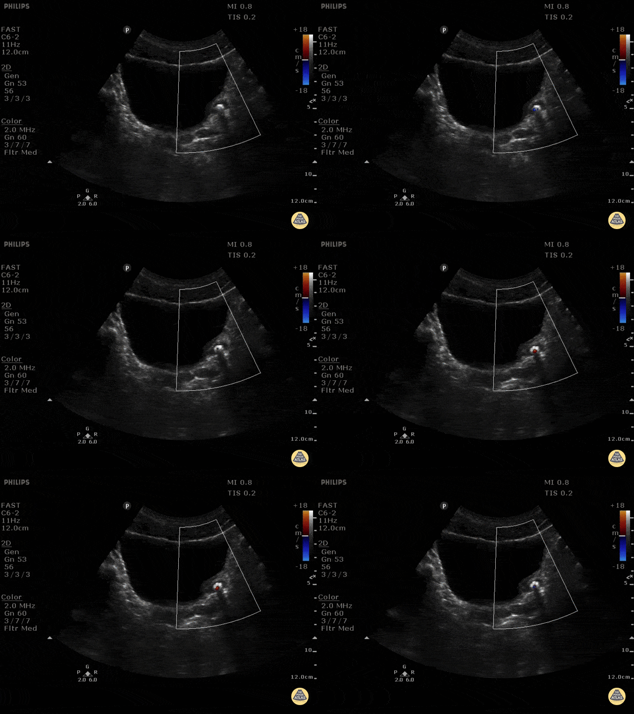
The Twinkle Artifact
Ultrasonographic color doppler twinkling artifact is a phenomenon that may aid in the detection of nephrolithiasis. Twinkling artifacts can be seen on color doppler ultrasounds when applied to stationary, highly echogenic objects, generating a false sense of movement on color doppler. The reason this occurs is unclear. Seen here is a left ureterovesicular junction stone with a positive twinkle artifact.
Maria Perez; Emergency Registrar; St Vincent’s Hospital; Melbourne - Australia

Assessing Hydronephrosis with Color Flow
Normal kidney - No hydro.
One of the common pitfalls in identifying hydronephrosis is not using color flow. You must use color flow doppler to ensure that what you're looking at is actually hydronephrosis, not simply vasculature.
Dr. Bryan Jarret - Kings County Emergency Medicine

Mild Hydronephrosis
Mild - grade two hydronephrosis with dilation of the renal pelvis and dilation of the some calyces. Grade one would not include dilated calyces.
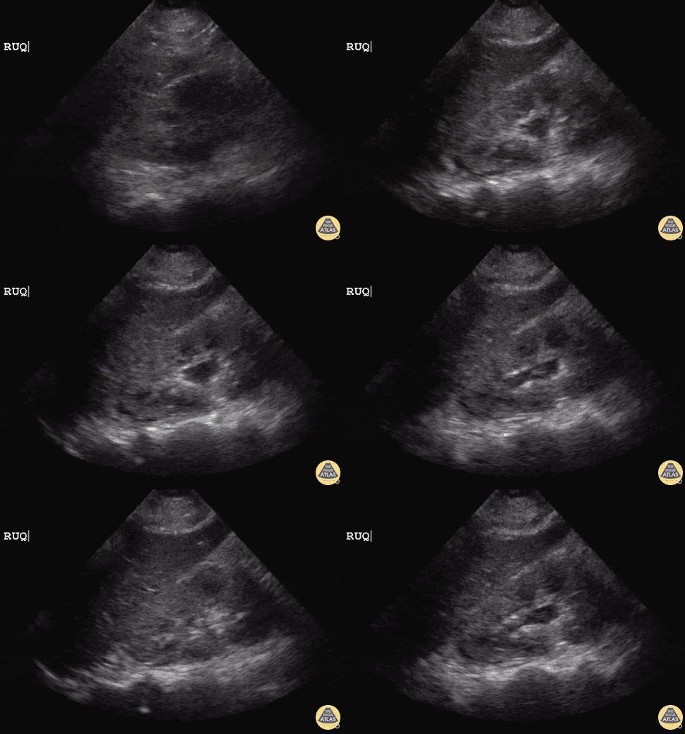
Mild Hydronephrosis
21 y/o female post-op emergency hysterectomy post uterine rupture with rising creatinine in surgical ICU.
POCUS revealed right-sided mild Grade I hydronephrosis with appreciable dilated major calyces and renal pelvis. Initial concern is for obstructive process or ureter injury.
Dr. Sathya Subramaniam, Pediatric EM Fellow - Kings County/SUNY Downstate
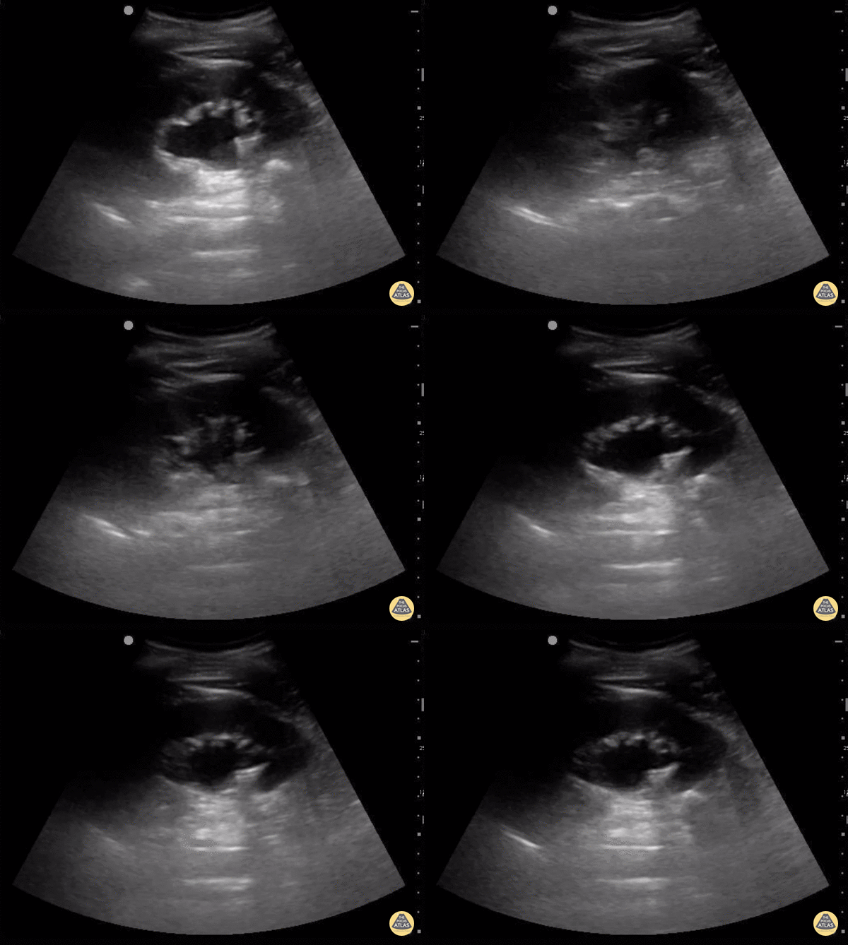
Moderate Hydronephrosis
The degree of hydronephrosis is determined by a grading system. Grade 0 (none) means there's no dilation of the renal pelvis.
Grade 1 (mild) means there's mild dilation of the renal pelvis without any dilation of the calyces. Grade 2 means there's moderate dilation of the renal pelvis that extends to a few calyces.
Grade 3 (moderate) means the renal pelvis dilation extends to all the calyces. Grade 4 (severe) means there's extension of dilation to all the calyces with the addition of thinning of the renal parenchyma.
In this clip, the renal pelvis calyces are dilated but there is no thinning of the renal parenchyma making this mild to moderate, grade 2.
Sukh Singh, MD
Caption by Matthew Riscinti, MD
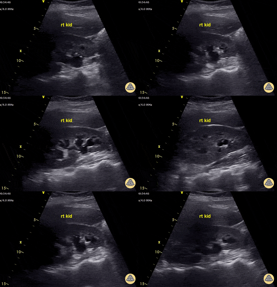
Hydroureter with Moderate Hydronephrosis
This young lady presented with clinical features of pyelonephritis - fever, rigors and right flank pain. Renal US shows moderate hydronephrosis and hydroureter. CT showed a 5.7mm right mid ureteric stone. Nephrostomy tube was placed to decompress obstructive uropathy.
Ultrasound is insensitive for pyelonephritis - most patients have normal scans. POCUS can be used to check for hydronephrosis, renal abscess, pyonephrosis or emphysematous pyelonephritis as these findings will alter management.
Images recorded by Dr. Khaled Taha
Submitted by Dr Cian McDermott
Mater University Hospital, Dublin, Ireland
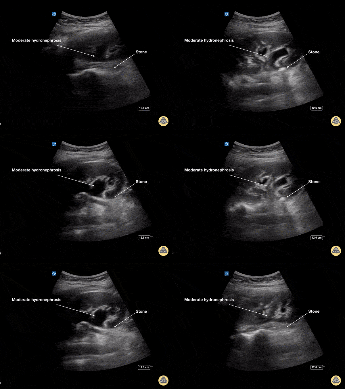
Moderate Hydronephrosis
This young female presented with colicky left flank pain worsening over the previous 24 hours. POCUS showed moderate hydronephrosis with rounding of the calyces of the left kidney collecting system. The stone is seen at the ureteropelvic junction (UPJ) as a hyperechoic structure with posterior acoustic shadowing measuring 7mm on CT. At ureteroscopy, the stone was retrieved, fragmented and a double J stent was placed
Dr Cian McDermott, Mater University Hospital, Dublin, Ireland
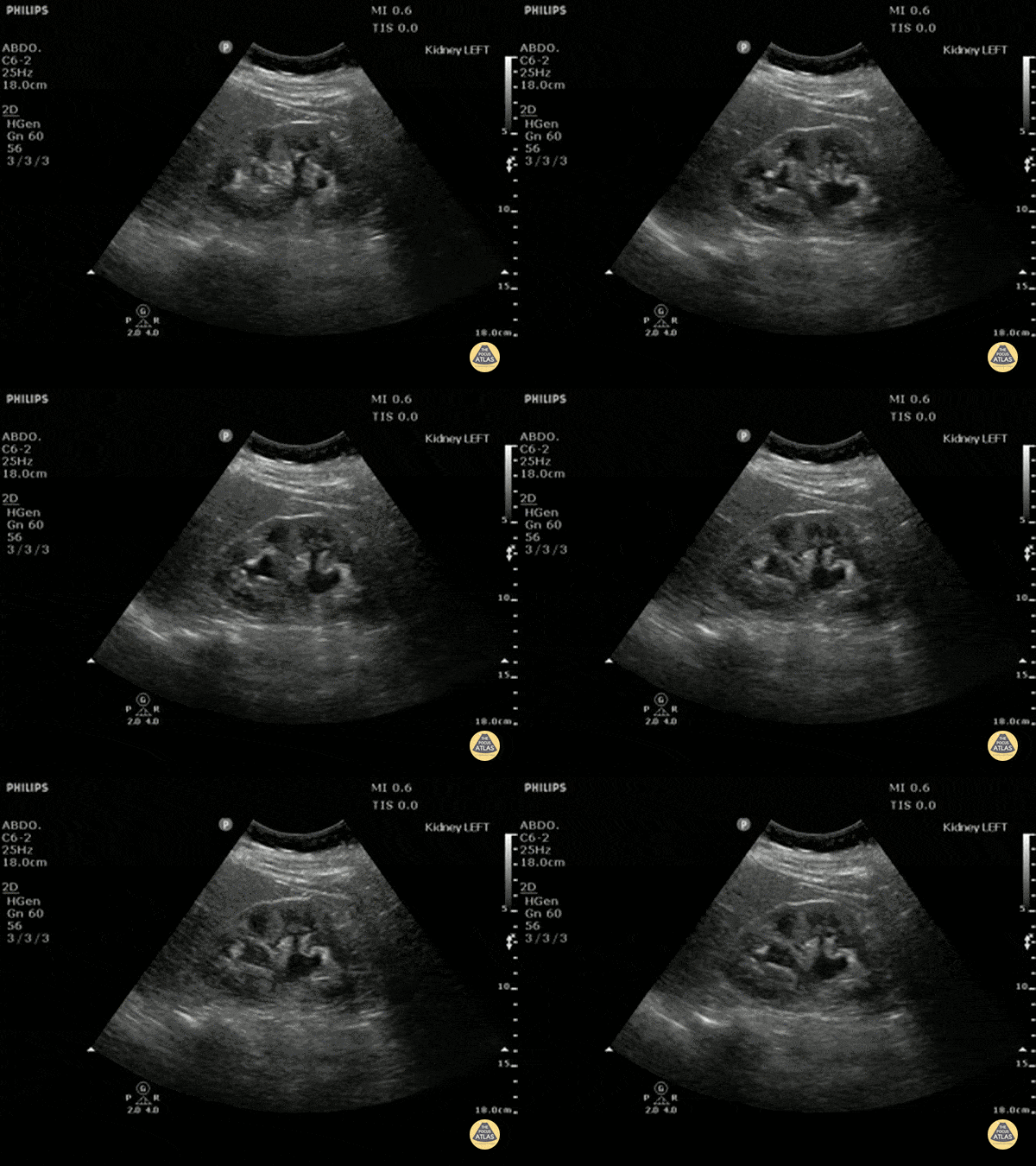
Moderate Hydronephrosis
Moderate (grade three) hydronephrosis can be appreciated here with dilation of both the renal pelvis and calcyces. The renal cortex is also thinned. There is not gross atrophy.
Dr. Justin Bowra et al. (Dr. Yogi)
Moderate Hydronephrosis
66 yo M with hx of congenital single kidney and prostate cancer presents with suprapubic discomfort x 1 week. Found to be in urinary retention.
POCUS allows grading of hydronephrosis based off of the severity of the dilation of the renal pelvis and calyces. Here we see dilated pelvis, ballooning calyces, and cortical thinning. This represents Grade 3, moderate hydronephrosis. Grade 4, severe, would demonstrate further atrophy and loss of borders, occurs with severe hydronephrosis.
Rushabh Shah, MD, MBA and Maria-Pamela Janairo, MD - Kings County/SUNY Downstate Emergency Medicine
Severe Hydronephrosis with Massive Hydroureter
Patient with urinary obstruction with severe hydronephrosis and absolutely massive hydroureter (CT confirmed).
Dr. Stephen Alerhand - US Fellow - Mt Sinai Hospital, NYC

Severe Hydronephrosis
In this patient with severe hydronephrosis, there is gross dilation of both the renal pelvis and calyces, which are ballooned. The cortex of the kidney has atrophied and is very thin. This is severe, or grade 4.
Dr. Justin Bowra et al. (Dr. Browne and Dr. Knights)
Severe Hydronephrosis in Prune Belly Syndrome
10 y/o with Prune Belly Syndrome presenting with suprapubic pain.
Bilateral severe grade IV hydronephrosis. Bear claw appearance of left kidney.
Prune Belly Syndrome is a rare disorder known for lack of abdominal muscles, cryptorchidism, and urinary tract malformations.
Dr. Sathya Subramaniam, Pediatric EM Fellow - Kings County/SUNY Downstate
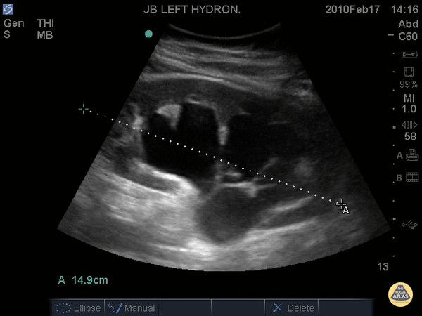
Severe Hydronephrosis
14.9cm long axis
In this patient with severe hydronephrosis, there is gross dilation of both the renal pelvis and calyces, which are ballooned. The cortex of the kidney has atrophied and is very thin.
Dr. Justin Bowra et al.

Left Renal Cyst
This simple renal cyst can be identified as an anechoic structure with well-defined, thin walls. Sometimes septations can be seen. Large cysts might even demonstrate posterior acoustic shadowing.
Dr. Justin Bowra et al.

Bladder Contracted with Indwelling Catheter
An echogenic foley catheter with the balloon inflated can be seen floating inside the anechoic urine within the bladder.
Dr. Justin Bowra et al.

Ureteric Jet
When an obstructing stone is suspected, measurement of the ureteric jets can be performed to see if the ureters are draining into the bladder. In this study, there is no left ureteric jet demonstrating obstruction of the right ureter, and likely an obstructing stone. Renal US is likely to demonstrate hydronephrosis of the right kidney.
Dr. Justin Bowra et al.

Ureterovesical Junction Stone Twinkle Artifact
Twinkling artifacts can be seen on color doppler ultrasounds when applied to stationary, highly echogenic objects, generating a false sense of movement on color doppler. The reason this occurs is unclear.
However, this is useful for kidney stones especially when they are in other echogenic environments such as the ureterovesicular junction. This scan demonstrates the twinkle artifact at the UVJ, thus a kidney stone that may have been missed otherwise.
Dr. Justin Bowra et al. (Dr. Sutijono)

Hydroureter with Stone
A dilated ureter is visualized posterior to the uterus in this transabdominal POCUS, indicating obstruction. The stone can be directly visualized as a hyperechoic structure in the ureter with shadowing.
Sukh Singh, MD

Large Prostate
In this transverse, transabdominal ultrasound one can see a full bladder and posterior to that a uniformly enlarged prostate suggesting urinary retention secondary to BPH.
Sukh Singh, MD

Renal Cysts
Renal cysts can be classified using the Bosniak Classification System based on CT imaging. A category I benign cyst is thin-walled without septations, calcifications, solidifications, nor contrast enhancement. A category II benign cyst is also thin-walled but may contain a few thin septa or calcifications. A category IIF cyst has a small risk of malignancy. It may have a slightly thicker wall, septa or calcifications, but no contrast enhancement. A category III cyst has a significant risk of malignancy and has irregular and thick septa which exhibit contrast enhancement. A category IV cyst (highest risk) has category 3 characteristics with the addition of contrast enhancing soft tissue components.
This clip shows septation of one of the cysts suggesting that it is AT LEAST a category 2 cyst.
Sukh Singh, MD
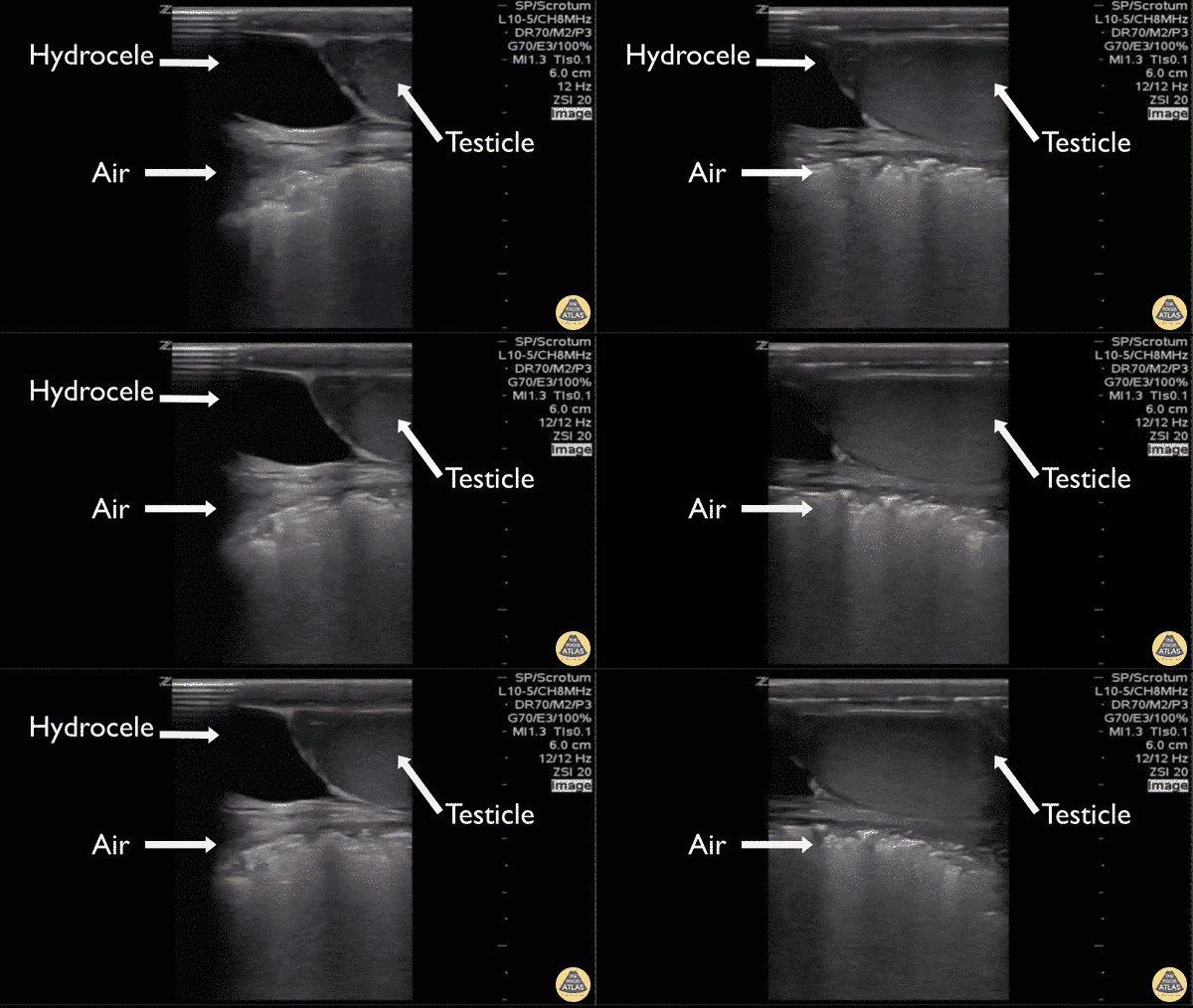
Fournier Gangrene
A patient presented for worsening, severe scrotal pain. Point-of-care ultrasound demonstrated a normal appearing testicle with an associated hydrocele. Significant ring down artifact is visualized posterior producing a “dirty shadow”. The patient was taken to the operating room where the ring down artifact was confirmed as significant subcutaneous air associated with a necrotizing infection.
By: Michael Schick DO, Emergency Physician
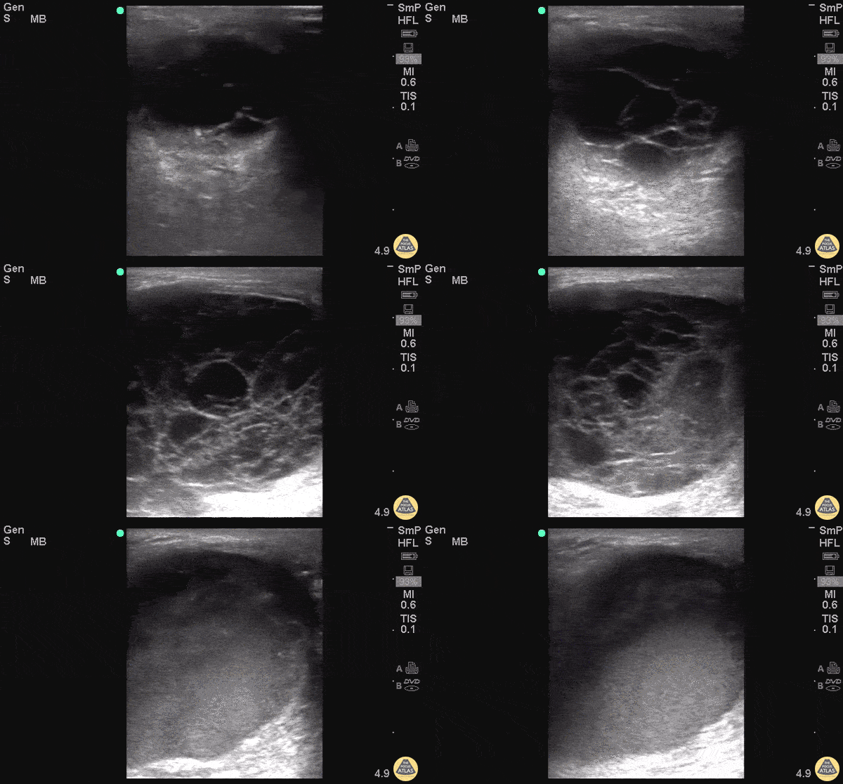
Pyocele
83 y/o M with R testicular swelling, tenderness, concern for epididymitis vs orchitis. POCUS with septated fluid collection concerning for pyocele.
Pycoeles are a urologic emergency that can lead to Fournier's and often require orchiectomy.
Dr. Tess Sexton - Kings County/SUNY Downstate EM
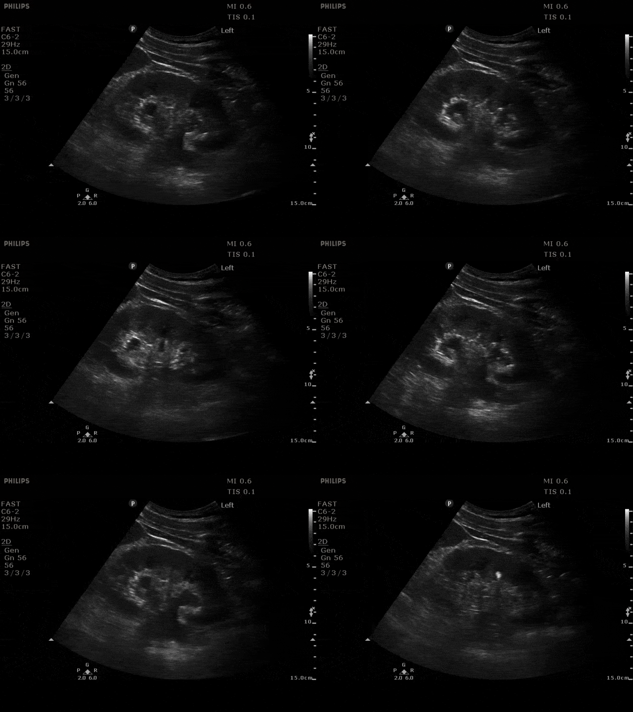
Mild Hydronephrosis
Mild - grade two hydronephrosis with dilation of the renal pelvis and dilation of the calyces. Grade one would not include dilated calyces.
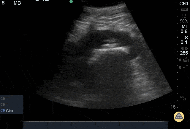
Staghorn Calculi
51yoM with history of staghorn calculi presenting with hematuria and left lower quadrant pain x 3 days. Ultrasound of left kidney in sagittal plane using a phased array 5-1MHz probe demonstrating a large hyperechoic structure in the renal pelvis with associated posterior acoustic shadowing consistent with staghorn calculi. Ultrasound serves as a low-cost, readily available method without radiation which can aid in the detection of renal or ureteral calculi. (Nicolau 2015)
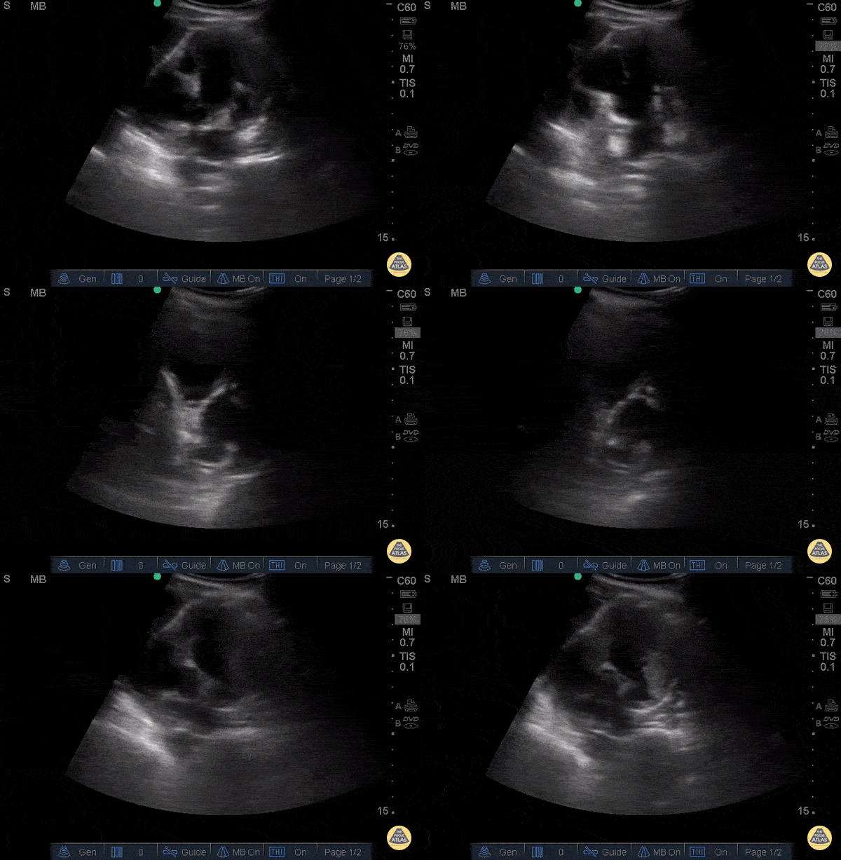
Severe Hydronephrosis - Congenital
16 yo F who is known to have non-neurogenic neurogenic bladder, HTN. Presented to ED with severe bilateral flank pain for 2 weeks, worsening, dull in nature, associated with decreased urination, nausea, vomiting multiple times of food contents. Exam showed severe diffuse abdominal tenderness with bilateral CVA tenderness with light touch. POCUS showed bilateral severe hydronephrosis (L>R) without bladder enlargement. Labs showed elevated creatinine from 1 into 3. Patient then transferred to outside hospital for Pediatric Urology.
Dr. Maan Al Dubayan - Kings County Emergency Medicine
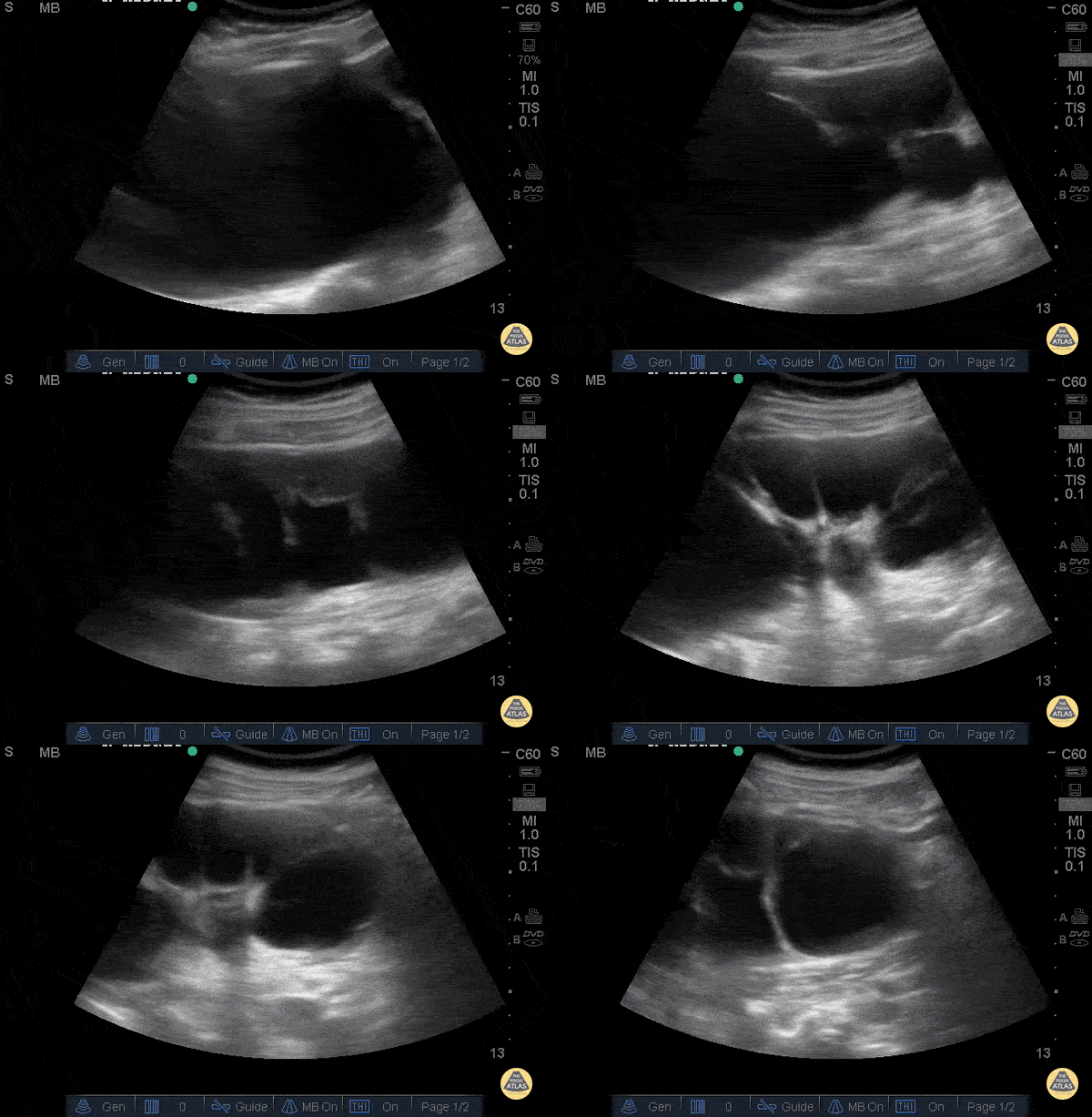
Severe Hydronephrosis
An 88 year old female with history of renal stones presented with flank pain and a nonfunctioning nephrostomy tube. Bedside US showed severe hydronephrosis. This is demonstrated by dilation of renal pelvis and ballooning of renal calyces. One calyx measured approximately 15cm. It was later confirmed via CT that the nephrostomy tube had been dislodged and coiled in the abdominal muscles.
Dr. Steven Greenstein, Dr. Maan Al Dubayan, Dr. Andrew Aherne - Kings County Emergency Medicine
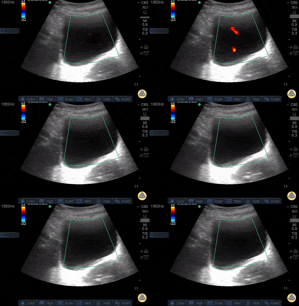
Decreased Right Ureteral Jet
An 88 year old female with history of renal stones presented with flank pain and a nonfunctioning nephrostomy tube. Bedside US showed severe hydronephrosis (see other image). Using color Doppler over the bladder, the ureteral jets were evaluated. The diagnosis of obstruction was made, due to the absence of the right jet.
Dr. Steven Greenstein, Dr. Maan Al Dubayan, Dr. Andrew Aherne - Kings County Emergency Medicine
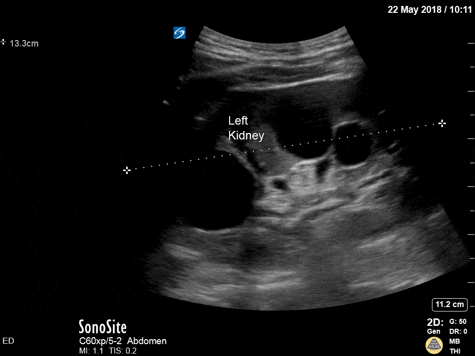
Polycystic Kidney
Hydronephrosis, right? Wrong! These are polycystic kidneys! Differentiate the two by looking for communication with the collecting system.
Dr. Michael Trauer
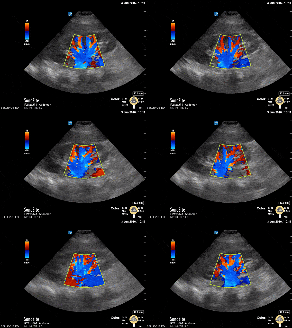
Kidney Doppler
This clip shows a kidney with color doppler overlay. Using color doppler is helpful in distinguishing hydronephrosis from prominent renal vasculature which can look similar in 2D mode. In this case, the color flow suggests that what may appear to be a dilated renal pelvis is likely just plump blood vessels.
Hannah Kopinksi and Dr. Lindsay Davis - NYU Emergency Medicine
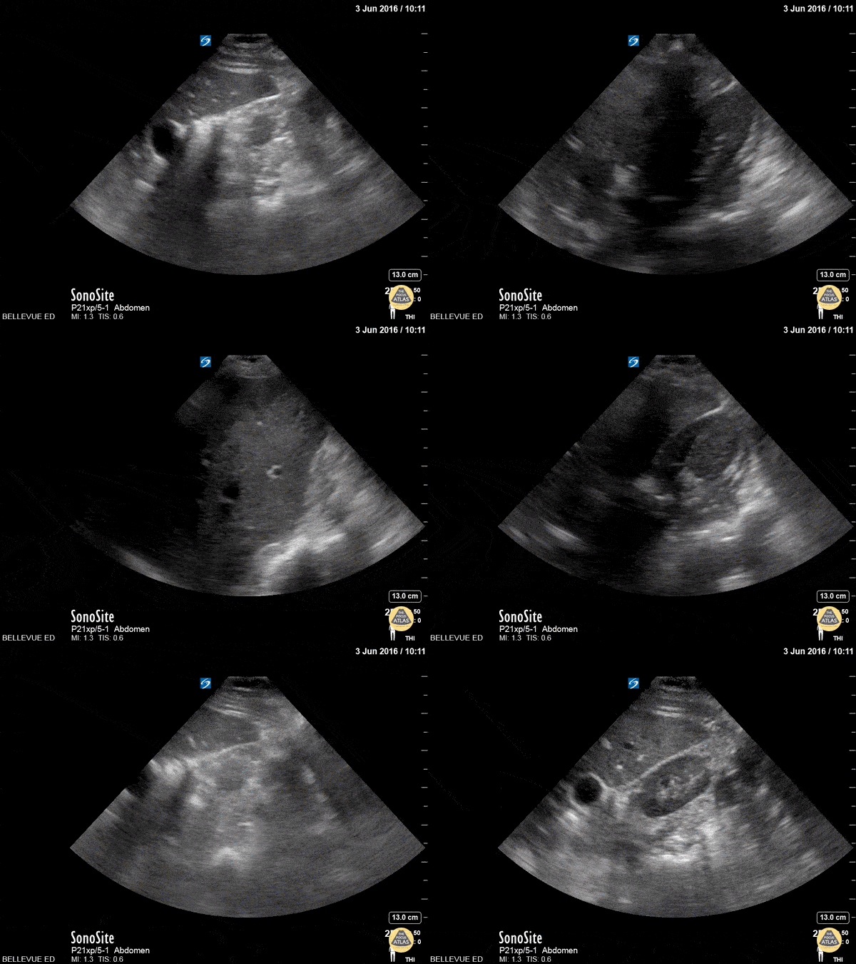
Kidney Transverse
This is a clip of the right upper quadrant structures in transverse view. The kidney has a hyperechoic center made up of the renal pelvis and calyces, surrounded by a hypoechoic cortex similar in echogenicity to the liver (seen to the left of the screen). Within the liver we see prominent anechoic vasculature. A dark rib shadow moves across the field as the sonographer fans.
Hannah Kopinksi and Dr. Lindsay Davis - NYU Emergency Medicine
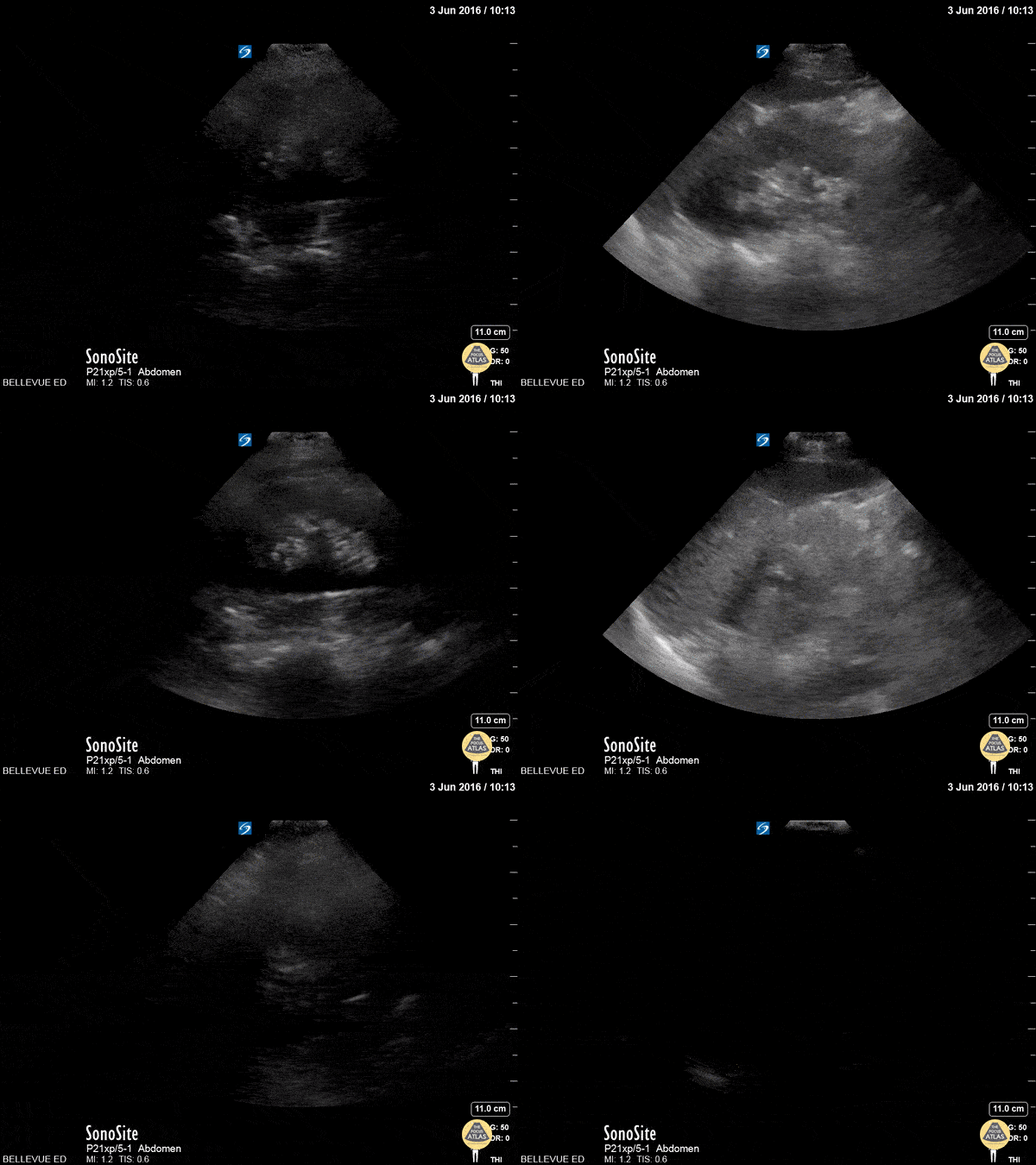
LUQ Kidney Long Axis
This clip fans through the left kidney in long axis. The hyperechoic central area is the renal pelvis and calyces, the darker hypoechoic area surrounding it is the renal cortex. The renal pyramids are visible as anechoic triangles within the medulla. Deep to the kidney we see the hyperechoic spine;on the left of the screen the spleen comes in and out of view.
Hannah Kopinksi and Dr. Lindsay Davis - NYU Emergency Medicine
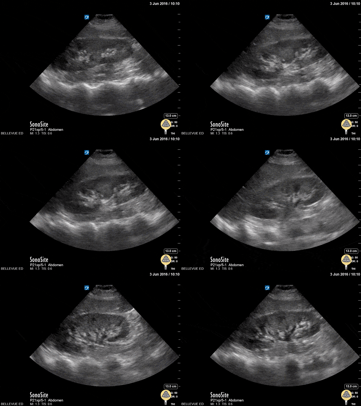
RUQ Kidney Long Axis
This clip fans through the right kidney in long axis. The hyperechoic central area is the renal pelvis and calyces, the darker hypoechoic area surrounding it is the renal cortex. The darker/anechoic spots between the cortex and the medulla are renal pyramids. Deep to the kidney we see the hyperechoic spine. On the left of the screen and superior to the kidney is the liver which has a similar echogenicity to the renal cortex.
Hannah Kopinksi and Dr. Lindsay Davis - NYU Emergency Medicine
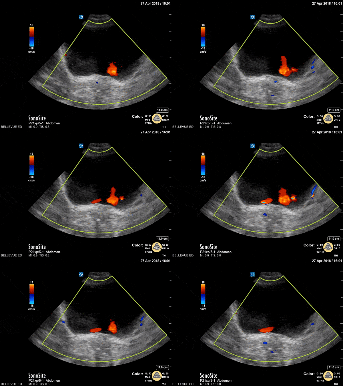
UVJ Jets
This is a clip of the bladder in transverse view with visible ureteral jets. We can use color doppler to help visualize the jets of urine flowing out of the ureterovesical junctions into the bladder. In this case we see the red UVJ jets bilaterally from both ureters, ruling out obstruction proximal to the UVJ.
Hannah Kopinksi and Dr. Lindsay Davis - NYU Emergency Medicine
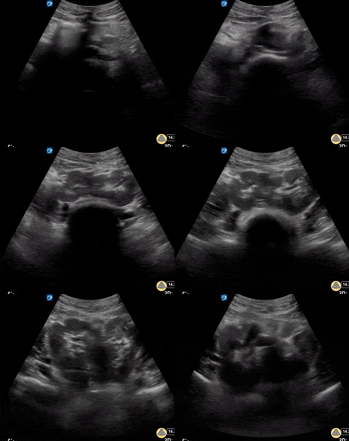
Horseshoe Kidney
Incidentally found horseshoe kidneys in pelvis joined at the isthmus.
Dr. Coneybeare

Bladder Diverticulum
An elderly male presented to the ED following a MVA. Imaging revealed a pelvic fracture. During a FAST exam, the transverse pelvic view revealed a bladder diverticulum.
Image courtesy of Robert Jones DO, FACEP @RJonesSonoEM
Director, Emergency Ultrasound; MetroHealth Medical Center; Professor, Case Western Reserve Medical School, Cleveland, OH
View his original post here

Moderate Hydronephrosis
30s M with no past medical history presented with acute onset right sided flank pain. POCUS demonstrated moderate hydronephrosis of the right kidney with evidence of hydroureter as well. Moderate hydronephrosis is seen here with distension of the renal pelvis as well as distension of most of the renal calyces, with intact renal cortical thickness. This patient had his symptoms controlled and was able to be discharged.
Dr. Mark Serpico, PGY3
Denver Health Residency in Emergency Medicine

Severe Hydronephrosis
30s M PMH HIV, latent TB p/w acute onset flank pain with dysuria. He was found to be febrile and tachycardic. Initial workup was consistent with sepsis due to pyelonephritis. Renal POCUS is shown here, demonstrating severe hydronephrosis, with distortion of the calyceal collecting system as well as thinning of the renal cortex. CT imaging of the abdomen/pelvis demonstrated ureteropelvic junction stenosis causing significant hydronephrosis. Urology was consulted and the patient was admitted for treatment of pyelonephritis as well as further workup of the renal abnormalities.
Dr. Nimish Bhatt, US Fellow
Denver Health Emergency Ultrasound Fellowship
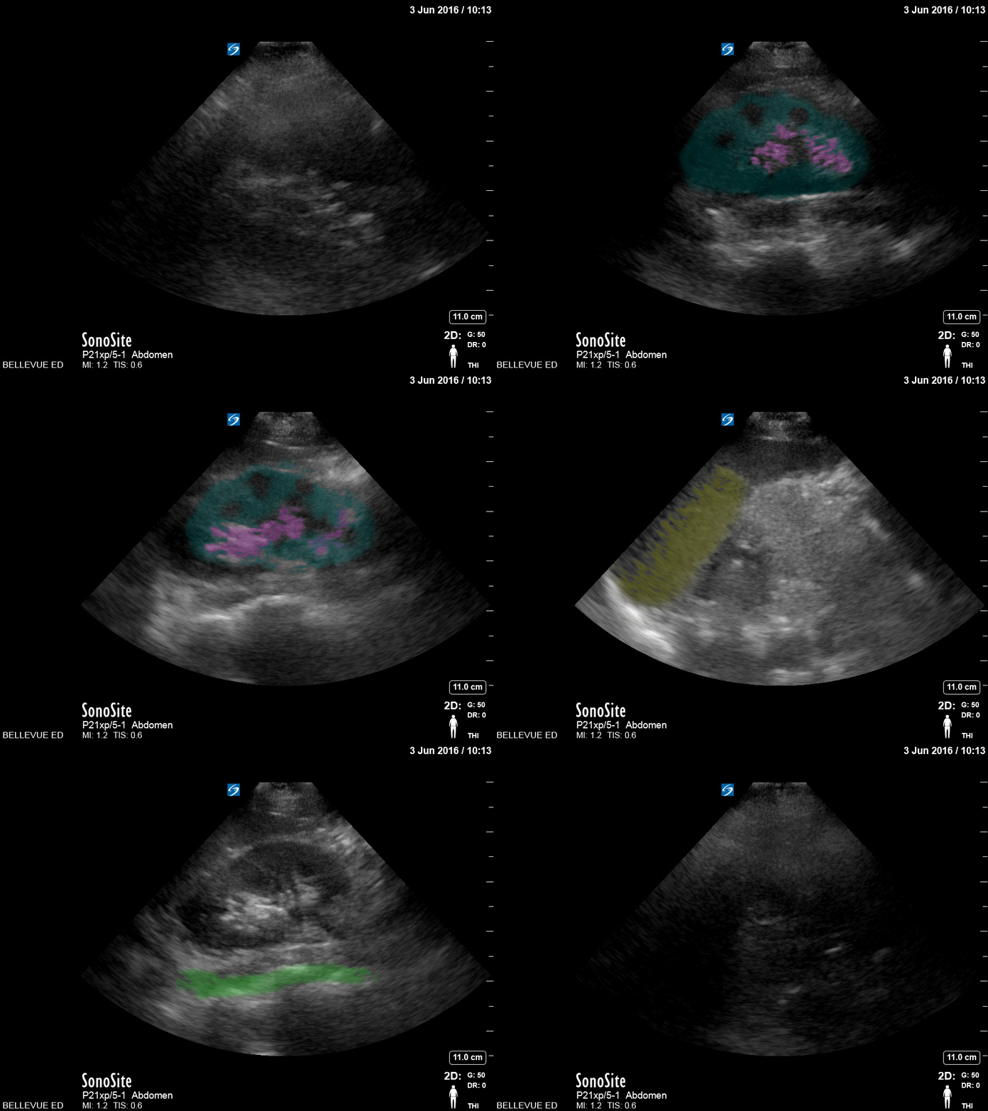
LUQ Kidney - Colorized
Left Kidney
Blue: Cortex, Pink: Medulla, Yellow: Spleen, Green: Spine

Subcapsular Renal Hematoma
This image demonstrates a subcapsular hematoma of the left kidney (left image) with intraperitoneal hemorrhage (right image).
Image courtesy of Cody McIlvain, MD.
Resident, Emergency Medicine; Denver Health Residency in Emergency Medicine, Denver, Colorado.
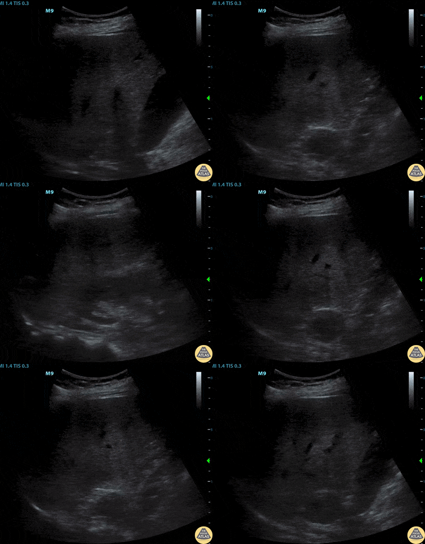
Pheochromocytoma
This image demonstrates an adrenal mass on the right kidney which was a known pheochromocytoma.
Image courtesy of Cody McIlvain, MD.
Resident, Emergency Medicine; Denver Health Residency in Emergency Medicine, Denver, Colorado.
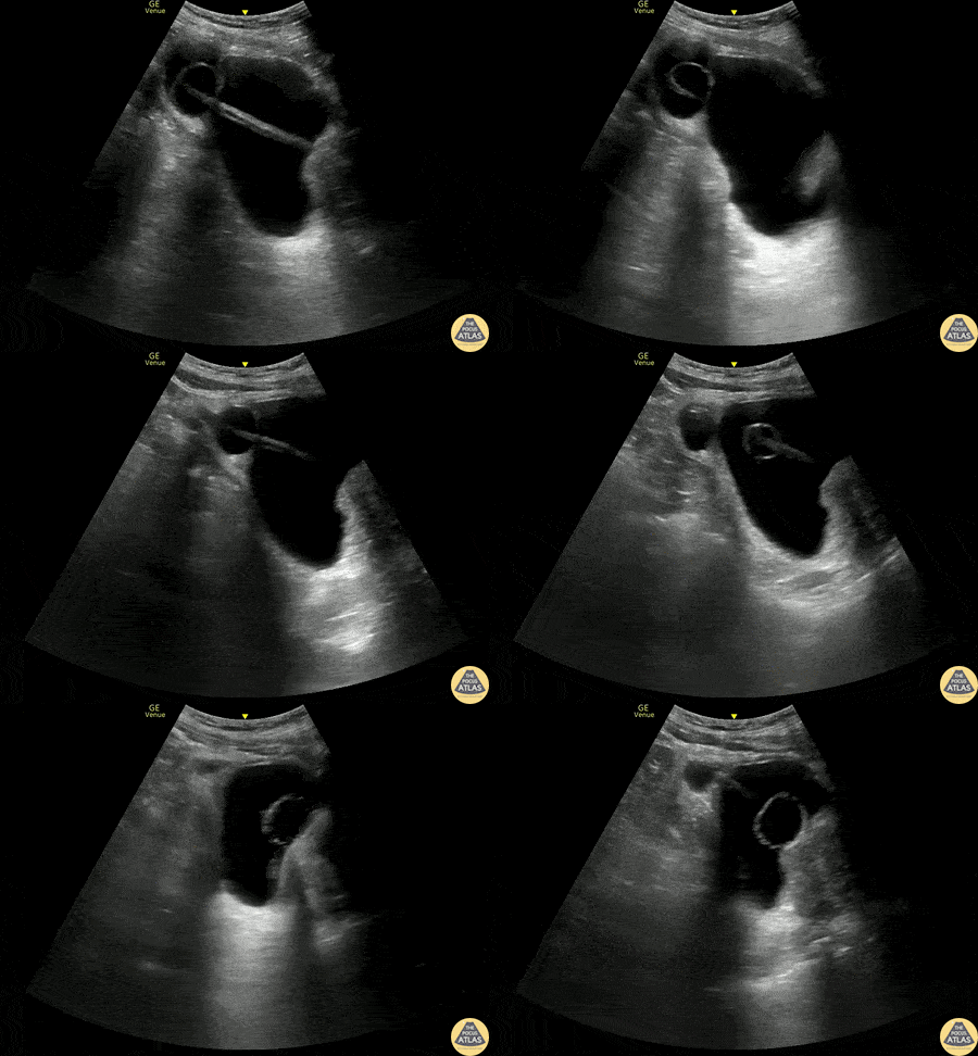
Bladder Diverticulum
60s M with past medical history of BPH with chronic indwelling foley catheter was referred to the ED after his foley catheter was noted to be not draining properly after replacement in urology clinic. POCUS demonstrated the foley balloon and distal catheter in a large bladder diverticulum, with resulting urinary retention. Under real time guidance, the balloon was deflated, retracted, and re-inflated in the bladder, and the bladder diverticulum and bladder both decompressed appropriately.
Molly Thiessen MD, Attending Physician, Denver Health Residency in Emergency Medicine
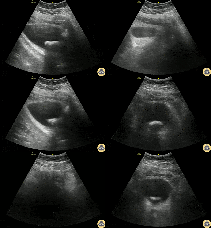
Bladder Stone
70s M with past medical history of BPH and multiple recent UTIs presented as a referral to the ED after an outpatient CT KUB showed a large bladder stone. POCUS was performed and demonstrates a >3cm bladder stone present which obstructs urinary outflow. The patient was admitted and taken for surgery the next day.
Phillip Breslow MD, Resident, Denver Health Residency in Emergency Medicine
Mike Heffler MD, Ultrasound Fellow, Denver Health Ultrasound Fellowship
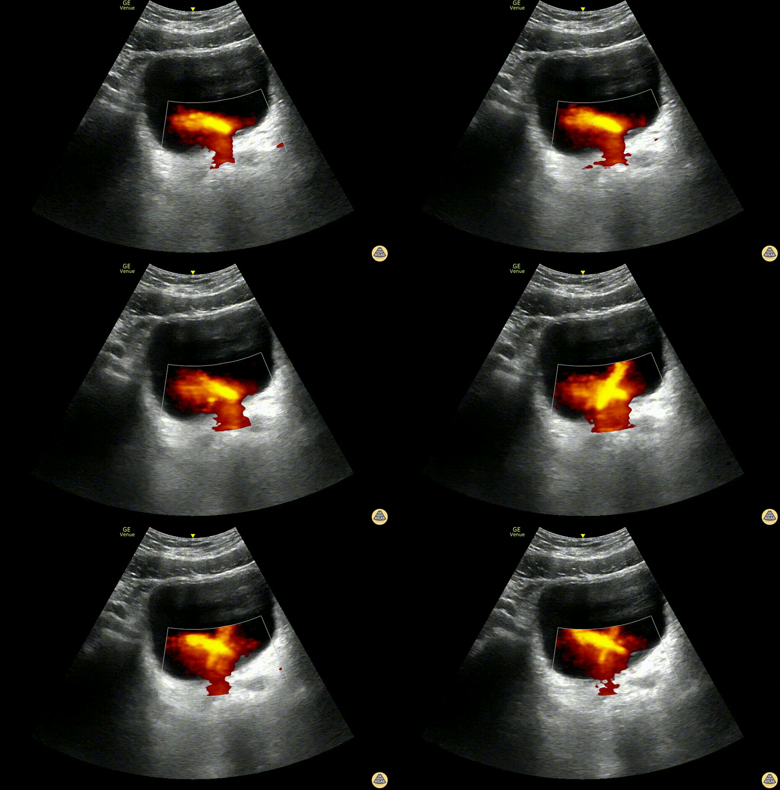
"Light Saber Sign” Ureteral Jets
A 48-year old male presented to the ED with lower abdominal pain and flank pain presented to the ED. A renal and bladder point-of-care ultrasound was done to evaluate for hydronephrosis which revealed distinct bilateral ureteral jets, which we dubbed the “light saber sign.”
Dr. Jannie Bolotnikov, PGY-1, Denver Health Emergency Medicine Residency
Dr. Fred Milgrim, Ultrasound Fellow, Denver Health Emergency Medicine


