Musculoskeletal

Fusobacterium Septic Joint
This patient presented to the emergency department for several days of ankle pain. Ultrasound of the joint revealed anechoic fluid within the joint, which normally suggests a non-exudative cause. However, this patient’s joint was eventually tapped which revealed thick purulent fluid that grew Fusobacterium, and a rare cause of septic joint.
Image courtesy of Robert Jones DO, FACEP @RJonesSonoEM
Director, Emergency Ultrasound; MetroHealth Medical Center; Professor, Case Western Reserve Medical School, Cleveland, OH
View his original post here

Monosodium urate crystals
Patient presented with great toe pain. POCUS performed and detected effusion in first metatarsophalangeal joint. Eventually determined to be monosodium urate crystals as seen in gout.
Image courtesy of Robert Jones DO, FACEP @RJonesSonoEM
Director, Emergency Ultrasound; MetroHealth Medical Center; Professor, Case Western Reserve Medical School, Cleveland, OH
View his original post here

Complex Left Ankle Effusion
This is an image of a left ankle demonstrating a complex effusion in a patient presenting with ankle pain and swelling. Arthrocentesis was performed confirming septic arthritis.
Michael Macias

Intra-Articular Air
This is an ultrasound clip from a patient who presented to the ED after sustaining a laceration near the knee. There was concern for violation of the knee joint so ultrasound was used to evaluate.
The probe is held in an sagittal orientation, just proximal to the patella, overlying the quadriceps tendon. A hyperechoic line with shadowing (similar in appearance to an A-line seen on lung US) can be seen deep to the quadriceps tendon confirming intra-articular air and therefore violation of the knee joint from the laceration. A small joint effusion can also be appreciated.
Clip courtesy of Dr. Daniel Mantuani and Highland Ultrasound
Twitter: @HGHED

US-Guided MTP Arthrocentesis
Patient with no history of crystallopathy presented with first metatarsophalangeal pain and swelling.
Longitudinal view of the MTP joint revealed an anechoic effusion containing echogenic debris. Dynamic US guided aspiration was performed using an in-plane approach. Synovial fluid analysis showed monosodium urate crystals. Gram stain and fluid culture were negative.
Learning point: Consider US guidance for arthrocentesis to increase success rate and maximize fluid yield especially in the case of a small effusion.
Michael Cover, MD
@michaelc0ver

Arthrocentesis and Joint Injection
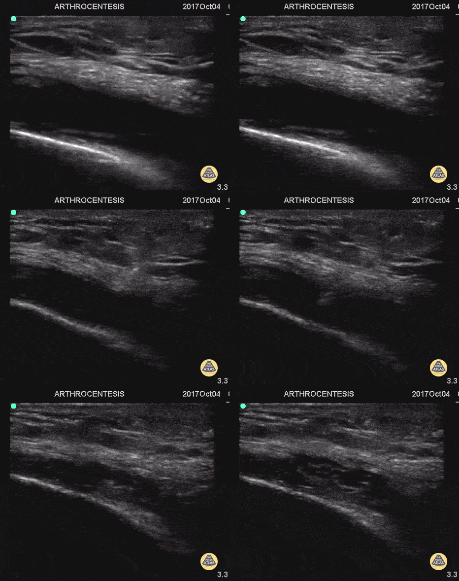
55 y/o with history of gout and osteoarthritis with an effusion. Join tapped and triamcinalone injected at the end.
Going to tap a joint and unsure of the best spot? Grab POCUS to find the biggest fluid pocket. Many prefer the in-place US guidance technique for big targets such as a joint and watching the needle enter the whole way.
Matthew Riscinti, MD - Kings County Emergency Medicine

Elbow Effusion (Traumatic)
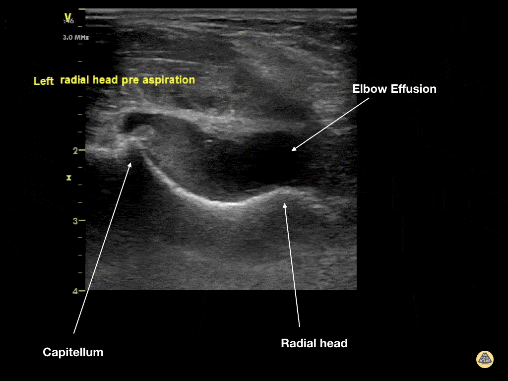
Aspiration of traumatic elbow effusions may be considered in the management of radial head fracture. Slide a linear transducer along the forearm towards the elbow until the radial head, effusion and capitellum are seen. Using an out of plane approach, insert a needle into the effusion. The syringe will fill itself under intrinsic pressure. Relief is often instantaneous and prolonged and the range of motion of the elbow will increase dramatically
Dr Cian McDermott, Emergency Physician, Mater University Hospital, Dublin, Ireland

Hip Effusion
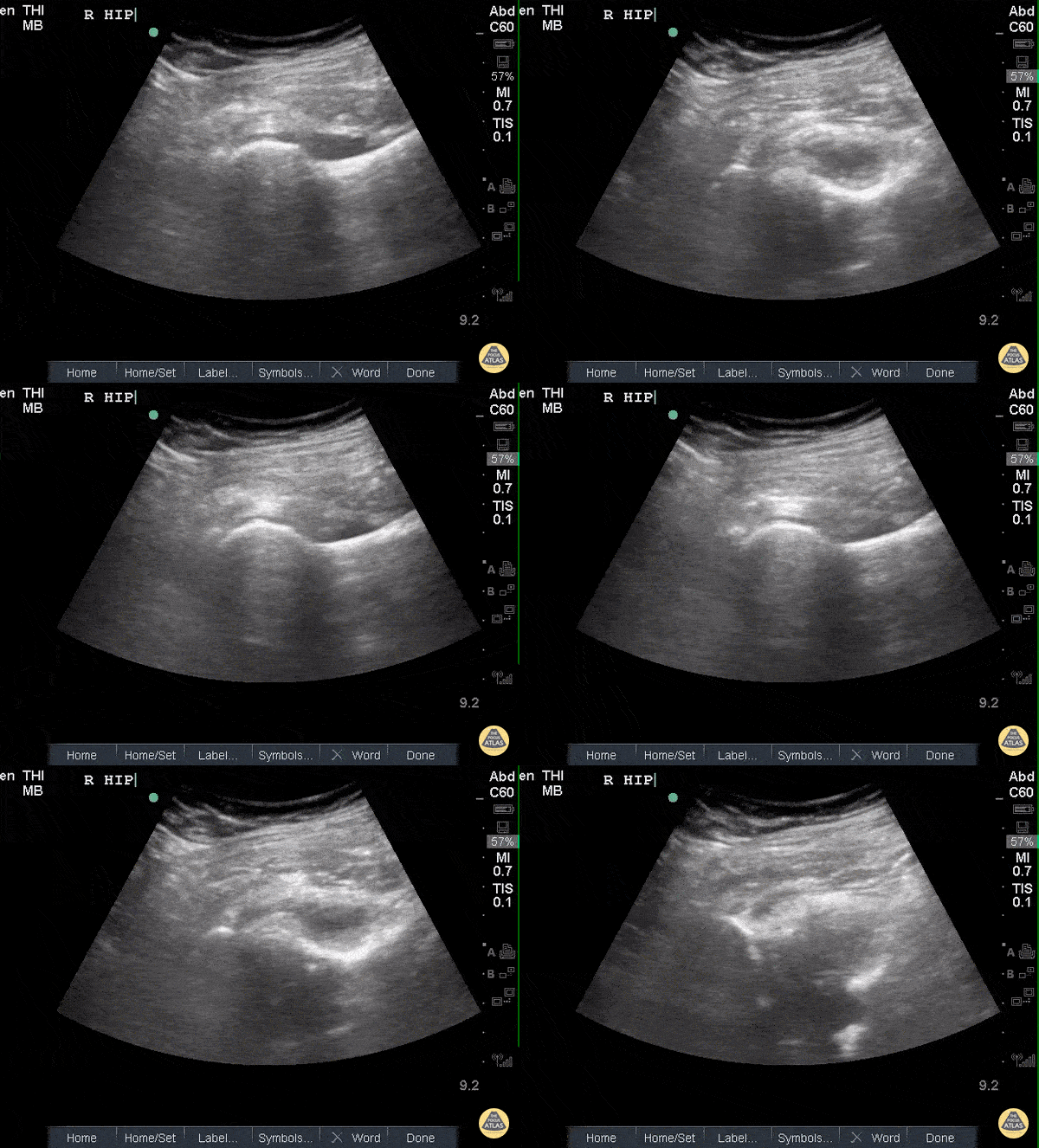
POCUS of the R hip shows an anechoic region adjacent to the femoral head and within the joint capsule consistent with an effusion.
Sukh Singh, MD

Arthrocentesis of Knee Effusion
40s F with prior history of contralateral trimalleolar ankle fracture s/p multiple surgeries presents 1 month of knee swelling and pain after starting levofloxacin. The symptomatic knee appeared swollen without warmth or erythema, ROM was preserved, and the patient was ambulatory. Point of care US demonstrated an effusion around the knee, so diagnostic and therapeutic arthrocentesis was performed as shown here. The linear probe was used in a transverse orientation just superior to the patella to view the suprapatellar bursa just deep to the quadriceps tendon. Using sterile technique, a needle was advanced under real-time, in-plane US guidance to enter the bursa and aspirate synovial fluid. Ultimately the patient did not have septic arthritis and was discharged with a compressive knee wrap with a plan for orthopedic surgery follow up.
Dr. Caleb Knight, PGY-2
Denver Health Residency in Emergency Medicine


