1
2
3
4
5
6
7
8
9
10
11

Metacarpal Head Fracture
34 y/o M presented with fall while skiing injuring closed fist against ice and was found to have a displaced/impact fracture of his third metacarpal head.
Video shows sagittal view scanning in the ulnar to radial direction at the third metacarpophalangeal joint (right is proximal). There is diffuse disruption of the cortex of the radial aspect of the metacarpal head and displaced/impaction fracture of the metacarpal head. Usually this surface should appear smooth. Articular cartilage can be seen as anechoic at the interface between the two bones. This defect was not visualized on initial or 2 week post x-ray, so ultrasound was able to guide the clinical team to appropriately splint and manage as a fracture.
Eben Alexander, DO
Devesh Patel, MD
Eastern Virginia Medical School
Comminuted Proximal Phalanx Fracture of Great Toe
Comminuted, dorsally angulated fracture of the proximal phalanx of the left great toe.
Clavicle fracture with hematoma
60 year old female with a subacute left clavicular fracture (occurred 2 weeks ago) presented with worsening pain at fracture site of onset while working with occupational therapy. Seen here is the left clavicle (hyperechoic structure) with noted fracture and mild heterogeneous (concern for bloody accumulation) edema around fracture site as observed in long axis view.
Kwasi Ampomah, DO, Eben Alexander IV, DO, Tariq Niazi, MD
EVMS PM&R
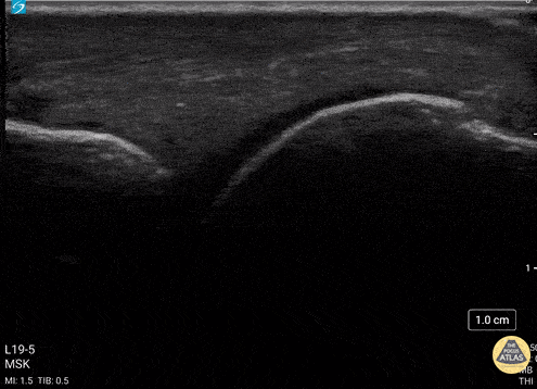
Rib Fracture
A middle aged man presented 1 week after sustaining a fall with direct injury to his left chest. He reported pain with inspiration and coughing; he localized pain to one specific area of his chest wall. Seen here is the image obtained when the linear probe was placed in the longitudinal plane to his area of point-tenderness. Notice the disruption of the hyperechoic cortex of the rib. Findings were confirmed in the transverse plan. The patient went on to have an anterior serratus nerve block for pain control related to his rib fracture.
Mandy Peach, MD @mandy_peach
Saint John Regional Hospital. NB, Canada
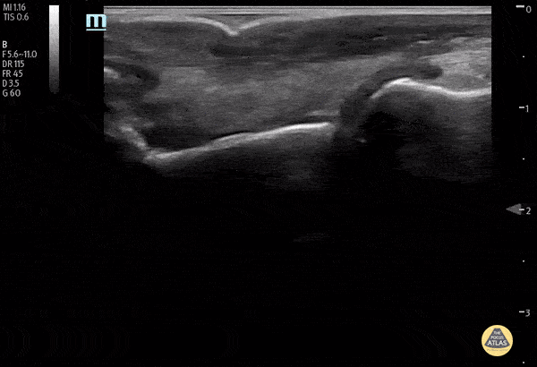
Rib Fracture
A rib fracture is seen here as disruption in the hyperechoic line or bony cortex. Also note the associated hypoechoic hematoma formation.
Aaron Inouye, PA-C, North Canyon Medical Center
@PAintheED
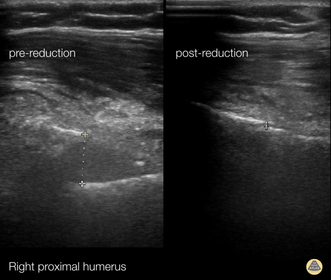
Fracture Reduction Monitoring
72 y/o female presents with right upper arm pain after a mechanical fall from standing.
A longitudinal coronal image was obtained at the right proximal humerus. Imaging showed displacement of bone fragments with hyperechoic lines 1.17cm apart. Pictures were obtained intermittently throughout reduction until displacement was reduced to 0.06cm.
This case demonstrates the utility of ultrasound in fracture reduction. Traditionally, care teams perform repeat X-rays until the fracture is reduced. In comparison, ultrasound can be quicker and reduce exposure to ionizing radiation.
Crozer Chester EM
Arthur Strzepka (MS4), Damarcus Ingram (MS4), Dr. Max Cooper
Posterior Fat Pad
Posterior fat pad aka Sail sign, is one of the common findings that we look for after a traumatic elbow injury that can indicate an underlying fracture. It represents hemarthrosis pushing the fat pad superiorly causing the triceps tendon to tilt. Plain films has been used as the initial modality of choice to look for sail sign but POCUS has been shown to be highly sensitive (97%) and specific (88%). It can be seen as anechoic fluid between the olecranon, humerus and fat pad. Source: Avci et. al. (PMID: 27645809). Also note the broken crystal on this image causing a dark artifact anteriorly.
Dr. Maan Al Dubayan, Steven Greenstein, and Matthew Riscinti - Kings County Emergency Medicine
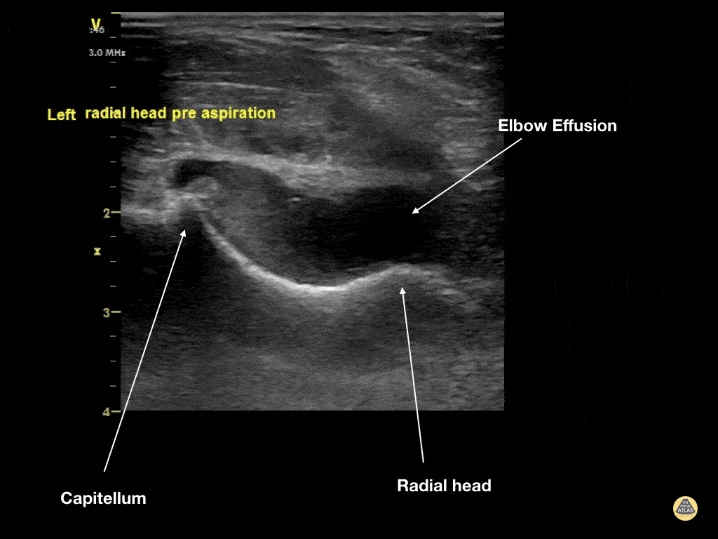
Elbow Effusion (Traumatic)
Aspiration of traumatic elbow effusions may be considered in the management of radial head fracture. Slide a linear transducer along the forearm towards the elbow until the radial head, effusion and capitellum are seen. Using an out of plane approach, insert a needle into the effusion. The syringe will fill itself under intrinsic pressure. Relief is often instantaneous and prolonged and the range of motion of the elbow will increase dramatically
Dr Cian McDermott, Emergency Physician, Mater University Hospital, Dublin, Ireland
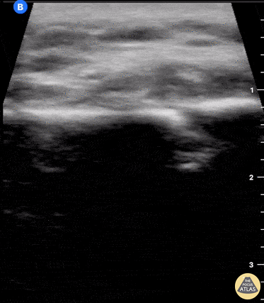
Metacarpal Fracture
Fractures can easily be diagnosed with POCUS especially in resource limited settings. Just remember... this could be painful so use A LOT of gel and try not to press hard or at all. Gently move the probe along the axis of the bones where you suspect a fracture.
The deepest and most hyperechoic horizontal line is the cortex and discontinuity in the lines represent fracture. Angulation and displacement can be measured. Two planes should be measured.
Sukh Singh, MD, Caption: Matthew Riscinti, MD
Rib Fracture
40 y/o M with polysubstance abuse, left-sided rib pain after a traumatic blow. Chest xray was equivocal. The patient was asked to "point to where it hurt", and the linear transducer revealed a displaced rib fracture. He complained of significant pain even after the resident gave two Percocet and was unwilling to leave the ED.
An intercostal nerve block, and that relieved the patient's pain and he went home.
Dr. Stephen Alerhand, Mt Sinai Hospital NYC
Thumb Fracture
30 year-old male ED resident who injured his thumb at some point while playing football versus the attendings in the annual flag football game. He figured the thumb had merely been sprained, and he kept playing in the game (and scoring touchdowns) while the residents dominated.
Two days later the swelling/ecchymoses seemed to worsen, he used the linear transducer in a water bath to diagnose a fracture of the base of the 1st metacarpal. An x-ray confirmed the diagnosis, and he underwent percutaneous pinning in the operation room the following week.
Dr. Stephen Alerhand, Mt Sinai Hospital, NYC