
Musculoskeletal

Comminuted Proximal Phalanx Fracture of Great Toe
Comminuted, dorsally angulated fracture of the proximal phalanx of the left great toe.

Sciatic Nerve Hematoma
Patient sustained a gun shot wound through left mid thigh.
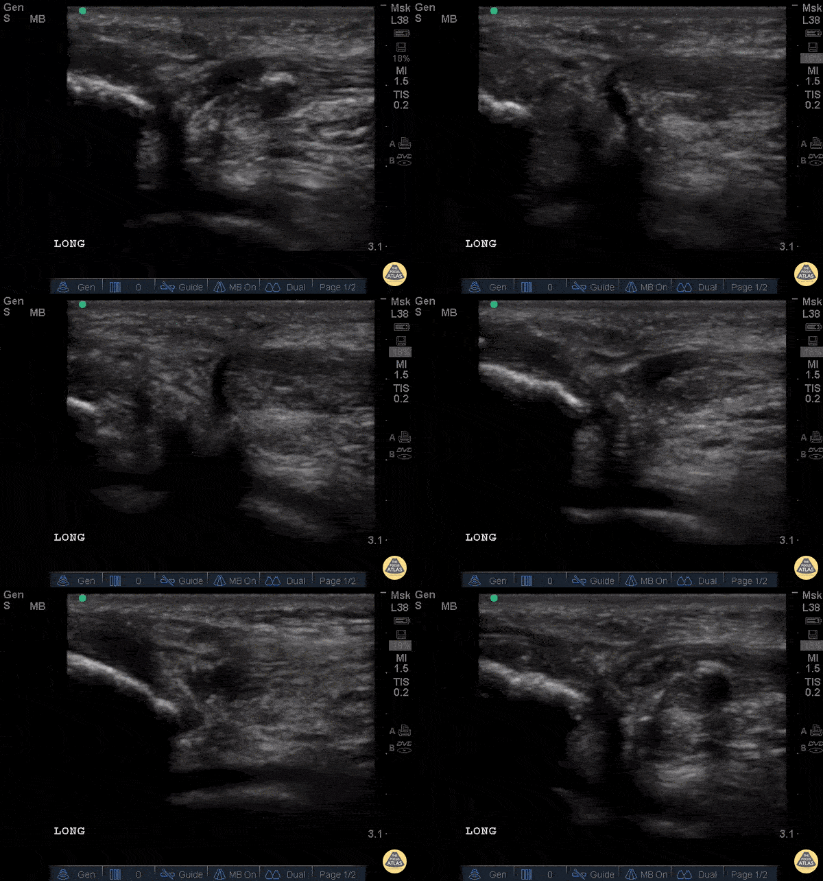
Image is long axis view of sciatic nerve mid thigh. Nerve is running distal to proximal from left to right at approximately 3cm depth. There is a hypoechoic fluid collection seen superficial to the nerve. The epineurium is intact and we can see smaller nerve fibers contained in it.
Patient had 0/5 plantar/dorsiflexion of ankle on admission, consistent with a sciatic nerve injury. The ultrasound exam on the sciatic nerve did not show any gross abnormality other than this fluid collection around the nerve. Within 7 days (4 days after this image) he regained strength in plantar/dorsiflexion in the ankle.
Mike Guju, MD @MichaelMGujuMD
Resident PM&R EVMS

Rib Fracture
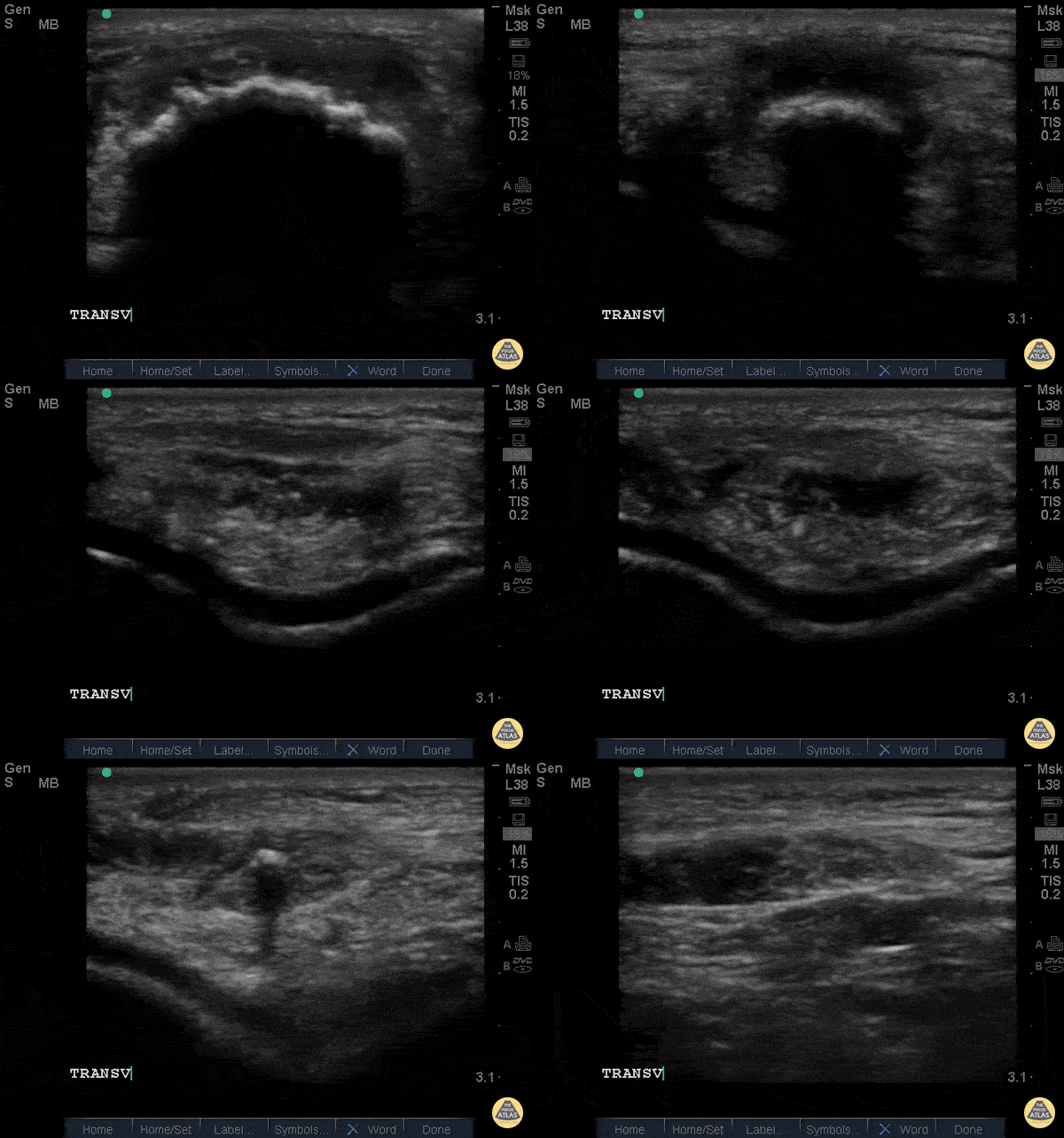
A middle aged man presented 1 week after sustaining a fall with direct injury to his left chest. He reported pain with inspiration and coughing; he localized pain to one specific area of his chest wall. Seen here is the image obtained when the linear probe was placed in the longitudinal plane to his area of point-tenderness. Notice the disruption of the hyperechoic cortex of the rib. Findings were confirmed in the transverse plan. The patient went on to have an anterior serratus nerve block for pain control related to his rib fracture.
Mandy Peach, MD @mandy_peach
Saint John Regional Hospital. NB, Canada

Distal Biceps Tendon Rupture
A 30-year-old male presented with acute-onset left anterior elbow pain after feeling a “pop” while weight-lifting. On exam, there was an obvious reverse popeye sign, swelling and mild ecchymosis. There was slight weakness in flexion and supination. Hook test was positive. A Butterfly IQ was used on an MSK soft tissue setting to assess the tendon in long and short axis via an anterior approach. Discontinuity of the tendon and surrounding anechoic fluid representing hematoma were noted. Orthopaedic surgery was consulted and reviewed ultrasound images. As a result, the patient had an urgent MRI and underwent expedited surgical repair.
Melanie Leclerc, MD CCFP(EM). @MelanieLecler19
David Lewis MB,BS FRCS FCEM CFEU PGDipSEM
Saint John Regional Hospital, New Brunswick, Canada

Shoulder Relocation
A 9-year-old female presented with left shoulder pain. She has a history of multiple dislocations and, as seen here on POCUS, is able to reduce the dislocation herself.
Julie Klensch, PEM Fellow & Paul Khalil, MD
University of Louisville/Norton Children's Hospital

ACL/LCL Injury
A 32-year-old male pedestrian struck by a jeep traveling at 30 mph was evaluated with POCUS for left knee injury. He sustained a left-sided rupture of ACL, LCL, and an avulsion fracture of his IT band. Pictured here is his left lateral knee US performed 10-days after the acute injury. You can appreciate the popliteus notch in the lateral femur, typically both the origin of the popliteus muscle and the plane the LCL traverses to the fibula; here only notable for diffuse edema.
Eben Alexander

Ligamentous Knee Injury
A 34-year-old male trauma patient was evaluated with POCUS for left knee swelling. He was appreciated to have multi-ligamentous injury of the left knee, with imaging notable for hypoechoic fluid within the suprapatellar plica (bursa) and evolving septation of the fluid.
Kwasi Ampomah

Achilles Tendon Injury
A 20s M presented with an ankle injury after landing a skateboard trick and feeling a painful pop in his posterior ankle. He had a positive Thompson test on exam. POCUS of the affected Achilles tendon was performed. This clip shows a long axis view of the Achilles tendon using the linear transducer, with inferior at the right of screen and superior at the left of screen. There is a relatively hypoechoic area (within red arrowheads) within the normally hyperechoic tendon, and the thickness is increased in this area as well, indicating focal injury to the tendon. Orthopedics was consulted, and the patient was placed into a splint in plantar flexion and discharged with outpatient follow up. MRI later confirmed a full thickness Achilles tendon tear and the patient was scheduled for surgery.
Jaimie Trenney, PA-C
Denver Heath Medical Center

Achilles Tendon Injury Short Ais
A 20s M presented with an ankle injury after landing a skateboard trick and feeling a painful pop in his posterior ankle. He had a positive Thompson test on exam. POCUS of the affected Achilles tendon was performed. This clip shows a short axis view of the Achilles tendon using the linear transducer. There is a relatively hypoechoic area (*) within the normally hyperechoic tendon, indicating focal injury to the tendon. Orthopedics was consulted, and the patient was placed into a short leg splint in plantar flexion and discharged with outpatient follow up. MRI later confirmed a focal full thickness Achilles tendon tear and the patient was scheduled for surgery.
Jaimie Trenney, PA-C
Denver Heath Medical Center

Nursemaid's Elbow
Nursemaid's pre and post reduction.
Nathan Jia, Orthopedic Resident

Ruptured Achilles Tendon
16-year-old male presented with acute onset sharp pain to his LE and inability to bare weight after having landed oddly while playing basketball. POCUS revealed a near-complete disruption of his Achilles Tendon.
Paul Khalil, MD. Assistant PEM POCUS director at University of Louisville/Norton Children’s
@Khalil3Paul

Rib Fracture
A rib fracture is seen here as disruption in the hyperechoic line or bony cortex. Also note the associated hypoechoic hematoma formation.
Aaron Inouye, PA-C, North Canyon Medical Center
@PAintheED
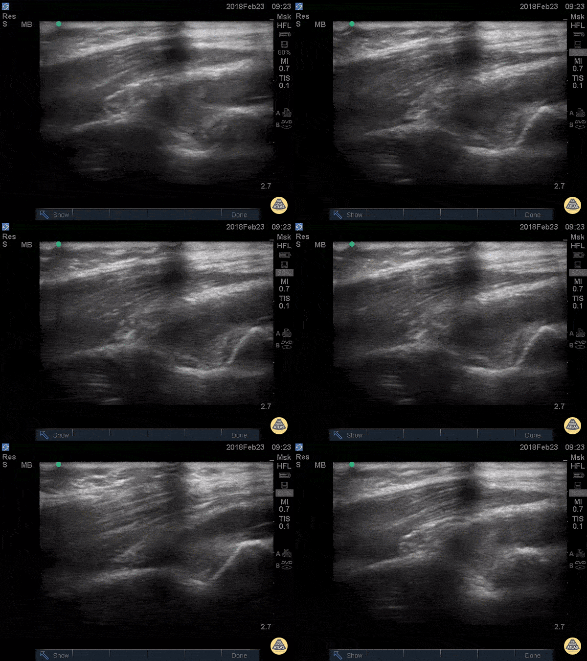
Posterior Fat Pad
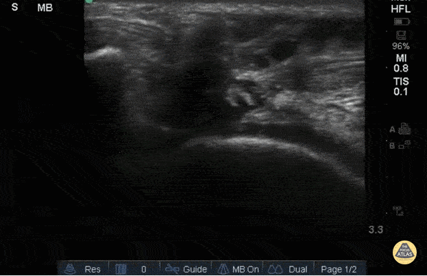
Posterior fat pad aka Sail sign, is one of the common findings that we look for after a traumatic elbow injury that can indicate an underlying fracture. It represents hemarthrosis pushing the fat pad superiorly causing the triceps tendon to tilt. Plain films has been used as the initial modality of choice to look for sail sign but POCUS has been shown to be highly sensitive (97%) and specific (88%). It can be seen as anechoic fluid between the olecranon, humerus and fat pad. Source: Avci et. al. (PMID: 27645809). Also note the broken crystal on this image causing a dark artifact anteriorly.
Dr. Maan Al Dubayan, Steven Greenstein, and Matthew Riscinti - Kings County Emergency Medicine
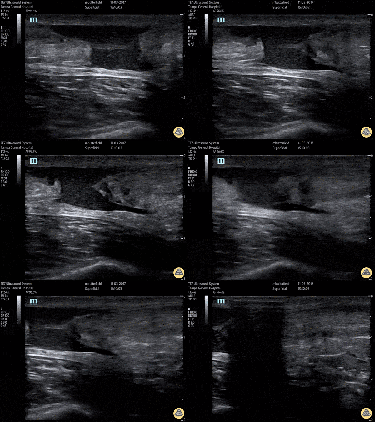
Achilles Rupture (Long Axis)
Full thickness tear of right achilles tendon after a skateboarding accident. (Long Axis)
Dr. Mike Butterfield
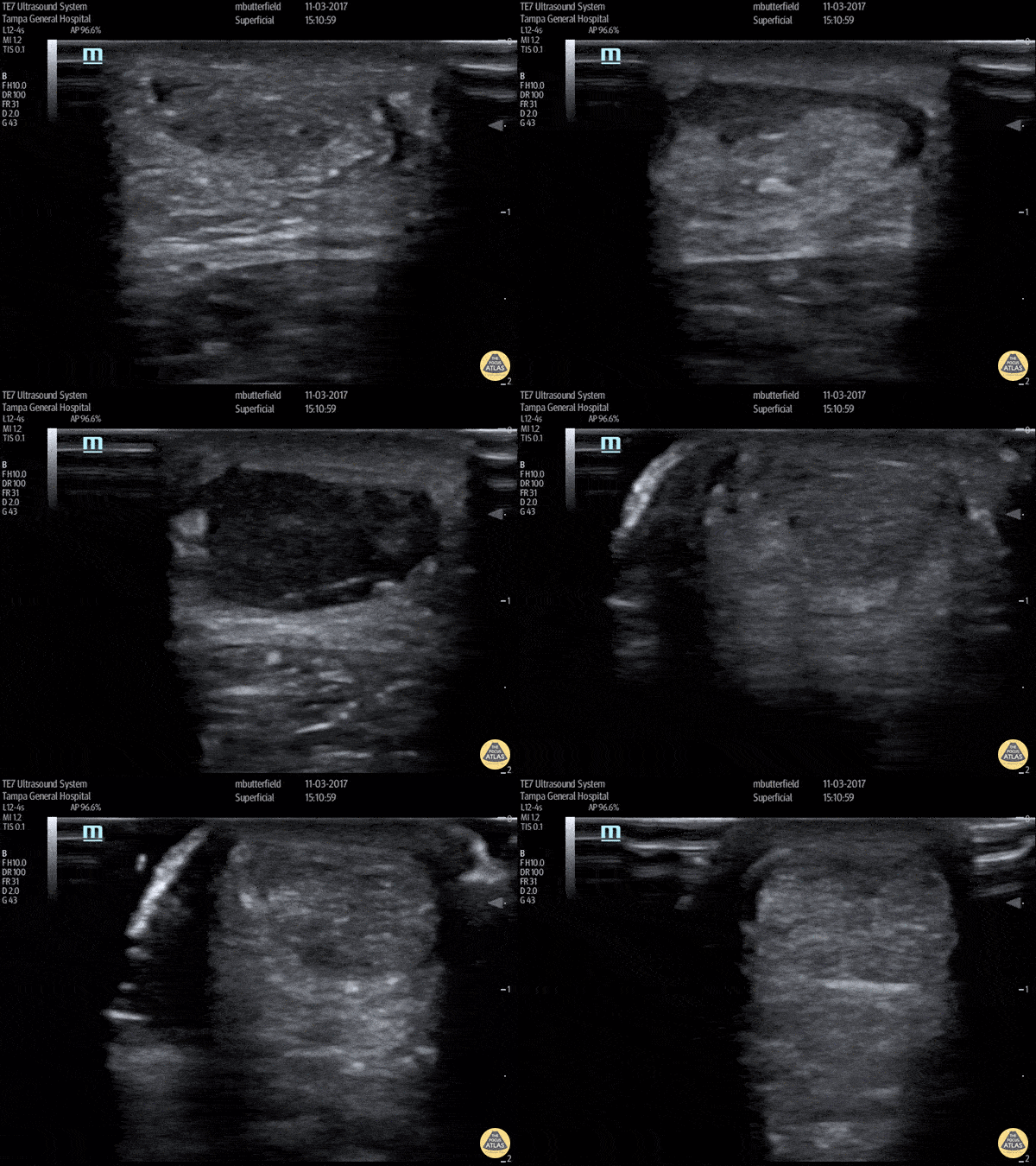
Achilles Tendon Rupture (Short Axis)
Full thickness achilles tendon rupture of the right leg after a skateboard accident. (Short Axis)
Dr. Mike Butterfield

Elbow Effusion (Traumatic)
Aspiration of traumatic elbow effusions may be considered in the management of radial head fracture. Slide a linear transducer along the forearm towards the elbow until the radial head, effusion and capitellum are seen. Using an out of plane approach, insert a needle into the effusion. The syringe will fill itself under intrinsic pressure. Relief is often instantaneous and prolonged and the range of motion of the elbow will increase dramatically
Dr Cian McDermott, Emergency Physician, Mater University Hospital, Dublin, Ireland
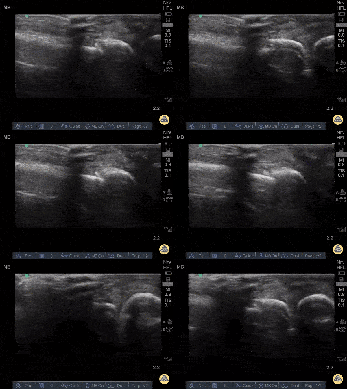
Metacarpal Fracture
Fractures can easily be diagnosed with POCUS especially in resource limited settings. Just remember... this could be painful so use A LOT of gel and try not to press hard or at all. Gently move the probe along the axis of the bones where you suspect a fracture.
The deepest and most hyperechoic horizontal line is the cortex and discontinuity in the lines represent fracture. Angulation and displacement can be measured. Two planes should be measured.
Sukh Singh, MD, Caption: Matthew Riscinti, MD
Patellar Tendon Rupture Longitudinal
34 y/o M presented with swelling and pain inferior to his knee following hearing a pop when he jumped playing basketball. Pt unable to extend leg and x-ray demonstrated a high riding patella. Longitudinal ultrasound showed a hyperechoic tendon that is not continuous between the patella and tibia, with an anechoic area of hemorrhage consistent with patellar tendon rupture.
Patellar tendon rupture can be diagnosed with H&P and POCUS can be used to confirm this diagnosis. In one study, diagnosis of tendon rupture by physical exam had a sensitivity of 100% and specificity of 76%, while diagnosis by POCUS had a sensitivity of 100% and specificity of 95%. Ultrasound is especially useful in patients who cannot cooperate with a physical exam, and serial ultrasound can also be used to monitor healing of a tendon rupture.
Caroline Rago - MS4, Dr’s Bryan Jarrett and Joshua Schechter - Kings County Emergency Medicine
Patellar Tendon Rupture Transverse
34 y/o M presented with swelling and pain inferior to his knee following hearing a pop when he jumped playing basketball. Pt unable to extend leg and x-ray demonstrated a high riding patella. Longitudinal ultrasound showed a hyperechoic tendon that is not continuous between the patella and tibia, with an anechoic area of hemorrhage consistent with patellar tendon rupture.
Patellar tendon rupture can be diagnosed with H&P and POCUS can be used to confirm this diagnosis. In one study, diagnosis of tendon rupture by physical exam had a sensitivity of 100% and specificity of 76%, while diagnosis by POCUS had a sensitivity of 100% and specificity of 95%. Ultrasound is especially useful in patients who cannot cooperate with a physical exam, and serial ultrasound can also be used to monitor healing of a tendon rupture.
Caroline Rago - MS4, Dr’s Bryan Jarrett and Joshua Schechter - Kings County Emergency Medicine
Right Patellar Tendon Rupture
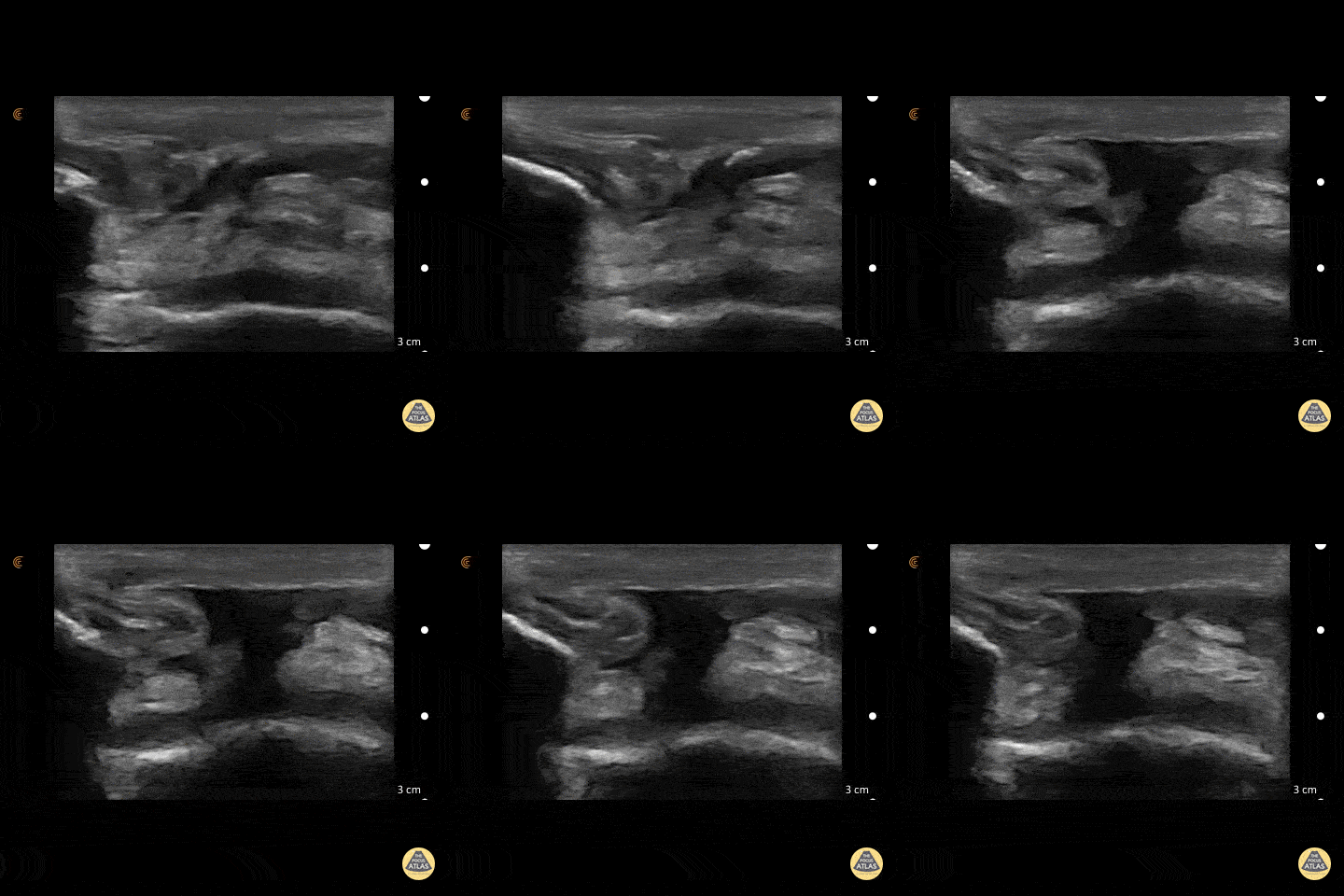
56yo M with right knee swelling after getting foot stuck under a pallet and falling backwards, found to have patella alta and right patellar tendon rupture. Longitudinal image using linear 13-6MHz probe along proximal (left) and distal (right) patellar tendon with hypoechoic fluid at site of tendon rupture. Dynamic ultrasound is useful in diagnosing tendon ruptures as the site and extent of rupture can be easily visualized, which facilitates triage to surgery, if indicated.
Dr. Jasmin Harounian
Patellar Tendon Rupture
50 y/o M presents with acute left anterior knee pain after fall. On exam, patient noted to have high riding patella.
Longitudinal sonogram of the infrapatellar region showed marked discontinuity of the normally hyperechoic linear patterned tendon. The discontinuity is replaced with an anechoic collection indicative of hemorrhage. Comparison to normal knee can be seen on the next post.
Of note, anisotropy may be encountered when utilizing ultrasound, leading to artifact. Depending on the angle of the insonating beam, a normally hyperechoic structure may be falsely viewed as hypoechoic due to poor return of echo. Ensure US probe remains perpendicular to the tendon to minimize this artifact.
Dr. Hannah Moreira, Dr. Tareq Azad, Dr. Kyle Kelson - Kings County/SUNY Downstate Emergency Medicine
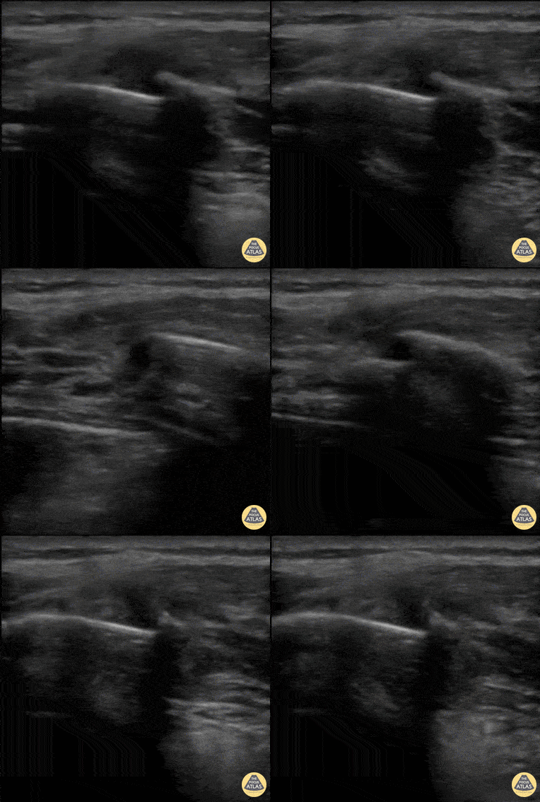
Rib Fracture
40 y/o M with polysubstance abuse, left-sided rib pain after a traumatic blow. Chest xray was equivocal. The patient was asked to "point to where it hurt", and the linear transducer revealed a displaced rib fracture. He complained of significant pain even after the resident gave two Percocet and was unwilling to leave the ED.
An intercostal nerve block, and that relieved the patient's pain and he went home.
Dr. Stephen Alerhand, Mt Sinai Hospital NYC
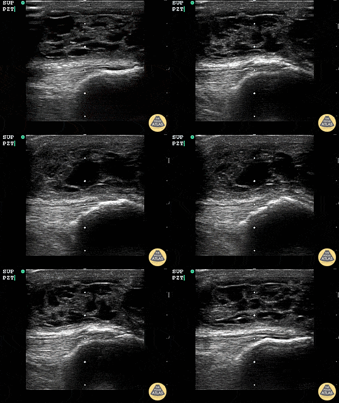
Suprapatellar Bursitis
Suprapatellar bursitis from repetitive trauma of playing on the floor with grandchildren. Presented with over a week of knee pain and swelling. Superficial involvement and septae are possible for abscess, however it is contained within the bursal space above the patella. Arthrocentesis revealed no infection, and conservative therapy yielded improvement.
Dr. Dustin Morrow
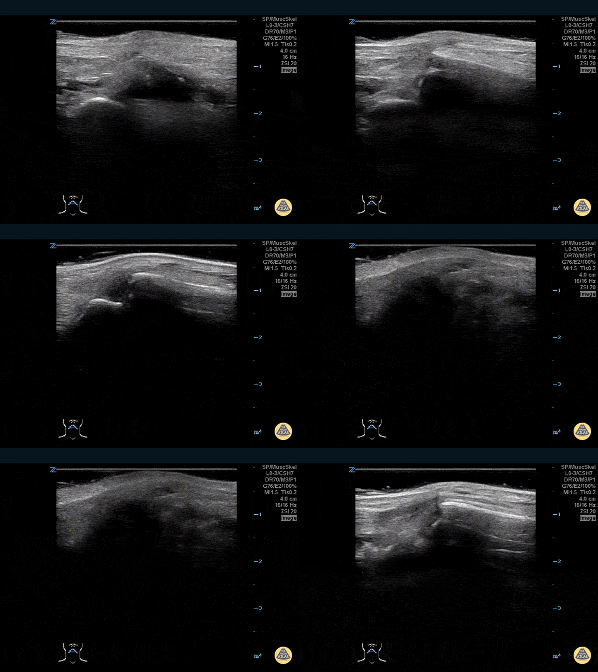
Thumb Fracture
30 year-old male ED resident who injured his thumb at some point while playing football versus the attendings in the annual flag football game. He figured the thumb had merely been sprained, and he kept playing in the game (and scoring touchdowns) while the residents dominated.
Two days later the swelling/ecchymoses seemed to worsen, he used the linear transducer in a water bath to diagnose a fracture of the base of the 1st metacarpal. An x-ray confirmed the diagnosis, and he underwent percutaneous pinning in the operation room the following week.
Dr. Stephen Alerhand, Mt Sinai Hospital, NYC

Subscapularis Tendon Tear
A middle aged male presented to the ED with shoulder pain after skiing crash. POCUS of the shoulder was performed, showing a subscapularis tendon tear. Here, the proximal humerus is shown in short axis, with the linear probe placed in transverse orientation at the anterior aspect of the shoulder. The biceps tendon is seen in the biceps groove, between the greater tuberosity (lateral or right of screen) and lesser tuberosity (medial or left of screen). The patient is asked to externally rotate the arm, which brings the subscapularis tendon into view, and a hypoechoic, thickened area is seen, indicating a tendon tear.
Dr. Matthew Riscinti
Denver Health Medical Center

Nearly Complete Achilles Tear
Healthy male in late-20s trying to do a backflip off a diving board. Still was able to plantarflex (very weak and with significant pain).
Submitted by Dr. Elias Jaffa


