
Thoracic Aortic Dissection & Aneurysm

Ascending Thoracic Aortic Aneurysm
A 70 y.o. male presented with altered mental status.
POCUS echocardiogram performed in the ED revealed an ascending thoracic aortic aneurysm measuring approximately 5 cm in diameter (parasternal long-axis view shown here). A chart review revealed that the patient does indeed have a history of TAA, and a comparison of our findings to a prior CTA demonstrates no significant increase in diameter. Nevertheless, this study demonstrates the utility of POCUS in the rapid and early detection of ascending aortic abnormalities.
Alex Schlangen, D.O. PGY-1 EM Resident at Central Michigan University; Andrew Namespetra, MB BCh BAO. @AndrewNamespet1 PGY-3 EM Resident at Central Michigan University

Aortic Dissection Flap in Arch of Aorta
A 65-year-old male presents with shortness of breath (no chest pain) and was found to have a dilated aortic root on CT pulmonary angiogram. POCUS (supra sternal view) showed a dissection flap in the arch of aorta; a finding subsequently confirmed on CT aortagram. Patient was sent for emergency surgical intervention.
Dr.Rajasutharsan Kathirgamanathan, Emergency Physician
The Northern Hospital, Melbourne, Australia
@raj_kathir007

Suprasternal View of Type A Dissection
Suprasternal notch view shows a mobile intimal dissection flap in the aortic arch.
Michael Cover, MD
@michaelc0ver

Type A Aortic Dissection - Double Valve Sign
Type A aortic dissection flap appears as a "second valve" on this parasternal long axis cardiac view.
Michael Cover, MD
@michaelc0ver

Descending Thoracic Aortic Dissection Seen on PSLA
This is a parasternal long axis view of an elderly male with PMH of hypertension and DM presenting with a dissection of the descending aorta (aka type B aortic dissection).
Image courtesy of Robert Jones, DO, FACEP @RJonesSonoEM
Director, Emergency Ultrasound; MetroHealth Medical Center; Professor, Case Western Reserve Medical School, Cleveland, OH
Find his original post here

Descending Thoracic Aorta Flap Seen on Apical 4 Chamber
33 yo male presented with chest/epigastric pain. POCUS is notable for acute aortic dissection as seen in the descending thoracic aorta flap as well as presence of a pericardial effusion.
Maxime Gautier
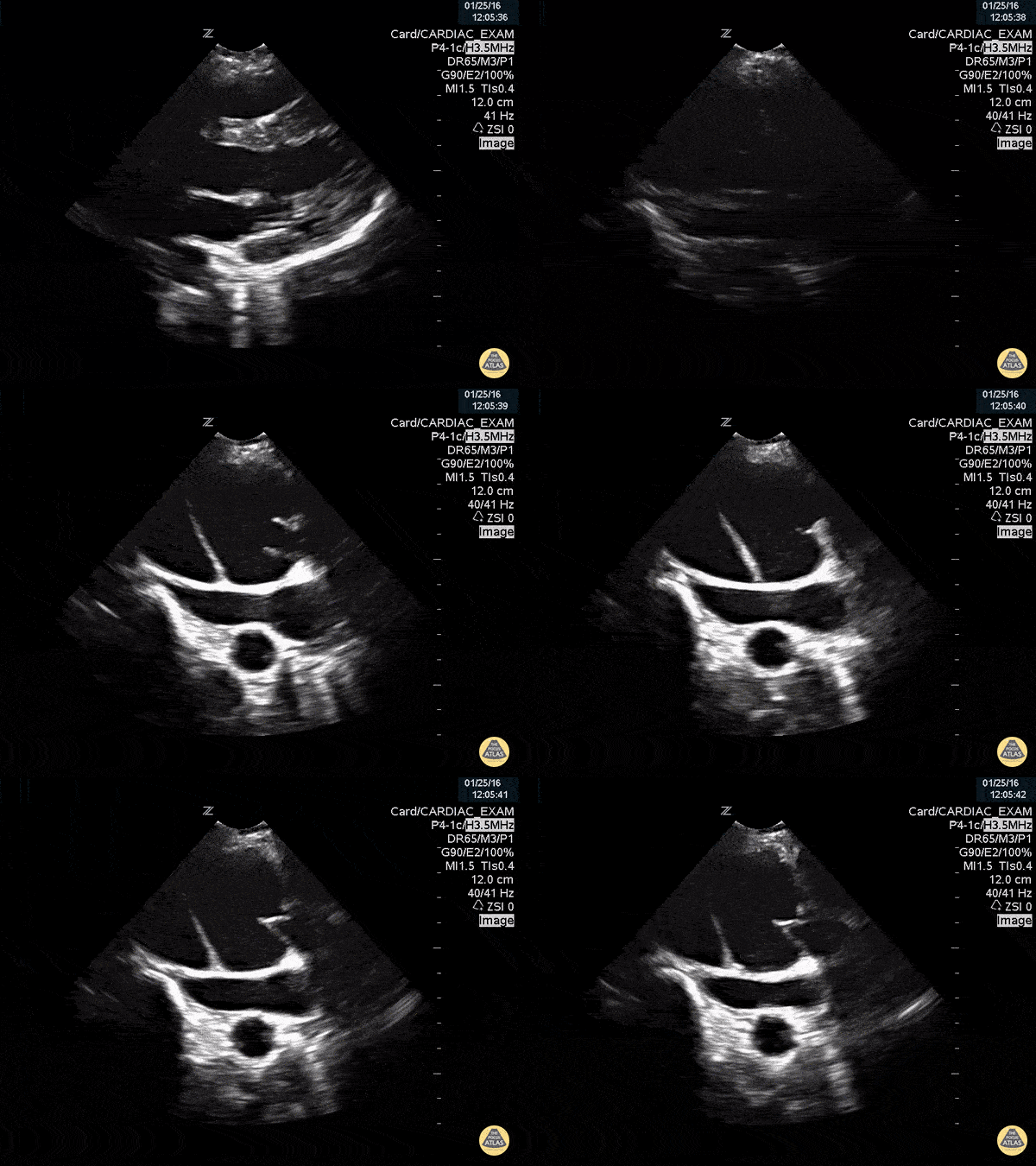
Aortic Dissection Flap Visualized in Proximal Aorta with Root Dilation
This is a parasternal long axis view demonstrating significant enlargement of the aortic root with an identified dissection flap located in the proximal ascending aorta.
Frances Russell, MD, RDMS
Assistant Professor of Emergency Medicine Division Chief, Ultrasound Fellowship Director, Ultrasound
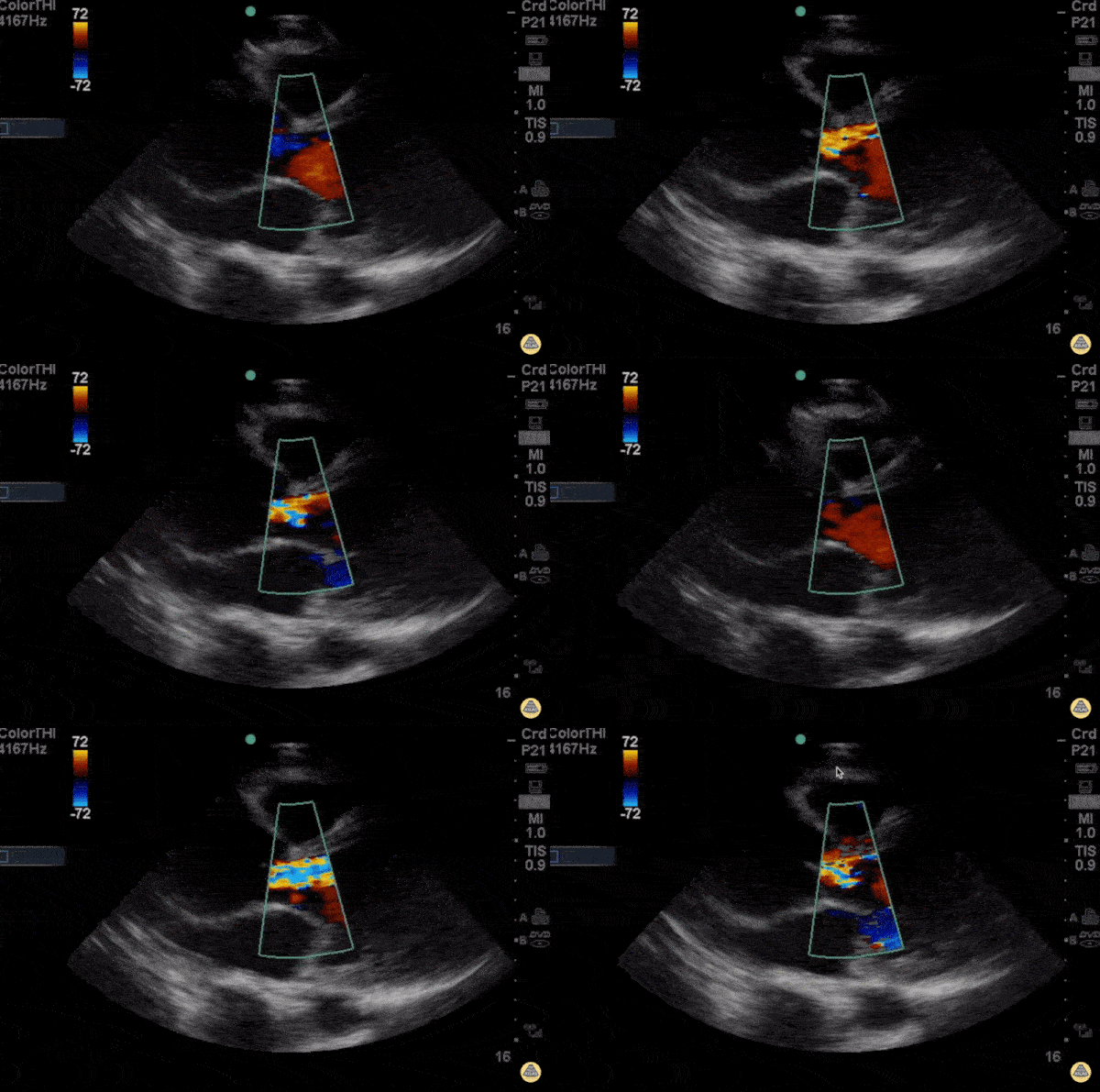
Breaking the rule of 3rds - Aortic Dissection with Root Dilation
This is a parasternal long axis view of a young patient presenting with 3 days of progressive dyspnea on exertion. He had no chest pain, a normal chest x-ray and and ECG with sinus tachycardia. Beside ultrasound got him to the OR in under 1 hour.
The rule of thirds states that in parasternal long, the right ventricular outflow tract, the aortic root, and left atrium should roughly be equal size. Any disparity between these can hint at pathology. In this case, the patient had severe and sudden aortic regurgitation and widening of the aortic root consistent with aortic dissection.
- Michael Macias, EM Resident Physician PGY-4, Northwestern University
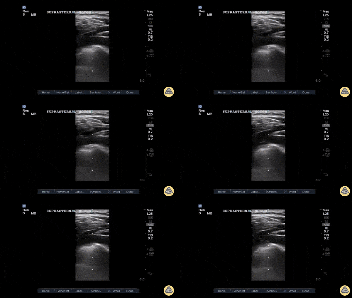
Type B Thoracic Aortic Dissection Flap on Suprasternal View
This is a suprasternal notch view demonstrating an aortic flap in a patient with a Stanford Type B thoracic aortic dissection.
This 40ish year old was a truck driver with untreated hypertension with sudden onset interscapular pain that migrated to his lumbar area. He stopped, lost strength in his right leg and was transported to our ED. The POCUS allowed the CV surgeon to prepare while the confirmatory CTA and standard treatment were performed. Suprasternal notch imaging with the linear or fine parts probe in a patient with suspicious signs/symptoms allows for a more rapid diagnosis of thoracic aortic dissection.
John E. Hipskind, MD, FACEP
Clerkship Director
ED, Kaweah Delta Hospital
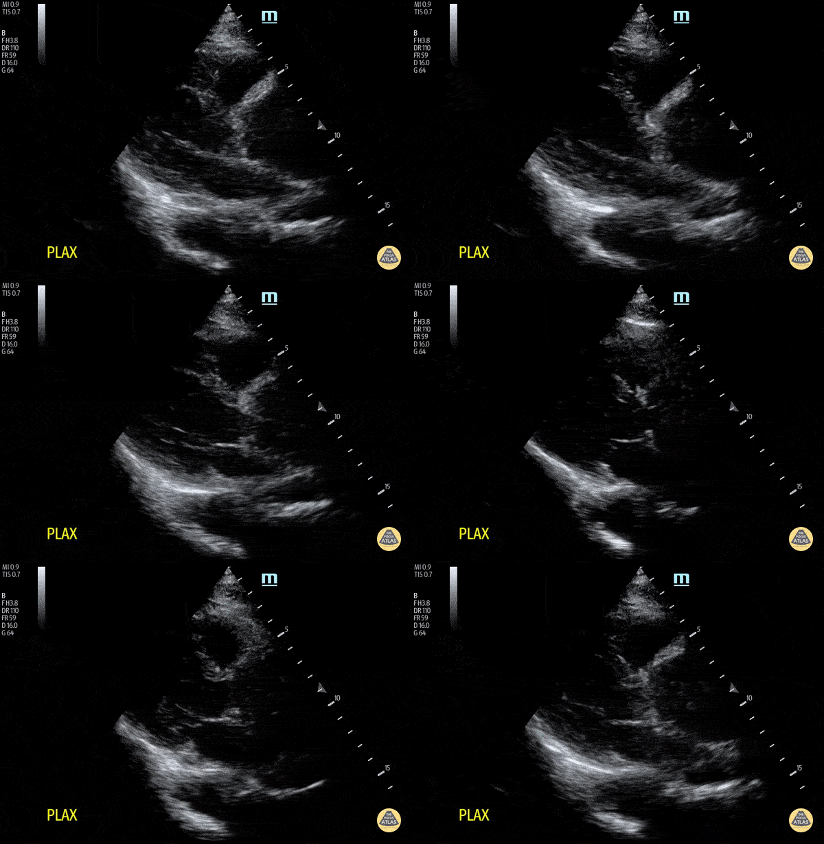
Aortic Root Dilation Due to Aortic Dissection
A dilated aortic root in a patient with chest pain radiating to the back. Note that the ratio of right ventricular outflow tract, aortic root and left atrium is not 1:1:1. Whilst you may not always see an intimal flap, a dilated aortic root and new aortic regurgitation may indicate aortic dissection. In this case, aortic dissection was subsequently confirmed on CT aortogram.
Dr. Cheng
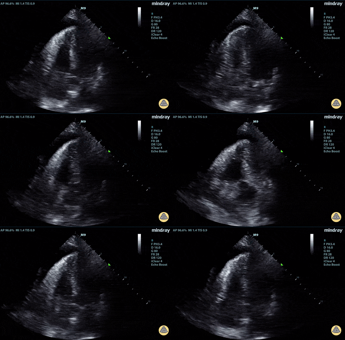
Aortic Dissection Causing Cardiac Tamponade
Elderly fellow who had a headache while bike riding, with some leg weakness. No chest or back pain. Stable for hours then came to hospital, suddenly hypotension and drowsy in ER
POCUS RUSH Exam performed lead to rapid diagnosis of Aortic Dissection with tamponade.
Right ventricular diastolic collapse can be seen.
Claire Heslop - Pediatric Emergency Medicine - University of Toronto Hospital for Sick Children
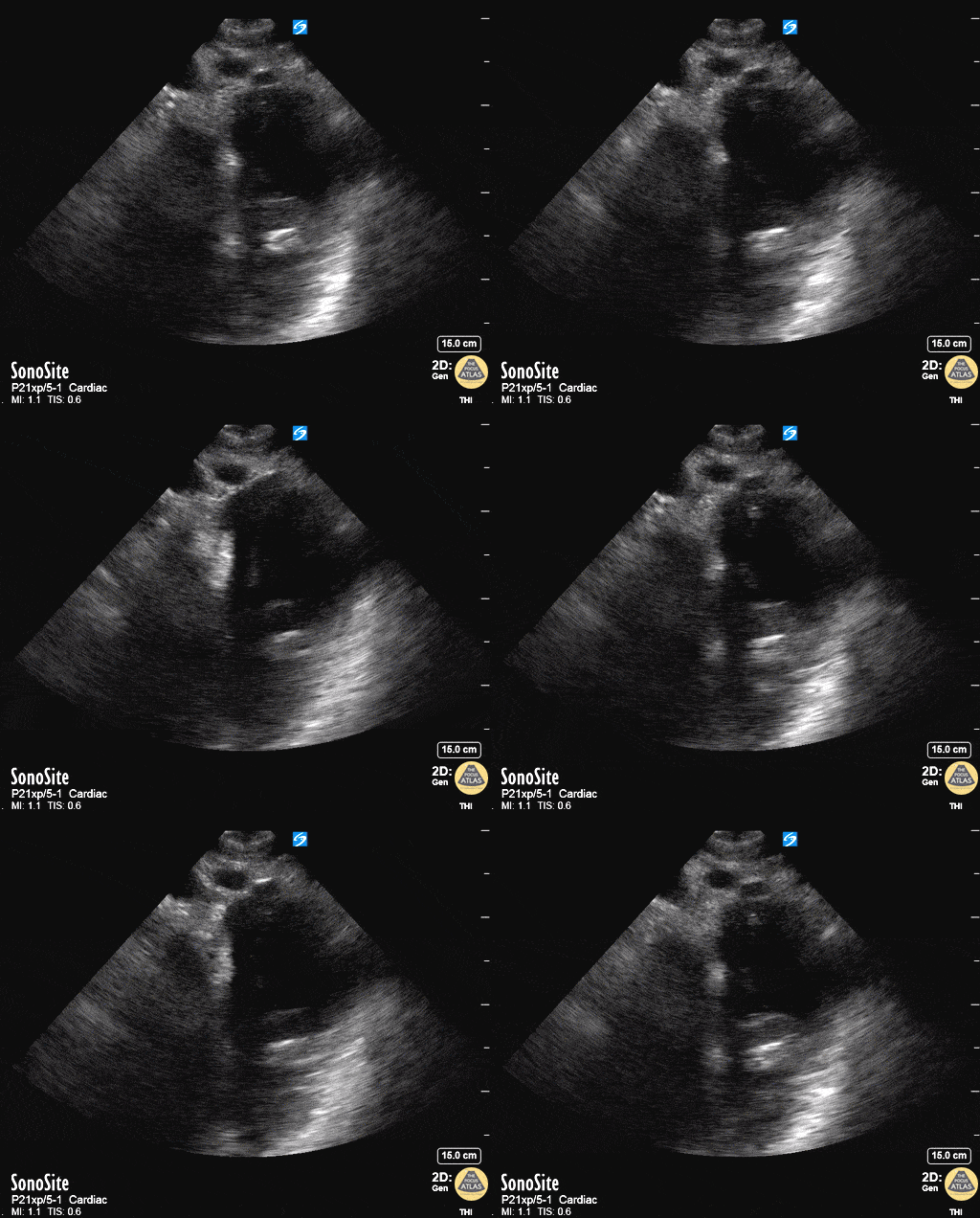
Aortic Dissection On Suprasternal View
50 year old mandarin speaking man complains of vague central chest pain. You pursue a routine cardiac workup which is fairly normal. Upon discharging him, the nurse tells you his systolic in now in the 80's.
After clenching the likely diagnosis of an Type A Aortic Dissection based on your echo, you confirm by placing the transducer in the patients suprasternal notch, transversely but with the probe marker rotated slightly toward the patient's hip and fan inferiorly. You see a grossly widened aorta and a dissection flap.
Dr. Matthew Riscinti - Kings County Emergency Medicine, Dr. Benjamin Clearly - NYU Langone Emergency Medicine
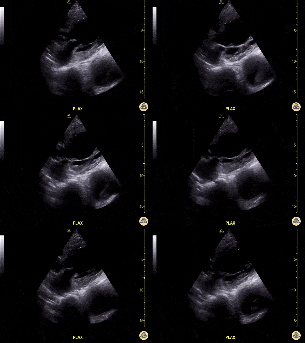
Thoracic Aorta Aneurysm with Intramural Thrombus
Don’t be distracted by the abnormal cardiac function in this clip…notice deep to the pericardium a thoracic aorta aneurysm is seen with moderate amount of intramural thrombus.
Image courtesy of Aventura Ultrasound; Their original Twitter posting can be found here.


