1
2
3
4
5
6
7
8
9
10
11
12
13
14

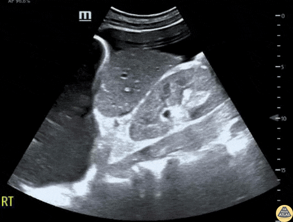
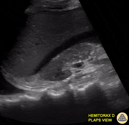
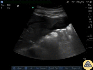
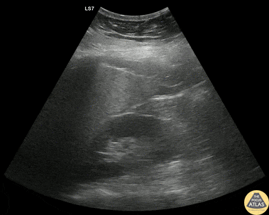
Pleural and Peritoneal Fluid in EFAST Exam
Patient presented to the emergency department with atraumatic acute onset of shortness of breath. An EFAST was performed to evaluate for pneumothorax and assess for free fluid in the abdomen, thorax, and pericardium.
A significant right pleural effusion was present. Additionally, US of RUQ demonstrated a liver which appeared small in size with prominent ascites. This demonstrates the utility of POCUS to identify free fluid in a quick and efficient manner as compared to other common studies such as a CT scan, even in non-traumatic patients.
Contributors: Lauren Lowes, DO; Krishna Patel, DO; Julia Tu, MS-4
Central Michigan University Residency of Emergency Medicine
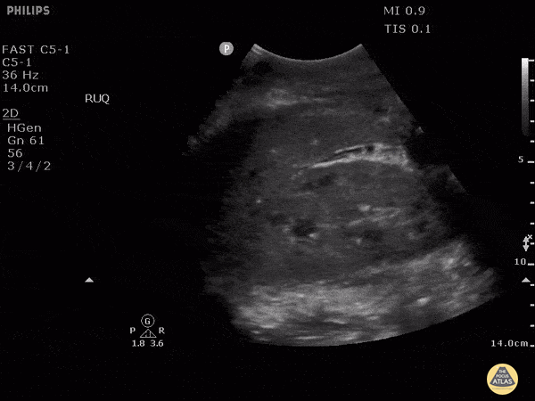
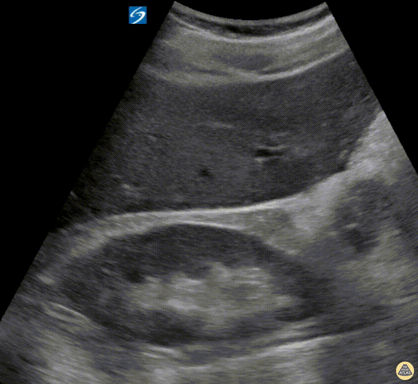
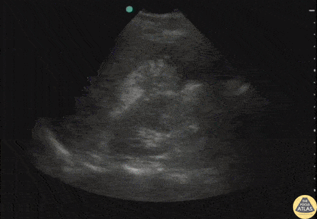
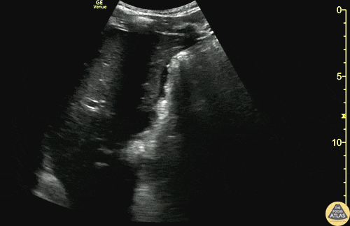
Hemoperotineum Or Not?
This is RUQ view from a patient who presented to the emergency department after falling from a roof and with low blood pressures. Although there appears to be perihepatic fluid, an interesting learning point form this case is whether or not we can discern if fluid observed is truly in the retroperotineal space or in the peritoneal cavity. Because the fluid is in direct contact with perinephric fat, this is actually in the retroperitoneal space and not in the peritoneal cavity. This may be difficult to distinguish in patients with very little perinephric fat.
Image courtesy of Robert Jones DO, FACEP @RJonesSonoEM
Director, Emergency Ultrasound; MetroHealth Medical Center; Professor, Case Western Reserve Medical School, Cleveland, OH
View his original post here
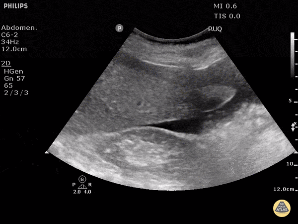
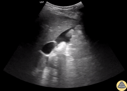
Hemoperitoneum
This clip was obtained in a patient presenting after a fall from roof. This hepatic window demonstrates an echogenic clot surrounded by a thin sliver of anechoic free fluid. This patient had massive acute hemoperitoneum. Remember that not all acute hemorrhage is anechoic!
Image courtesy of Robert Jones DO, FACEP @RJonesSonoEM
Director, Emergency Ultrasound; MetroHealth Medical Center; Professor, Case Western Reserve Medical School, Cleveland, OH
View his original post here
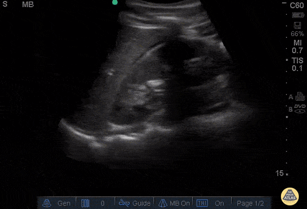
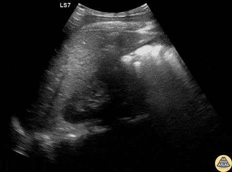
RUQ Free Fluid
A patient presented with LUQ trauma but the perisplenic window was negative for free fluid. Morison’s pouch was positive for free fluid emphasizing the importance of scanning all regions for free fluid in trauma.
Image courtesy of Robert Jones DO, FACEP @RJonesSonoEM
Director, Emergency Ultrasound; MetroHealth Medical Center; Professor, Case Western Reserve Medical School, Cleveland, OH
View his original post here
Positive FAST
Seen here is a subtle positive RUQ FAST scan in a trauma patient — a pertinent reminder to never stop simply after evaluating Morison's Pouch. Always also trace anteriorly to the lowest part of the liver.
Hjalti Már Björnsson
@hjaltimb
Free Fluid in Morrison's Pouch
This 30-year-old male was brought to our ED after a fall from scaffolding with evidence of trauma to his right thoracoabdominal region. E-FAST performed in the RUQ revealed both the presence of free fluid within Morrison's pouch and an ipsilateral hemothorax. Appreciate the presence of “spine sign” as an additional ultrasonographic indicator of free fluid within the pleural space.
Renato Tambelli, Emergency Physician Hospital das Clínicas de Marília, Brazil.
@R_Tambelli // @JediPocus
Positive FAST - RUQ - Morison's Pouch
Blunt trauma patient with POSITIVE FAST scan. The liver can been seen floating in free fluid with the kidney posteriorly. The fluid is in Morison's pouch. Be sure to visualize the tip of the liver to complete the evaluation.
Dr. Justin Bowra
Positive FAST - RUQ
30 y/o pedestrian struck by car, hemodynamically unstable, tachycardic. FAST performed after primary survey revealed free fluid in all four abdominal views of the FAST exam
Free fluid in Morison’s pouch of the RUQ view. This is the most sensitive view to detect free fluid in trauma.
Dr. Catharine Bon - Kings County Emergency Medicine
Positive FAST - RUQ
Blunt trauma patient with POSITIVE FAST scan. The liver can been seen floating in fluid with adjacent bowel.
Dr. Justin Bowra
Ruptured Viscus From Blunt Trauma
Pt struck by car while riding bike. Perihepatic view on FAST exam revealed a perforated viscus (gastric rupture). Note the free air within the fluid.
Image courtesy of Robert Jones DO, FACEP @RJonesSonoEM
Director, Emergency Ultrasound; MetroHealth Medical Center; Professor, Case Western Reserve Medical School, Cleveland, OH
View his original post here.
+FAST in Morrison's Pouch from Splenic Injury
20s M presented with abdominal and back pain several hours after a fall off a ladder, and on FAST exam, had free fluid seen here in Morrison’s pouch, as well as a large amount of free fluid seen in the pelvis/lower abdomen. He was slightly tachycardic but normotensive, so underwent CT of the abdomen/pelvis, which demonstrated a significant spleen injury which was managed by IR embolization.
Dr. Greg Wiener, PGY3
Denver Health Residency in Emergency Medicine
RUQ +FAST
A teenage male patient presented to the ED after a helmeted mountain bike crash, and due to mechanism, underwent a bedside FAST exam, which was positive in the RUQ as well as suprapubic views. CT demonstrated a grade 4 spleen laceration as well as multiple buckle rib fractures, and he was admitted for observation.
Dr. Gabe Siegel, PGY-2 and Dr. Michael Kidon, PGY-4
Denver Health Residency in Emergency Medicine
Subtle RUQ +FAST
30s F presented with worsening abdominal pain in the setting of a known ectopic pregnancy. FAST exam was very subtly positive in the RUQ, with more free fluid seen in the pelvic/suprapubic view. This clip shows the RUQ view, and a trace amount of free fluid is seen at the liver tip, highlighting the importance of complete visualization of this area when viewing the RUQ. This patient remained hemodynamically stable, and so was managed expectantly rather than with surgical exploration.
Dr. Lindsay Howe, PGY-3
Denver Health Residency in Emergency Medicine
Subtle +RUQ FAST from GSW
20s M presented as a walk in after sustaining a GSW to the hip. He was initially hemodynamically unstable, however responded well to transfusion of blood products and vitals normalized. FAST exam of the RUQ is shown here, with a trace amount of free fluid seen along the liver tip. FAST was also positive in the LUQ and pelvic views. Plain films demonstrated a retained missile in the abdomen. As he was hemodynamically stable, CT of the abdomen and pelvis was obtained, showing hemoperitoneum and free air concerning for bowel and vascular injury. The patient was taken emergently to the OR, where exploratory laparotomy demonstrated a pelvic hematoma and hemoperitoneum from an iliac vein injury, as well as multiple areas of small bowel injury. His injuries were repaired, he recovered well, and was discharged within days of his injury.
Dr. Ian Eisenhauer, PGY1
Denver Health Residency in Emergency Medicine