ddxof: Ultrasound in Ectopic Pregnancy
Another #FOAMEd knowledge bomb from our partner ddxof
Check out the original post here: ddxof: Ultrasound in Pregnancy
This would make a great intro for any GYN ultrasound student learner, or even third year medical student in OBGYN. We will paraphrase here but be sure to check out the original post for the evidence based details.
From their post: 27 year old female syncopizes, comes in from EMS hypotensive. She is lethargic and has a remarkably tender abdomen. You perform your rush exam, starting with a FAST which shows free fluid. HCG is positive. Large bore IV's, uncrossmatched blood, and get them to the OR with GYN STAT.
Algorithm for the Evaluation of Suspected Ectopic Pregnancy
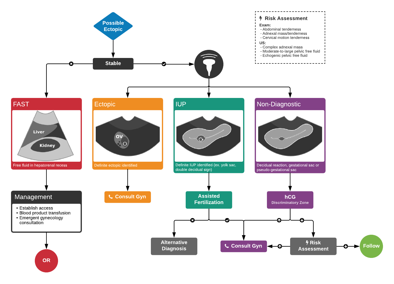
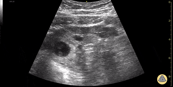
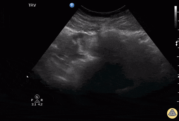
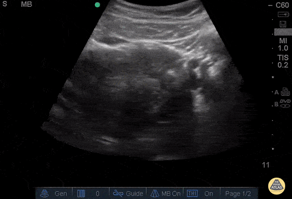
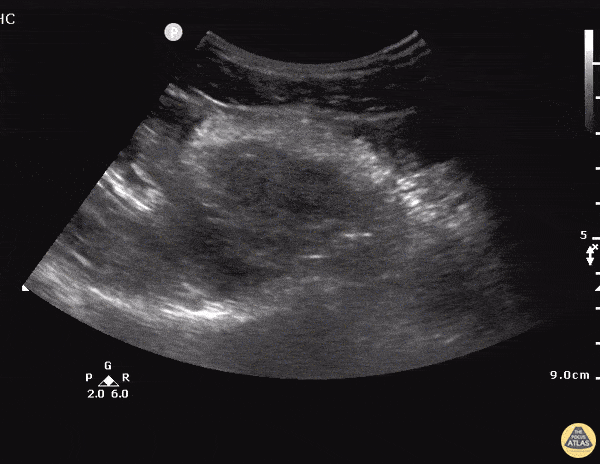
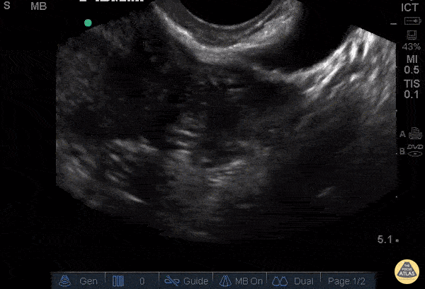
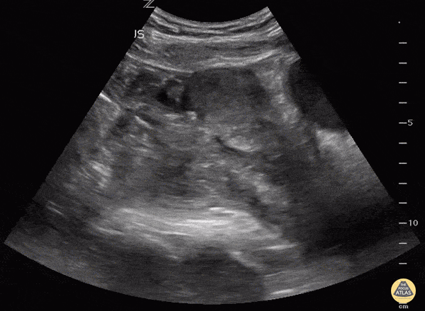
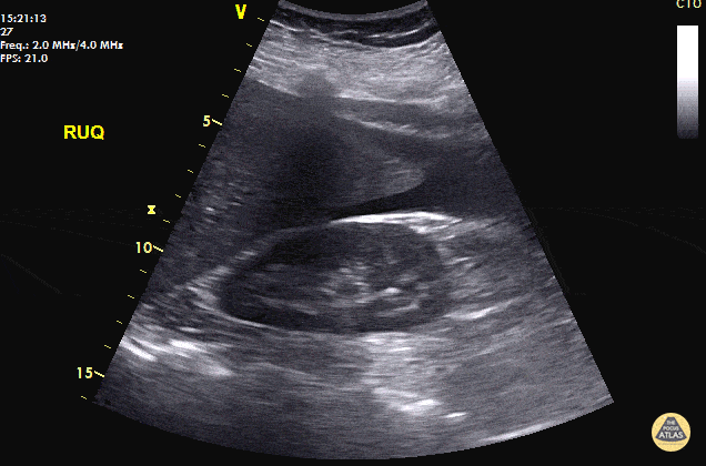
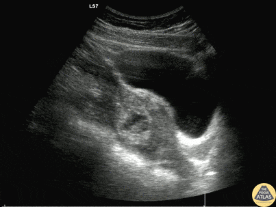
What do these things look like on ultrasound? That is where our GYN Atlas comes in. Here are some of our favorite ectopic images hand picked for you.
30s female currently 7 week pregnant with lower abdominal pain and tenderness to palpation to lower abdomen. POCUS showed a gestation sac containing a yolk sac and fetal pole outside of uterus. Also noted is a thickened endometrial stripe (to the right of the gestational sac). No free fluid was noted in the RUQ. The patient was taken to the OR for definitive management of ectopic pregnancy.
Dimitri Livshits DO, Ultrasound Fellow, Kings County/SUNY Downstate; Jane Belyavskaya MD, Ultrasound Fellow, Kings County/SUNY Downstate; Farnam Kazi MD, Ultrasound Faculty, Kings County/SUNY Downstate;
Notice to the left of the screen the presence of an adnexal mass separate to the ovaries which in this case indicates a right tubal ectopic pregnancy. Also noted here within the endometrium is an outer echogenic layer, middle hypoechoic layer and an inner hyperechoic stripe, which makes up the classic trilaminar pattern that is usually observable during the proliferative phase of menstruation.
Image courtesy of Robert Jones DO, FACEP @RJonesSonoEM
Director, Emergency Ultrasound; MetroHealth Medical Center; Professor, Case Western Reserve Medical School, Cleveland, OH
View his original post here
29 y/o female with intermittent lower abdominal and vaginal pain and nausea x3 days. LMP 3.5 weeks prior. Transabdominal ultrasound demonstrates pregnancy in cornu of the uterus. Symptoms progressed over 24 hours and patient became hypotensive and was taken to OR for exploratory laparotomy and found to have ruptured cornual ectopic with hemoperitoneum.
Stacey Frisch, MD Juliana Jaramillo, MD Stephan Rinnert, MD - Kings County/SUNY Downstate Emergency Medicine
24 y/o F presents after a brief syncopal episode. Endorses heavy vaginal bleeding for 2 weeks. On exam, tachycardic and tender to palpation in lower abdomen. Urine HCG was positive.
Transabdominal longitudinal sonogram view of the pelvis showed a large hypoechoic collection suggestive of free pelvic fluid in the proximity of a solid hyperechoic mass in the left adnexal. Just inferior to the mass is the uterus a small amount of hypoechoic fluid in the endometrium but no clear intrauterine pregnancy.
GYN was consulted immediately and the ultimate operative note for this patient described a ruptured cornual ectopic pregnancy. A cornual pregnancy or interstitial pregnancy is a type of ectopic pregnancy located outside of the uterine cavity in the distal fallopian tube as it penetrates into the muscular wall of the uterus. This type of ectopic pregnancy has the potential to grow to larger sizes than standard tubal ectopic pregnancies and carries a higher mortality risk.
Dr. Tareq Azad and Dr. Scott Kendall - Kings County Emergency Medicine
24 y/o F presents after a brief syncopal episode. Endorses heavy vaginal bleeding for 2 weeks. On exam, tachycardic and tender to palpation in lower abdomen. Urine HCG was positive.
Transabdominal tranverse sonogram view of the pelvis showed again a hyperechoic mass in the left adnexal abbuting an empty uterus, ovaries in view, and a significant amount of free fluid beneath these structures.
GYN was consulted immediately and the ultimate operative note for this patient described a ruptured cornual ectopic pregnancy. A cornual pregnancy or interstitial pregnancy is a type of ectopic pregnancy located outside of the uterine cavity in the distal fallopian tube as it penetrates into the muscular wall of the uterus. This type of ectopic pregnancy has the potential to grow to larger sizes than standard tubal ectopic pregnancies and carries a higher mortality risk.
Dr. Tareq Azad and Dr. Scott Kendall - Kings County Emergency Medicine
31 y/o G1P0 presenting at unknown gestational age for pelvic pain and vaginal bleeding. Described increasing vaginal bleeding for 7 days and intermittent sharp lower abdominal and pelvic pain, mostly left sided, 6/10 in severity. Pelvic exam was significant for cervical motion tenderness and left adnexal tenderness.
Pelvic ultrasound shows left adnexal complex heterogeneous structure and associated free fluid in the pelvis. Patient underwent diagnostic laparoscopy with left salpingectomy for left tubal ectopic pregnancy.
Stacey Frisch, MD Aleksandr Gleyzer, MD - Kings County/SUNY Downstate Emergency Medicine
Always make sure the "IUP" is actually in the uterus by observing the uterus superior to the bladder in the sagittal view. This young lady could have been diagnosed easily with IUP and her abdomen pain dismissed as pain with pregnancy instead of a 10 week ectopic.
Matt Rutz, MD
@IUEM_Ultrasound
Indiana University School of Medicine
Department of Emergency Medicine
Division of Ultrasound
40 y/o F with abdominal pain, syncopal, +HCG. POCUS reveals free fluid in RUQ at inferior pole of the kidney and caudal tip of liver. Patient taken immediately to the OR and 800mL of blood evacuated. A left fallopian ectopic pregnancy was found.
Always look for free fluid at the inferior pole of the kidney. POCUS saves lives.
Dr. Cian McDermott - Dublin, Ireland
A 20s F presented with syncope in the setting of multiple days of abdominal pain which acutely worsened. She arrived hypotensive, tachycardic, and on FAST exam, had free fluid in the right and left upper quadrants as well as suprapubic window. Serum beta-hCG testing was positive. A detailed examination of the adnexa is shown here, demonstrating a ruptured ectopic pregnancy. This patient received blood via massive transfusion protocol and was taken emergently to the OR, where an exploratory laparotomy demonstrated an ectopic pregnancy and more than 2L of hemoperitoneum.
Dr. Will Dewispelaere, PGY2, and Dr. Greg Wiener, PGY4
Denver Health Residency in Emergency Medicine