The Evidence Atlas: Pulmonary
Jump To Clinical Application: Pulmonary Edema Pneumothorax Pleural Effusion Pneumonia
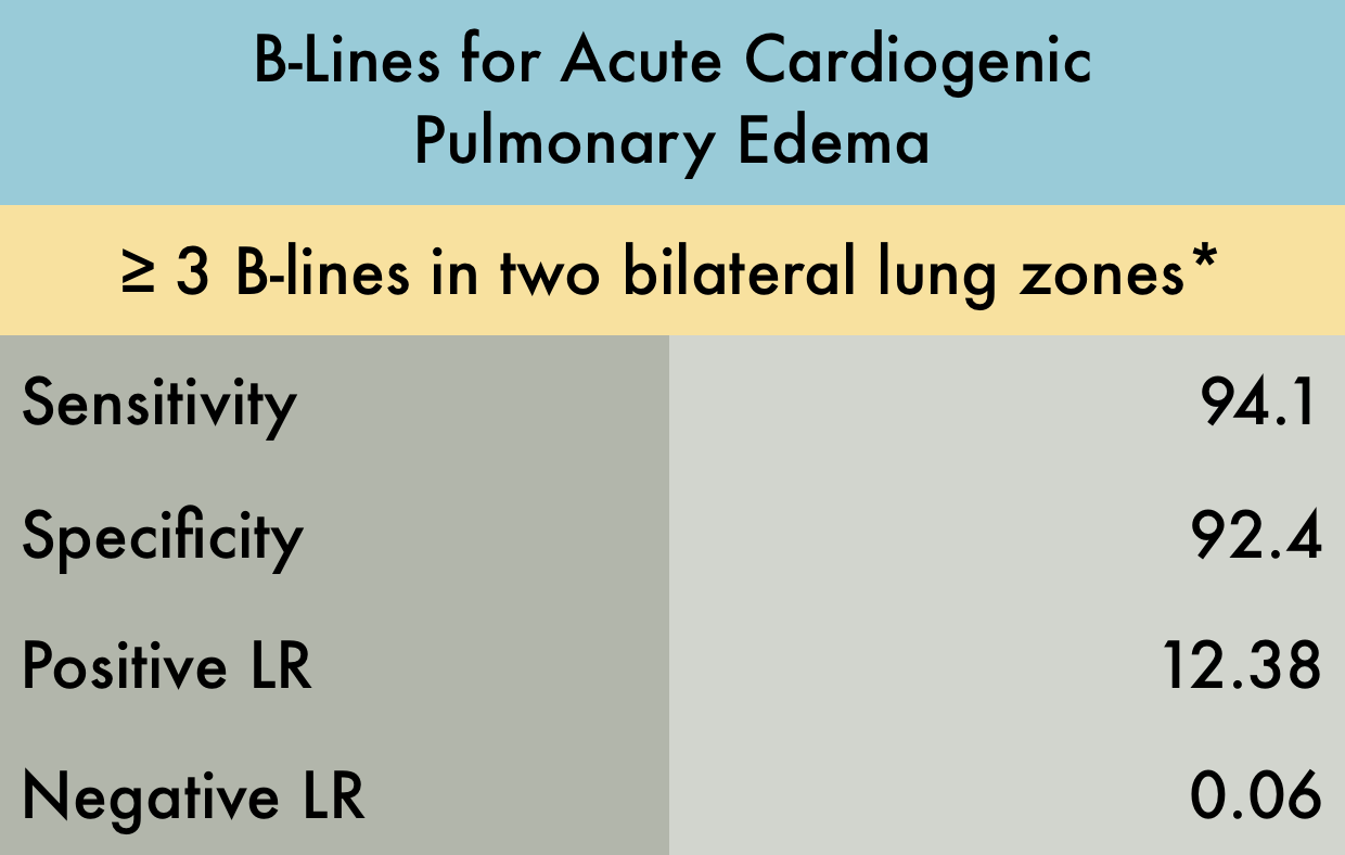
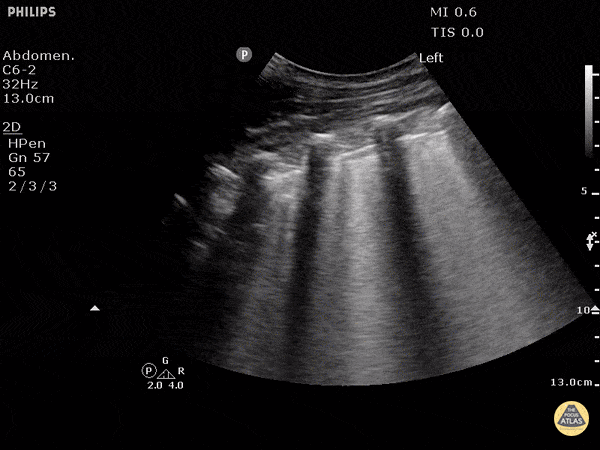
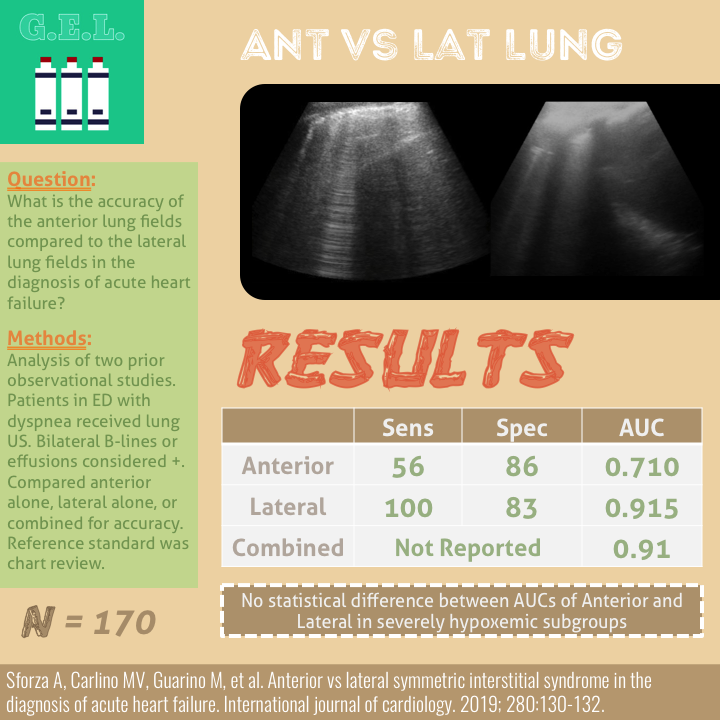
Pulmonary Edema
Featured
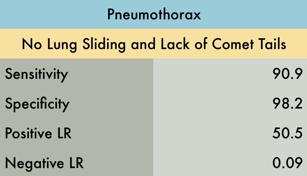
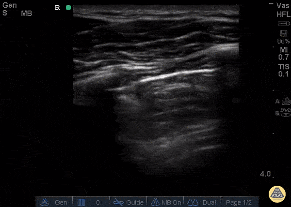
Pneumothorax
Featured
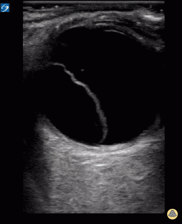
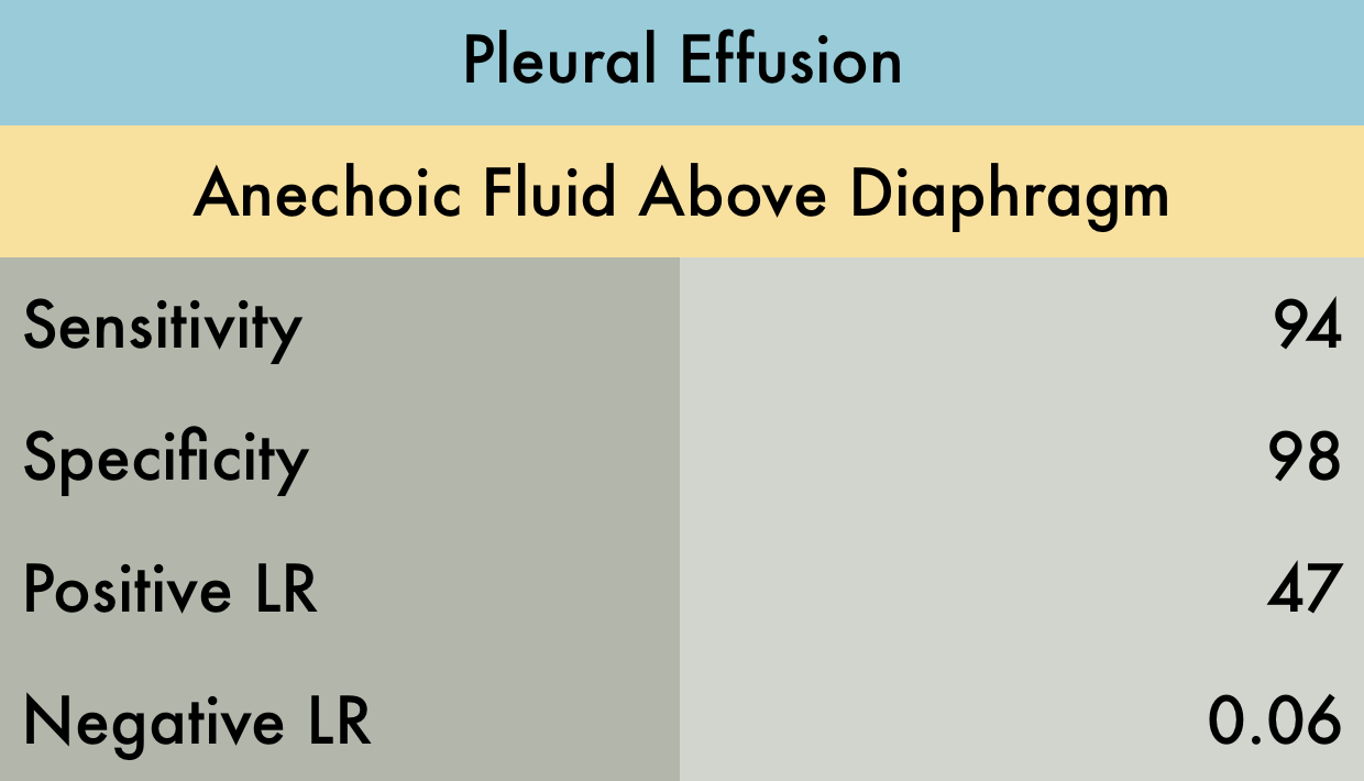
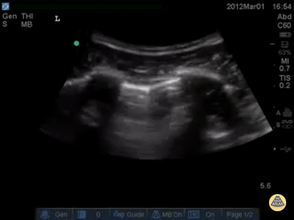
Pleural Effusion
Featured
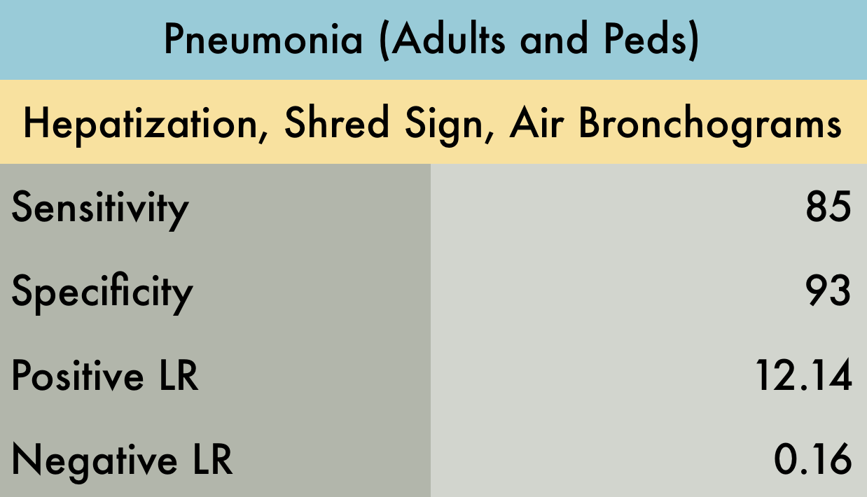
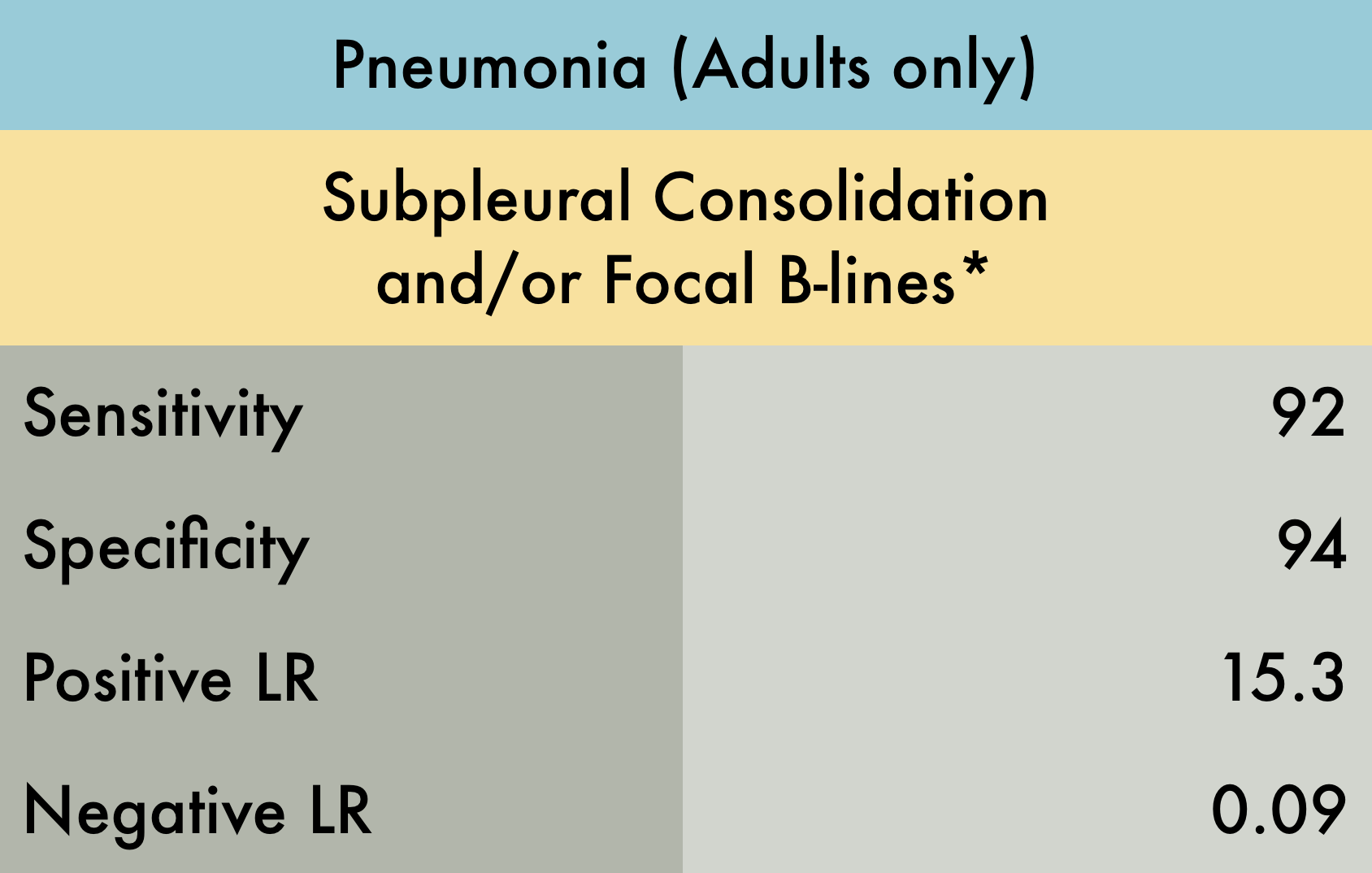
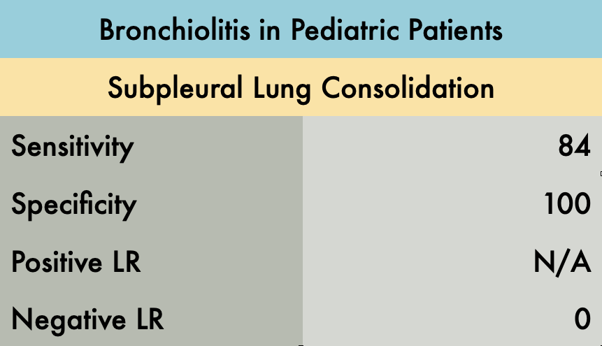
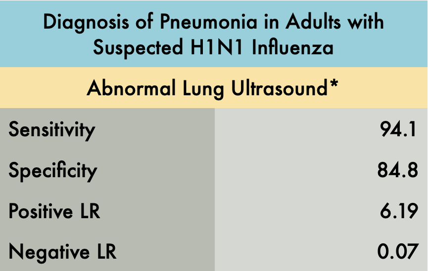
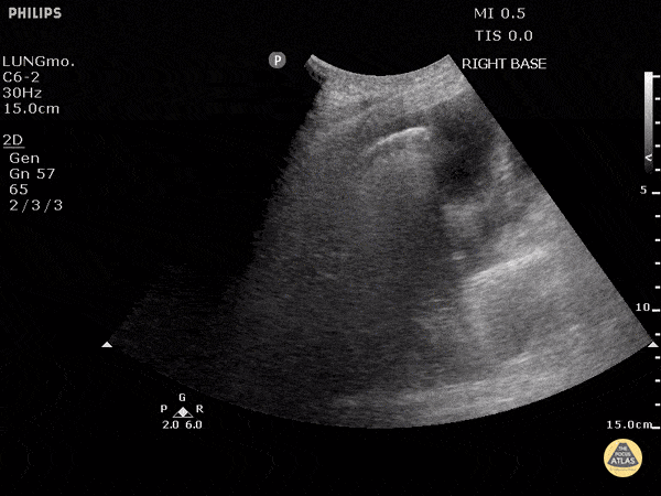
Pneumonia
Featured
For more evidence check out our friends at US G.E.L.
Perfect your Pulmonary POCUS Technique with 5 Minute Sono