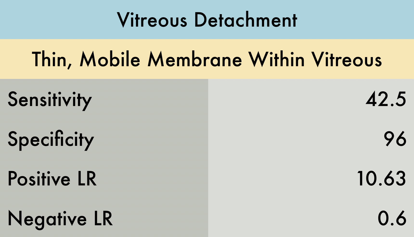
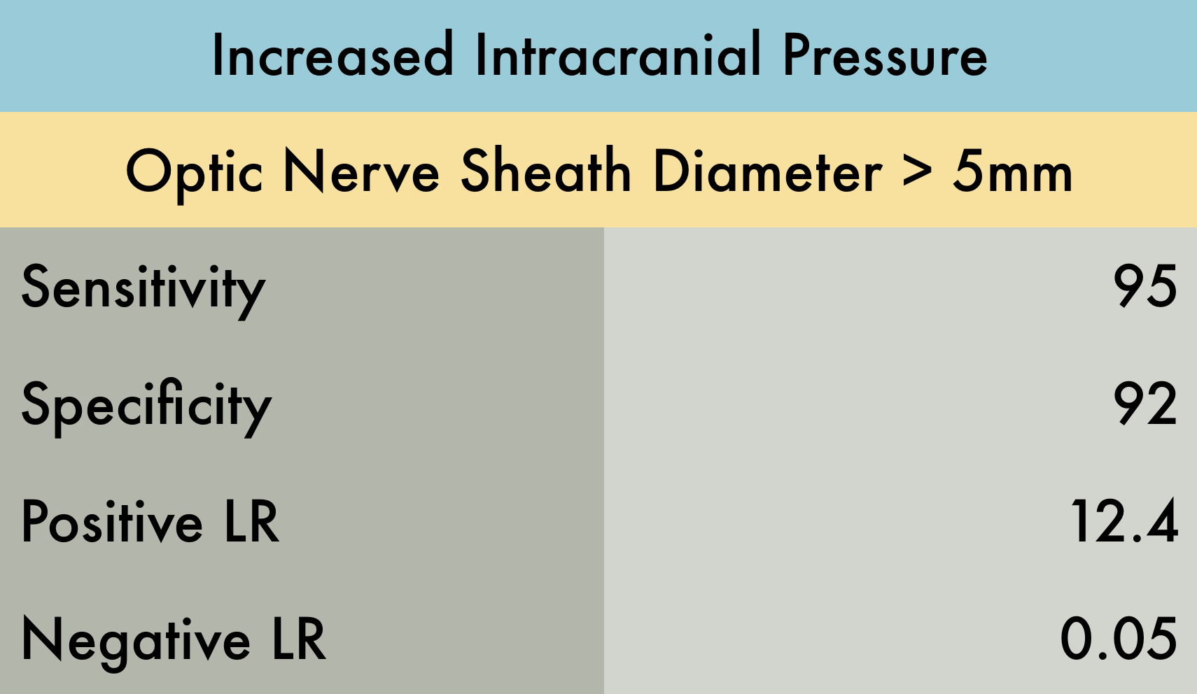
The Evidence Atlas: Orbital
Featured

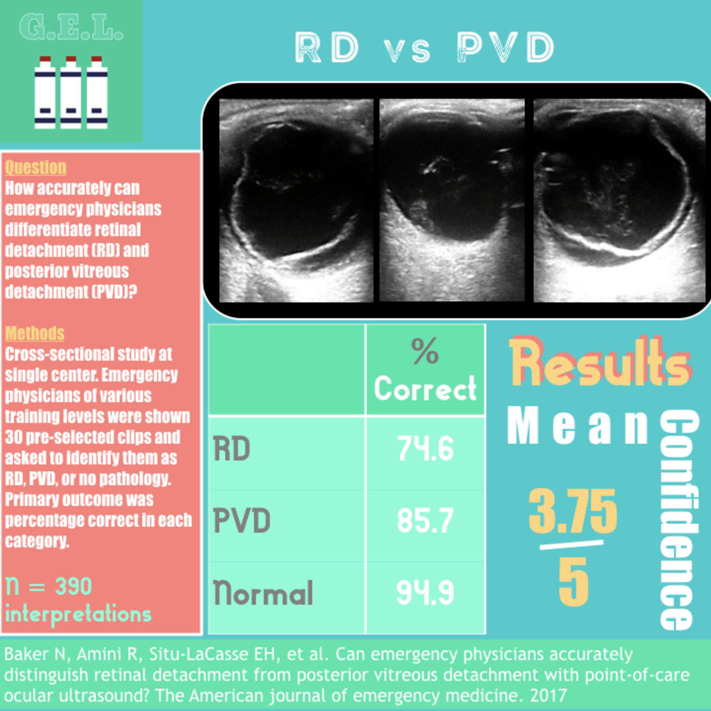
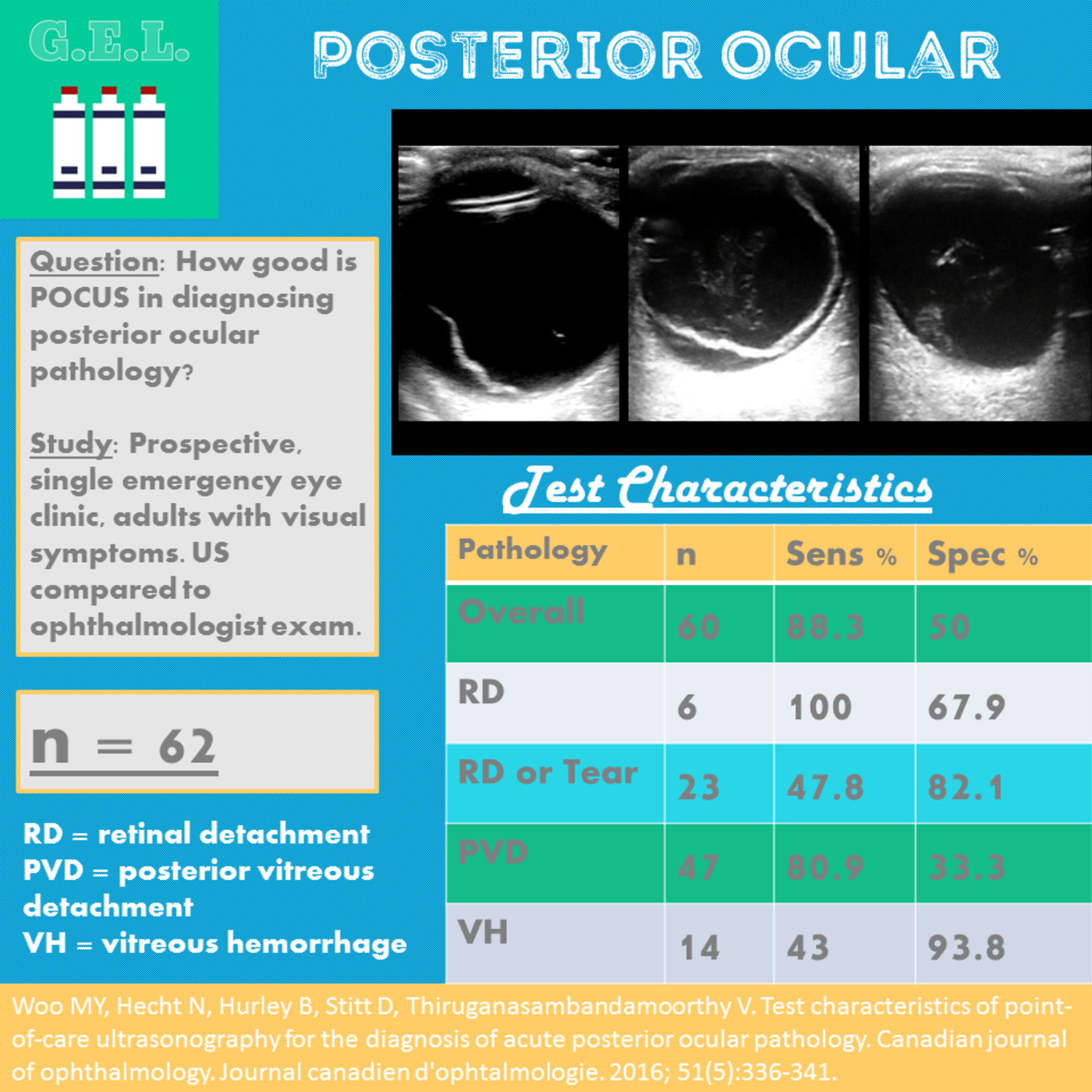
For more evidence check out our friends at US G.E.L.
Perfect your Orbit POCUS Technique with 5 Minute Sono